INTRODUCTION
Chronic inflammation, measured with biomarkers such as interleukin-6 (IL-6), neutrophils, and C-reactive protein (CRP), has been the focus of numerous studies in chronic kidney disease (CKD). These in-flammatory markers have been found to be predictors of all-cause and
cardiovas-cular mortality independent of traditional risk factors for cardiovascular disease (CVD) and are thus associated with ather-osclerotic cardiovascular disease (1–4). Age-controlled CVD mortality rates are about 10 to 20 times higher in dialysis pa-tients than those in the general population (5). It has been suggested that
inflamma-tory mediators might be causal in the ac-celerated atherosclerosing process ob-served in this patient population (5,6).
Macrophage migration inhibitory fac-tor (MIF), a cytokine discovered in 1966, is recognized as a pleiotropic proinflam-matory molecule (7,8). The actions of MIF in inflammation are numerous, in-cluding in monocytes and macrophages, T and B lymphocytes, endothelial cells, and eosinophilic neutrophils. MIF has been identified as a proinflammatory cy-tokine in sepsis as well as in autoim-mune diseases such as rheumatoid arthritis, and systemic lupus erythemato-sus (9). MIF has been implicated in atherogenesis and atheroma formation as
(MIF) Concentrations in Chronic Kidney Disease (CKD) Are
Associated with Markers of Oxidative Stress and Endothelial
Activation
Annette Bruchfeld,
1Juan J Carrero,
2Abdul R Qureshi,
2Bengt Lindholm,
2Peter Barany,
1Olof Heimburger,
1Maowen Hu,
3Xinchun Lin,
3Peter Stenvinkel,
1and Edmund J Miller
3,41Division of Renal Medicine, Karolinska University Hospital, CLINTEC, Karolinska Institute, Stockholm, Sweden; 2Baxter Novum,
CLINTEC, Karolinska Institute, Stockholm, Sweden; and 3Feinstein Institute for Medical Research, Manhasset, New York, United States of America; and 4Albert Einstein College of Medicine, Bronx, New York, United States of America
Chronic kidney disease (CKD) carries an increased risk of cardiovascular disease (CVD). Macrophage migration inhibiting factor (MIF) is a proinflammatory cytokine implicated in the pathogenesis of sepsis, autoimmune disease, atherogenesis, and plaque instability, and is a known cardiac depressant. This post-hoc, cross-sectional study examined whether MIF serum con-centrations are elevated in CKD patients. Our study included CKD 3–5 patients with moderate to severe renal dysfunction (n = 257) (mean age SD; 55 ±12 years) and 53 controls (60 ±12 years). Serum MIF concentrations, measured by enzyme-linked im-munosorbent assay (ELISA), were studied in relation to glomerular filtration rate (GFR), presence of CVD, outcome and inflam-matory and oxidative stress markers. MIF was significantly elevated in CKD patients compared with controls (CKD: median 676 [range 118–8275 pg/mL] controls: 433 [142–4707] pg/mL; P = 0.008). MIF was also associated with 8-hydroxy-2-deoxyguanosine (8-OH-dG) levels (rho = 0.26; P = 0.001), a marker of oxidative stress, and ICAM-1 levels (rho = 0.14; P = 0.02), a marker of en-dothelial activation. However, the elevated MIF concentrations were neither correlated with glomerular filtration rate (GFR) nor inflammatory markers such as CRP, IL-6, and TNF. When combining MIF and IL-6 as a marker of inflammation, a significant in-crease in risk for CVD was found, but when analyzing all-cause mortality, this did not differ significantly with regard to mortality from inflamed patients with low MIF levels. The data suggest that increased serum MIF levels found in CKD is not caused pri-marily by poor renal function, but is associated with markers of oxidative stress and endothelial activation and may play a role in vascular disease associated with CKD.
© 2009 The Feinstein Institute for Medical Research, www.feinsteininstitute.org Online address: http://www.molmed.org
doi: 10.2119/molmed.2008.00109
Address correspondence and reprint requests toAnnette Bruchfeld, Division of Renal
Medicine, K56, Karolinska University Hospital at Huddinge, Department of Clinical Science, Interventions and Technology (CLINTEC), Karolinska Institutet, 141 86 Stockholm, Sweden. Phone: 0046-8-58582685; Fax:0046-8-7114742; E-mail: [email protected].
well as instability of human artherioscle-rotic plaques (10,11). In recent sponta-neous atherosclerosis animal models, aortic inflammation was reduced and neointimal plaques were stabilized after administration of anti-MIF antibody (12,13). The concept of anti-MIF therapy in artheriosclerotic and inflammatory disease in CKD is therefore of consider-able interest. The availconsider-able literature on the role of MIF in renal patients is, how-ever, quite limited. One study reported increased circulating MIF levels in vas-culitis patients and another study exam-ined MIF in the urine of transplanted pa-tients (14,15). However, these studies were small and did not control for the in-fluence of renal function. The aim of this current study was, therefore, to deter-mine whether circulating MIF levels were elevated in CKD 3–5 patients with a wide range of GFR.
MATERIALS AND METHODS
A total of 257 CKD patients were in-vestigated as part of an ongoing prospec-tive study in part described previously (16). The CKD 3–5 group (n = 257) con-sisted of 190 CKD 5 patients prior to the start of dialysis therapy (GFR <15 mL/ min) and 67 CKD 3–4 patients (GFR 15–60 mL/min) and these were com-pared with 53 controls. These controls were recruited from a population-based group in the Stockholm region and were used for comparative analyses of bio-chemical and metabolic parameters. The control group was comprised of individ-uals who agreed to participate, as volun-teers, in response to an invitation sent to 1,000 randomly selected individuals in the Stockholm region by Statistics Swe-den. No additional exclusion criteria other than unwillingness to participate in the study were applied in the selection of the control group. The CKD exclusion criteria were clinical signs of acute infec-tion, acute vasculitis, or liver disease at the time of evaluation, or unwillingness to participate in the study.
The causes of kidney failure in the CKD 3–5 group were diabetes mellitus, n = 76 (30%); chronic glomerulonephritis,
n = 59 (23%); polycystic kidney disease, n = 26 (10%); nephrosclerosis, n = 27 (10%); vasculitis, n = 4 (2%); and 65 pa-tients (25%) could not be classified.
After an overnight fast, venous blood samples were drawn and the sera stored at –70°C for future biochemical analyses. High-sensitivity CRP (hs-CRP), serum al-bumin, HbA1c, and hemoglobin (Hb)
were analyzed subsequently. The hs-CRP was analyzed using an immunonephelo-metric procedure (Behring AG, Marburg, Germany), whereas the remaining bio-chemical analyses were carried out using routine methods at the Department of Clinical Chemistry at Karolinska Univer-sity Hospital at Huddinge. GFR was esti-mated by the mean of creatinine and urea clearances from a 24 h collection of urine, or iohexol-clearance. Routine laboratory tests including CRP were analyzed by standard methods. TNF and IL-6 were analyzed in serum by immunometric as-says on an Immulite Analyzer (DPC, Los Angeles, CA, USA) according to the in-structions of the manufacturers. Measure-ments of soluble vascular cell adhesion molecule-1 (sVCAM-1) and intracellular vascular cell adhesion molecule (sICAM-1) were performed using commercial kits (R&D Systems Europe Ltd, Abington, UK); 8-OH-dG concentration was deter-mined by a competitive ELISA kit (Japa-nese Institute for the Control of Aging, Fukuroi, Shizuoka, Japan). MIF concen-trations were measured by commercial ELISA (R&D Systems, MN, USA) and studied in relation to inflammatory and oxidative stress markers as well as GFR.
STATISTICAL ANALYSIS
Results are expressed as mean and standard deviation (SD; normally dis-tributed variables) or median and range (non-normal distribution) unless other-wise indicated, with P<0.05 indicating significance. Comparisons between groups for nominal variables were made using the chi-square test. Spearman’s rank correlation test was used for con-tinuous and ordinal variables. Determi-nants of CVD and odd ratios (ORs) with 95% confidence intervals (CI) were
stud-ied with a multinomial logistic regres-sion model. Kaplan–Meier survival anal-ysis was performed and adjusted rela-tive risk of all-cause mortality was calculated from a Cox proportional haz-ards model. The statistical analysis was performed using statistical software SAS version 9.1.4 (SAS Institute Inc., Cary, NC, USA).
The study protocol was approved by the Ethics Committee of Karolinska Uni-versity Hospital Huddinge, Stockholm, Sweden, and informed consent was ob-tained from each subject.
RESULTS
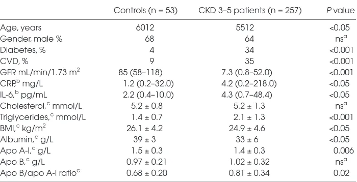
MIF Plasma Levels in CKD Patients General characteristics of the patients versus controls are summarized in Table 1. Median (and range) serum MIF levels in CKD 3–5 patients were significantly higher than in the controls (676 [118–8275] versus 433 [414–4707] pg/mL, respec-tively) shown in Figure 1A. Median MIF levels did not differ significantly between CKD 3–4 and CKD 5 patients. The sensi-tivity of the assay was 31.25 pg/mL.
There were no differences in MIF lev-els among patients with or without clini-cal cardiovascular disease, diabetes mel-litus, gender, or protein-energy wasting as assessed by SGA (Subjective Global Assessment)—data not shown.
Univariate and Multivariate Correlates for MIF Levels
MIF levels showed no significant cor-relation with GFR, CRP, IL-6, or TNF and primary kidney disease. When ana-lyzing the largest subgroup of diabetic patients separately, we found no further association between MIF and clinical and laboratory parameters including BMI and HbA1c. However, there were
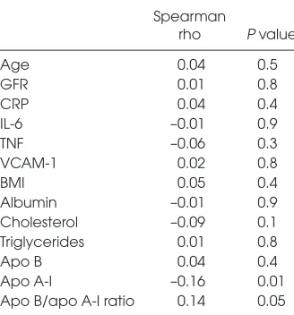
atherogenic lipoprotein particles, and apolipoprotein A-I (apo A-I), which re-flects the corresponding concentration of the anti-atherogenic HDL also were investigated. Apo A-I was correlated negatively with MIF; apo B/apo A-I ratio was correlated positively with MIF, although apo B alone was not cor-related significantly (Table 2).
We tested the hypothesis that there might be a different risk pattern for MIF in the presence or absence of inflamma-tion. In this study, inflammation was de-fined according to the median levels of IL-6 (4.3 pg/mL). In this context, the con-tribution of low or high MIF (defined by the median of 676 pg/mL) was studied to predict CVD, which was recorded from patient charts and included coro-nary artery disease, such as myocardial infarction and angina pectoris, stroke, and significant peripheral vascular dis-ease. IL-6 was used as an inflammatory marker because IL-6 is possibly the most reliable predictor of cardiovascular dis-ease and mortality in patients with end stage renal disease (4). Thus, in a logistic regression model, which included factors known to affect CVD such as gender, di-abetes, and age (Table 3), the group with inflammation and high MIF was the only one that significantly predicted CVD whereas the absence of inflammation
and high MIF was associated negatively with CVD.
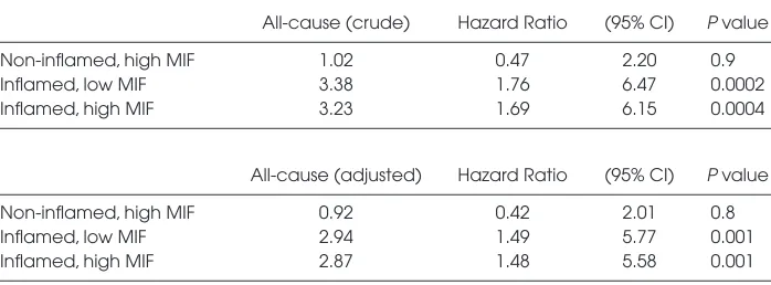
Finally, we assessed all-cause mortality risk of median 35 (range 1–72) months. In the presence of inflammation, groups with high or low MIF exhibited a similar probability of dying, both crude and after adjustment for age, sex, CVD, and diabetes mellitus (DM) (Table 4).
DISCUSSION
The main finding of the current study indicated serum MIF concentrations were elevated significantly in CKD 3–5 com-pared with controls. MIF levels were also associated positively with 8-hydroxy-2-deoxyguanosine (8-OH-dG) levels and ICAM-1 levels. However, we observed no correlation between MIF and GFR or inflammatory markers such as CRP, IL-6, or TNF.
CKD is characterized as a state of chronic inflammation and accelerated atherosclerosis with important repercus-sions for morbidity and mortality inde-pendent of traditional risk factors (1–4). MIF, a proinflammatory cytokine of the innate immune system, has been impli-cated in atherogenesis and atheroma for-mation as well as plaque instability in human artheriosclerotic disease (8–14). To the best of our knowledge this is the first study that addresses circulating MIF
and the role of renal function, and possi-ble associations to CVD in CKD.
Since CKD is associated with inflam-mation, it was somewhat surprising that MIF in this population was not associ-ated with common inflammatory mark-ers. This differs from previous CKD studies that have identified GFR as an important predictor of elevated circulat-ing pro-inflammatory cytokines such as IL-10, IL-6, TNF, and HMGB-1 (17–19). We found this to be untrue for MIF, sug-gesting the primary cause of increased serum MIF may not be due to poor renal function in this population.
The association between MIF and 8-OH-dG levels as a marker of oxidative stress, and ICAM-1 levels as a marker of endothelial activation raises the question of a possible role for MIF in vascular dis-ease and associated myocardial damage in CKD. However, in the stepwise regres-sion analysis, elevated levels of MIF alone did not predict the risk of CVD, al-though increased MIF in the presence of elevated concentrations of IL-6 was linked to an increased likelihood of CVD.
No previous study has examined cir-culating MIF in CKD with a wide range of GFR. A recent paper by Zaza et al. studied specific genomic patterns associ-ated with systemic microinflammation in dialysis patients. By microarray analysis the gene for MIF was upregulated and directly associated with CRP levels (20). This was a genomic association and no further data with regard to circulating levels or cardiovascular events were available. In the current study, some weak but significant associations with MIF were also found in the lipoprotein variables apo A-I and the ratio apo B/ apo A-1, which may further indicate vas-cular involvement (21). Data from the global INTERHEART study suggests that apo B/apo A-1 is superior to any of the cholesterol ratios for estimation of the risk of cardiovascular events in the gen-eral population (22). Recently, a clinical study of anti-TNF therapy in rheumatoid arthritis (RA) suggested a link between clinical improvement, CRP, and MIF re-duction, and an improved apo B/apo A-1
Table 1.General characteristics.
Controls (n = 53) CKD 3–5 patients (n = 257) P value Age, years 6012 5512 <0.05 Gender, male % 68 64 nsa
Diabetes, % 4 34 <0.001
CVD, % 9 35 <0.001
GFR mL/min/1.73 m2 85 (58–118) 7.3 (0.8–52.0)
<0.001 CRP,bmg/L 1.2 (0.2–32.0) 4.2 (0.2–218.0) <0.05
IL-6,bpg/mL 2.2 (0.4–10.0) 4.3 (0.7–48.4)
<0.05 Cholesterol,cmmol/L 5.2 ±0.8 5.2 ±1.3 nsa
Triglycerides,cmmol/L 1.4
±0.7 2.1 ±1.3 <0.001 BMI,ckg/m2 26.1 ±4.2 24.9 ±4.6 <0.05
Albumin,cg/L 39
±3 33 ±6 <0.05
Apo A-I,cg/L 1.5 ±0.3 1.4 ±0.3 0.006
Apo B,cg/L 0.97
±0.21 1.02 ±0.32 nsa
Apo B/apo A-I ratioc 0.68 ±0.20 0.81 ±0.34 0.02 aNot significant.
ratio (23). This potential cardiovascular benefit in RA, which is recognized as an inflammatory disease with accelerated atherogenesis, could be of interest in other conditions with a similar cardio-vascular risk profile such as CKD and lupus. In the current study, the combina-tion of elevated IL-6 and MIF was signif-icantly associated with an increased risk of CVD. A reasonable speculation is that the systemic chronic inflammation found in CKD may amplify atherosclero-sis in the presence of atherogenic and in-flammatory factors, such as MIF, elicit-ing oxidative stress and endothelial activation. MIF, on the other hand, is known to induce proinflammatory me-diators in both acute and chronic inflam-matory conditions (8,9). Understanding of shared inflammatory mechanisms may lead to earlier detection of cardio-vascular risk and conceivably preven-tion of complicapreven-tions.
The current study has several limita-tions. The number of patients and con-trols was limited, and MIF values were measured post hocby single determina-tions. Moreover, as atherosclerosis is ac-celerated in CKD, serial measurements of MIF, over time, paired with clinical data and dialysis vintage would, perhaps, have been more useful to evaluate any predictive value in this patient popula-tion. Data on therapy were not included,
Table 2.Spearman rank correlations for MIF
in CKD 3–5 patients.
Spearman
rho P value Age 0.04 0.5 GFR 0.01 0.8 CRP 0.04 0.4 IL-6 –0.01 0.9 TNF –0.06 0.3 VCAM-1 0.02 0.8 BMI 0.05 0.4 Albumin –0.01 0.9 Cholesterol –0.09 0.1 Triglycerides 0.01 0.8 Apo B 0.04 0.4 Apo A-I –0.16 0.01 Apo B/apo A-I ratio 0.14 0.05
Figure 1.(A) MIF is elevated in CKD 3–5 patients as compared with healthy individuals.
which is another weakness of the study as medication might influence MIF val-ues. Information regarding MIF gene polymorphism is not yet available in CKD. However, in one study of RA pa-tients, a correlation between disease severity, genetic functional variants, and circulating levels of MIF has been shown (24). Recent data also suggest that CD74, the MIF-receptor, is upregulated by high concentrations of glucose and TNF-αin glomerular podocytes, and may play a role in the pathogenesis of diabetic nephropathy (25). In the diabetic pa-tients, we did, however, not find any clear association between MIF and clini-cal and laboratory parameters.
We could not find an effect on survival stratified by MIF levels alone. Again, the patient sample was possibly too small, but as CVD, which dominates mortality in CKD, is multifactorial, other risk fac-tors may be stronger predicfac-tors of out-come in this population. Moreover, pa-tient diagnosis relies on papa-tient charts. As clinical CVD is not always recognized and confirmed by appropriate clinical in-vestigations, we may have underesti-mated the true prevalence of CVD in our patient population (26). Finally, even though high MIF combined with ele-vated IL-6 was a significant predictor of CVD in a logistic regression model, ele-vated IL-6 with low MIF levels predicted
all-cause mortality equally, which raises the question of disparities regarding the influence of MIF in this setting.
In conclusion, although circulating serum levels of MIF are elevated signifi-cantly in CKD patients compared with controls, no association with GFR or common inflammatory markers was found. Instead, our data suggest an asso-ciation with markers of oxidative stress and endothelial activation with possible implications for a role in vascular processes in this population. Undoubt-edly, further studies are needed to shed light on the involvement of MIF as a pos-sible active regulator in CVD associated with CKD.
ACKNOWLEDGMENTS
This study was supported by Karolinska Institutet research funds, MEC (EX2006-1670), the Swedish Heart and Lung Foun-dation, the Swedish Medical Research Council, and research grants from the ERA-EDTA.
DISCLOSURE
We declare that the authors have no competing interests as defined by Molec-ular Medicine, or other interests that might be perceived to influence the re-sults and discussion reported in this paper.
REFERENCES
1. Stenvinkel P, et al. (2005) IL-10, IL-6, and
TNF-alpha: central factors in the altered cytokine net-work of uremia—the good, the bad, and the ugly.
Kidney Int. 67:1216–33.
2. Avesani CM, et al. (2006) Inflammation and
wast-ing in chronic kidney disease: partners in crime.
Kidney Int. Suppl. 70:S8–S13.
3. Panichi V, et al. (2004) Interleukin-6 is a stronger
predictor of total and cardiovascular mortality than C-reactive protein in haemodialysis pa-tients. Nephrol. Dial. Transplant. 19:1154–60.
4. Honda H, et al. (2006) Serum albumin, C-reactive
protein, interleukin 6, and fetuin a as predictors of malnutrition, cardiovascular disease, and
mor-tality in patients with ESRD. Am. J. Kidney Dis.
47:139–48.
5. Foley RN, Parfrey PS, Sarnak MJ. (1998) Clinical epidemiology of cardiovascular disease in chronic renal disease. Am. J. Kidney Dis. 32(5 Suppl 3): S112–9.
6. Go AS, Chertow GM, Fan D, McCulloch CE, Hsu CY. (2004) Chronic kidney disease and the risks
Table 3.Significant predictors of CVD in a multinomial logistic regression CKD 3–5 patients.
95% CI
Parameter Odds ratio Lower Upper P value Intercept of CVD <0.0001 Diabetes mellitus, presence 2.35 1.26 4.39 0.007 Sex, men 2.21 1.16 4.22 0.01 Age, 50–65 years 4.61 1.97 10.79 0.0004 Age,>65 years 10.60 4.41 25.48 <0.0001 Non inflamed and high MIF 0.83 0.33 2.06 0.7 Inflamed and low MIF 2.11 0.89 4.99 0.09 Inflamed and high MIF 2.39 1.02 5.63 0.04 The model included CVD as the dependent variable and all factors significantly associated with the dependent variable in univariate analysis. Inflamed patients were defined as those with IL-6 >4.3 pg/mL (median), and high MIF was defined as those with
>676 pg/mL (median); Pseudo r2= 0.22; P<0.001; ages 20–49. Non-inflamed patients with
low MIF were considered reference groups.
Table 4.Crude and adjusted relative risk of all-cause mortality in CKD3-5 patients.
All-cause (crude) Hazard Ratio (95% CI) P value Non-inflamed, high MIF 1.02 0.47 2.20 0.9 Inflamed, low MIF 3.38 1.76 6.47 0.0002 Inflamed, high MIF 3.23 1.69 6.15 0.0004
of death, cardiovascular events, and hospitaliza-tion. N. Engl. J. Med. 351:1296–305.
7. David JR. (1966) Delayed hypersensitivity in
vitro: its mediation by cell-free substances formed
by lymphoid cell-antigen interaction. Proc. Natl.
Acad. Sci. U. S. A. 56:72–7.
8. Zernecke A, Bernhagen J, Weber C. (2008) Macrophage migration inhibitory factor in
car-diovascular disease. Circulation. 117:1594–602.
9. Morand EF. (2005) New therapeutic target in flammatory disease: macrophage migration in-hibitory factor. Intern. Med. J. 35:419–26.
10. Alexander Schmeisser, et al. (2005) The
expres-sion of macrophage migration inhibitory factor
1α(MIF 1α) in human atherosclerotic plaques is
induced by different proatherogenic stimuli and
associated with plaque instability. Atherosclerosis
178:83–94.
11. Yao-Zhong Kong, et al. (2005) Macrophage
migra-tion inhibitory factor induces MMP-9 expression: implications for destabilization of human
athero-sclerotic plaques. Atherosclerosis178:207–15.
12. Anke Burger-Kentischer, et al. (2006) Reduction
of the aortic inflammatory response in sponta-neous atherosclerosis by blockade of macrophage
migration inhibitory factor (MIF). Atherosclerosis
184:28–38.
13. Schober A, et al. (2004) Stabilization of
atheroscle-rotic plaques by blockade of macrophage migra-tion inhibitory factor after vascular injury in
apolipoprotein E-deficient mice. Circulation
109:380–5.
14. Becker H, et al. (2006) Relationship between serum
levels of macrophage migration inhibitory factor and the activity of antineutrophil cytoplasmic
an-tibody-associated vasculitides. Clin. Rheumatol.
4:1–5.
15. Brown FG, et al. (2001) Urine macrophage
migra-tion inhibitory factor concentramigra-tions as a diag-nostic tool in human renal allograft rejection.
Transplantation. 71:1777–83.
16. Stenvinkel P, et al. (1999) Strong association
be-tween malnutrition, inflammation, and
athero-sclerosis in chronic renal failure. Kidney Int.
55:1899–911.
17. Descamps-Latscha B, et al. (1995) Balance between
IL-1 beta, TNF-alpha, and their specific inhibitors in chronic renal failure and maintenance dialysis. Relationships with activation markers of T cells,
B cells, and monocytes. J. Immunol. 154:882–92.
18. Pecoits-Filho R, et al. (2003) Associations between
circulating inflammatory markers and residual
renal function in CRF patients. Am. J. Kidney Dis.
41:1212–8.
19. Bruchfeld A, et al. (2008) High Mobility Group
Box Protein-1 correlates with renal function in
Chronic Kidney Disease (CKD). Mol. Med.
14:109–15.
20. Zaza G, et al. (2008) Dialysis-related systemic
mi-croinflammation is associated with specific genomic patterns. Nephrol. Dial. Transplant. 23:1673–81. 21 Thompson A, Danesh J. (2006) Associations
be-tween apolipoprotein B, apolipoprotein AI, the apolipoprotein B/AI ratio and coronary heart
disease: a literature-based meta-analysis of pro-spective studies. J. Intern. Med. 259:481–92.
22. McQueen MJ, et al. (2008) INTERHEART study
investigators. Lipids, lipoproteins, and apolipo-proteins as risk markers of myocardial infarction in 52 countries (the INTERHEART study): a
case-control study. Lancet372:224–33.
23. Wijbrandts CA, et al. (2008). Sustained changes in
lipid profile and macrophage migration in-hibitory factor (MIF) levels after anti-TNF
ther-apy in rheumatoid arthritis. Ann. Rheum. Dis.
2008, Aug 22. [Epub ahead of print]
24. Radstake TR, et al. (2005) Correlation of
rheuma-toid arthritis severity with the genetic functional variants and circulating levels of macrophage
migration inhibitory factor. Arthritis Rheum.
52:3020–9.
25. Sanchez-Niño MD,et al. (2008) The MIF receptor
CD74 in diabetic podocyte injury. J. Am. Soc.
Nephrol. 2008 Oct 8. [Epub ahead of print] 26. Stenvinkel P, Pecoits-Filho R, Lindholm B. (2003)
Coronary artery disease in end-stage renal
dis-ease: no longer a simple plumbing problem. J. Am.