P R I M A R Y R E S E A R C H
Open Access
Transcriptome sequencing of gingival
biopsies from chronic periodontitis patients
reveals novel gene expression and splicing
patterns
Yong-Gun Kim
1,2†, Minjung Kim
3†, Ji Hyun Kang
4, Hyo Jeong Kim
4, Jin-Woo Park
1, Jae-Mok Lee
1, Jo-Young Suh
1,
Jae-Young Kim
2,4, Jae-Hyung Lee
3,5*and Youngkyun Lee
2,4*Abstract
Background:Periodontitis is the most common chronic inflammatory disease caused by complex interaction between the microbial biofilm and host immune responses. In the present study, high-throughput RNA sequencing was utilized to systemically and precisely identify gene expression profiles and alternative splicing.
Methods:The pooled RNAs of 10 gingival tissues from both healthy and periodontitis patients were analyzed by deep sequencing followed by computational annotation and quantification of mRNA structures.
Results:The differential expression analysis designated 400 up-regulated genes in periodontitis tissues especially in the pathways of defense/immunity protein, receptor, protease, and signaling molecules. The top 10 most up-regulated genes wereCSF3,MAFA, CR2,GLDC,SAA1,LBP,MME,MMP3,MME-AS1, andSAA4. The 62 down-regulated genes in periodontitis were mainly cytoskeletal and structural proteins. The top 10 most down-regulated genes wereSERPINA12,MT4,H19,KRT2,DSC1,PSORS1C2,KRT27,LCE3C,AQ5, andLCE6A. The differential alternative splicing analysis revealed unique transcription variants in periodontitis tissues. The EDB exon was predominantly included in
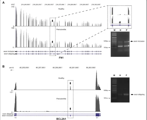
FN1, while exon 2 was mostly skipped inBCL2A1.
Conclusions:These findings using RNA sequencing provide novel insights into the pathogenesis mechanism of periodontitis in terms of gene expression and alternative splicing.
Keywords:Periodontitis, Transcriptome sequencing, Alternative splicing, Gene expression profile
Introduction
Periodontitis is a chronic inflammatory disease of peri-odontium, characterized by massive destruction of both soft and hard tissues surrounding the teeth [1]. The current concept for the periodontal diseases involve com-plex interaction between the microbial biofilm and host immune responses that leads to the alteration of bone and connective tissue homeostasis [2, 3]. Understanding the
molecular mechanisms underlying the pathogenesis as well as development of efficient therapeutics is further-more important since periodontitis is linked to other metabolic and/or systemic diseases including diabetes, cardiovascular diseases, and rheumatoid arthritis [4–6].
The analysis of transcriptome by microarrays has been a valuable tool to study the changes in gene expression pro-files in gingival tissues of periodontitis patients [7–9]. How-ever, recent advances in the high-throughput RNA sequencing technology revolutionarily enhanced our under-standing on the complexity of eukaryotic transcriptome [10, 11]. RNA sequencing has several key advantages over the hybridization-based microarray techniques. First of all, direct sequencing enables an unbiased approach compared * Correspondence:jaehlee@khu.ac.kr;ylee@knu.ac.kr
†Equal contributors
3
Department of Life and Nanopharmaceutical Sciences, Kyung Hee University, Seoul 02447, Korea
2Institute for Hard Tissue and Bone Regeneration, Kyungpook National
University, Daegu 41940, Korea
Full list of author information is available at the end of the article
with the microarrays that depends on the predetermined genome sequences. Secondly, RNA sequencing is highly accurate in detecting gene expression with very wide dy-namic detection ranges with low background. Thus, RNA sequencing is not only useful to precisely determine gene expression profiles but also particularly powerful to detect novel transcription variants via alternative splicing [10].
In the present study, we analyzed the pooled transcrip-tome from gingival tissues of periodontitis patients and compared with that of healthy patients. The large sum of novel information on the gene expression profiles as well as novel transcripts through alternative splicing would provide not only insights into the pathogenesis of periodontitis but also basis for the development of bio-markers and therapeutic targets.
Materials and methods
Periodontitis patient characteristics and gingival tissue samples
Gingival tissue samples were collected from chronic peri-odontitis patients or healthy individuals. On the basis of clinical and radiographic criteria, the periodontitis-affected site had a probing depth of ≥4 mm, clinical attachment level of ≥4 mm, and bleeding on probing. A total of 10 gingival samples were collected from 9 periodontal healthy patients who visited Kyungpook National University Hospital. Similarly, a total of 10 periodontitis tissue samples were obtained from 4 periodontitis patients with pocket depth of 4~6 mm and 3 severe periodontitis patients with pocket depth of 7 mm or deeper. The patient characteris-tics are given in Additional file 1: Table S1. All patients were non-smoking and did not have untreated metabolic/ systemic diseases nor associated with infection/auto-immune diseases at the time of tissue collection. The size of 3-mm2 gingival biopsies were obtained from the mar-ginal gingiva during periodontal flap surgery and immedi-ately stored in RNAlater solution (Thermo Fisher Scientific, Waltham, MA) at −70 °C after removal of blood by brief washing in phosphate-buffered saline. The study was ap-proved by the institutional review board of the Kyungpook National University Hospital with informed consent from all patients.
Isolation of RNA and RNA sequencing
Frozen tissues were disrupted in the lysis solution of mirVana RNA isolation kit (Thermo Fisher Scientific) using disposable pestle grinder system (Thermo Fisher Scientific). After RNA extraction, the same amount of total RNA isolated from each individual sample (1 μg) was pooled into 2 groups (healthy and periodontitis) and used for further analysis. The integrity of pooled total RNA was analyzed by Agilent 2100 Bioanalyzer (Agilent Technologies, Santa Clara, CA). After purification of mRNA molecules by poly-T oligo-attached magnetic
beads followed by fragmentation, the RNA of approxi-mately 300-bp size was isolated using gel electrophor-esis. The cDNA synthesis and library construction was performed using the Illumina Truseq RNA sample prep-aration kit (Illumina, San Diego, CA), following the manufacturer’s protocol. The PCR-amplified cDNA tem-plates on a flow cell was loaded and sequenced in the HiSeq 2000 sequencing system (Illumina) in the paired-end sequencing mode (2 × 101 bp reads).
Sequencing data analysis
All sequencing raw reads were aligned to the human gen-ome reference hg19 using the GSNAP alignment tool (2013-11-27) [12]. Only uniquely and properly mapped read pairs were used for further analysis. The differentially expressed genes between gingival tissues from periodontal healthy patients and periodontitis patients were identified using the DESeq R package [13]. Differentially expressed genes were defined as those with changes of at least 2-fold between samples and at a false discovery rate (FDR) cutoff of 5 % based on DESeq adjustedpvalues. The analysis of alternative splicing events was performed using MATS software [14]. The differences in the alternative splicing in genes were considered significant when the inclusion dif-ference between samples was equal or greater than 5 % at a 10 % FDR. Each alternative splicing change of the skipped exon vent was manually inspected in UCSC genome browser using the sequencing data. The functional classifica-tion analysis of differentially expressed genes was performed using the PANTHER tools (http://www.pantherdb.org). The GO term and KEGG pathway enrichment analysis was per-formed as described previously [15]. Briefly, the fraction of genes in a test set associated with each GO category was cal-culated and compared with that of control set comprised of randomly chosen genes of the same number and length of the test genes. The random sampling was repeated 100,000 times for the calculation of empirical pvalue. The signifi-cance of enriched GO terms or KEGG pathways were deter-mined by the pvalue cutoff, which was 1/total number of GO terms considered.
Validation of differentially expressed genes and alternative splicing events
From the pooled RNA samples, 1μg of RNA was reversed transcribed using the Superscript II Reverse Transcriptase (Thermo Fisher Scientific). Quantitative real-time PCR analysis was performed by the addition of 1μg of cDNA and SYBR green master mix in MicroAMP optical tubes using the AB 7500 system (Thermo Fisher Scientific). The expression of genes relative to that ofHPRT1was deter-mined by the 2–ΔΔCT
10 s at 98 °C, 30 s at 60 °C, and 1 min at 72 °C. The primers for the detection of alternative splicing were de-signed by the PrimerSeq software [17] in order that the PCR product to span the region of exon inclusion/skip-ping, enabling the differentiation of alternative splicing events by product size. The primer sequences for the real-time RT-PCR analysis of selected genes and those for the RT-PCR detection of alternative splicing events of FN1 and BCL2A1 gene were provided in the supplemental tables (Additional file 2: Table S2 and Additional file 3: Table S3).
Results
RNA sequencing results
Total RNA was extracted from 10 healthy gingival tissue samples and 10 chronic periodontitis-affected gingival tissues as described above. Then, cDNAs synthesized from the pooled RNA samples of both groups were sequenced using the Illumina HiSeq 2000 system, which generated approximately 80 million pairs of reads of 101 bp in size. When compared with the reference sequence of Genome Reference Consortium GRCh37 (hg19), more than 90 % of read pairs were uniquely mapped on the human genome (Table 1). Gene annotation using the Ensembl (re-lease 75) identified that a total of 36,814 genes have at least 1 read mapped on the exonic regions. Among these, 4800 genes were unique to the periodontitis tissue sample, while 2811 transcripts were detected only in healthy gingival sample.
Identification and classification of differentially expressed genes between periodontitis and healthy gingiva
The differential expression of genes between periodon-titis and healthy gingival samples was analyzed by DESeq package [13]. By applying the cutoff of at least twofold change in the number of reads with 5 % FDR, we found a total of 462 genes differentially expressed between the samples (Fig. 1a, volcano plot). While 400 genes were up-regulated in the periodontitis tissue sample, 62 genes were down-regulated compared with the healthy control (Additional file 4: Table S4). Previously, Davanian et al. reported the discovery of 381 genes up-regulated in the periodontitis-affected gingival tissues by RNA sequencing [18]. Notably, 182 genes among them were also found to be up-regulated in the present study (Additional file 5: Figure S1), demonstrating an overlap between the two sets of gene lists when analyzed by a hypergeometric test (p< 2.2e−16) [19].
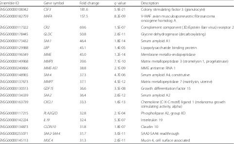
The top 20 up-regulated genes listed in Table 2 included cytokines and immune response-related genes (CSF3,
CR2, LBP, CXCL1, and IL19), serum amyloid proteins (SAA1, SAA4, and SAA2), and proteases (MME, MMP3,
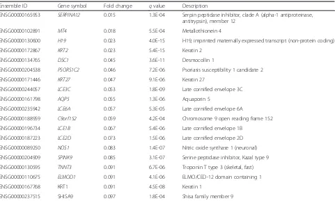
MME-AS1, and MMP7). The 20 most down-regulated genes (Table 3) included peptidase inhibitors (SERPINA12 and SPINK9) and structural proteins (KRT2, KRT27,
LCE3C,LCE6A,LCE1B,LCE2D, andKRT1).
To classify the differentially expressed genes into func-tionally related subgroups, we utilized the PANTHER classification system (http://pantherdb.org). As a result, the 462 differentially expressed genes between periodon-titis and healthy gingival tissues were segregated into 20 different classes of proteins. When we compared the com-position of these protein classes, there was a significant difference in the number of genes between periodontitis and healthy gingival samples in 6 protein classes. In the periodontitis tissue, genes classified as defense/immunity protein, receptor, protease, and signaling molecules were significantly enriched (Fig. 1b). On the other hand, genes in the categories of cytoskeletal protein and structural protein were predominant in healthy tissue sample com-pared with periodontitis. Furthermore, functional annota-tion of GO and KEGG pathway enrichment analyses as previously described [15] revealed enhanced immune responses in the periodontal tissues, including NOD-like receptor signaling, cytokine and chemokine activities, re-sponse to lipopolysaccharide, Jak-STAT signaling pathway, and B cell receptor signaling pathway (Additional file 6: Table S5 and Additional file 7: Table S6).
Validation of differentially expressed genes between periodontitis and healthy gingiva by quantitative real-time PCR analysis
To validate the differential gene expression results by RNA sequencing analysis, we selected 10 up-regulated or down-regulated genes in periodontal tissue and assessed their expression by quantitative real-time RT-PCR ana-lysis. Figure 1c shows that the examination of differential gene expression by both methods is significantly concord-ant, with the Pearson’s correlation coefficient (R) value of 0.81 (p= 0.005). Since the current study design employed pooling of samples, we further validated the variations in gene expression in individual samples of healthy and peri-odontitis patients. The real-time RT-PCR analyses for se-lected genes (Additional file 8: Figure S2) mostly repeated the RNA sequencing results, showing significant reduction in NOS1, CHP2, CDON, and MT4. Similarly, significant
Table 1Summary of RNA sequencing read mapping results
Number of total sequencing pairs Number of unique pairs Number of unmapped pairs Percentage of uniquely mapped pairs
Periodontitis tissue 87,118,086 80,778,080 6,340,006 92.72
elevation was observed in ICAM1, MMP13, LYN,
CSF3, MMP3, LBP, and CXCL2 while the expression of IL6 and IL19 only slightly increased. However, a large individual variation was observed in SERP1 and
KRT2 expression.
Alternative splicing events in periodontitis and healthy gingival tissues
More than 90 % of human genes are alternatively spliced through different types of splicing [20, 21]. To identify the differential splicing events between the healthy and
Fig. 1Differential gene expression between periodontitis-affected and healthy gingival tissues.aA volcano plot shows the differentially expressed genes.Red dotsrepresent the significantly up-regulated genes andblue dotsstand for the significantly down-regulated genes in periodontitis-affected gingival tissues. Thex-axis represents the log2-transformed gene expression in periodontitis tissues (P) divided by that in healthy gingival tissues (H).
They-axis is the adjustedpvalue (−log2) by Benjamini-Hochberg correction.bProtein functional classification in differentially expressed genes was
performed using the PANTHER tool. Thegreen arrowsindicate protein functional classes that show significantly different composition (more than 7 % composition difference) between healthy and periodontitis tissues.cThe expression of selected genes in RNA sequencing data was validated by a real-time RT-PCR analysis. Thex-axis indicates the−ΔΔCt values and they-axis represents log2(fold changes) obtained by RNA sequencing. The linear regression was
performed with Pearson’s correlation coefficient (R) and the correspondingpvalue based on the gene expression values by both methods
Table 2Top 20 up-regulated genes in periodontitis tissues
Ensemble ID Gene symbol Fold change qvalue Description
ENSG00000108342 CSF3 181.6 5.9E-21 Colony stimulating factor 3 (granulocyte)
ENSG00000182759 MAFA 157.5 8.2E-09 V-MAF avian musculoaponeurotic fibrosarcoma
oncogene homolog A
ENSG00000117322 CR2 69.6 1.5E-07 Complement component (3D/Epstein Barr virus) receptor 2
ENSG00000178445 GLDC 50.8 2.6E-11 Glycine dehydrogenase (decarboxylating)
ENSG00000173432 SAA1 46.4 1.8E-14 Serum amyloid A1
ENSG00000129988 LBP 45.1 1.4E-05 Lipopolysaccharide binding protein
ENSG00000196549 MME 45.0 1.2E-14 Membrane metallo-endopeptidase
ENSG00000149968 MMP3 39.6 7.1E-10 Matrix metallopeptidase 3 (stromelysin 1, progelatinase)
ENSG00000240666 MME-AS1 38.8 2.1E-09 MME antisense RNA 1
ENSG00000148965 SAA4 37.3 4.7E-06 Serum amyloid A4, constitutive
ENSG00000137673 MMP7 37.1 4.5E-12 Matrix metallopeptidase 7 (matrilysin, uterine)
ENSG00000130513 GDF15 36.6 3.5E-08 Growth differentiation factor 15
ENSG00000134339 SAA2 36.4 2.6E-12 Serum amyloid A2
ENSG00000163739 CXCL1 33.3 1.6E-13 Chemokine (C-X-C motif) ligand 1 (melanoma growth stimulating activity, alpha)
ENSG00000117215 PLA2G2D 32.8 2.1E-04 Phospholipase A2, group IID
ENSG00000142224 IL19 32.4 5.3E-07 Interleukin 19
ENSG00000134873 CLDN10 31.8 1.8E-07 Claudin 10
ENSG00000255071 SAA2-SAA4 31.7 3.0E-11 SAA2-SAA4 readthrough
periodontitis gingival tissues, the inclusion level of alterna-tive spliced exons was compared using the MATS tool [14] based on a statistical model that calculates the differ-ence in the isoform ratio of a gene. The MATS analysis of RNA sequencing data revealed 183 significantly differen-tial alternative splicing events in 155 genes with a cutoff of 5 % inclusion difference and 10 % FDR (Table 4 and Additional file 9: Table S7). The GO and KEGG pathway enrichment analyses for the determination of the bio-logical relevance of those differentially spliced genes showed significant difference in the pathways including RNA splicing regulation, substrate adhesion-dependent cell spreading, response to wound healing, and positive regula-tion of cell migraregula-tion (Addiregula-tional file 10: Table S8 and Additional file 11: Table S9).
Among the genes that exhibited prominently novel in-cluded exons was FN1 that encodes one of the major extracellular matrix protein fibronectin [22]. Fibronectin structure consists of 2 nearly identical ~250-kDa
glycoprotein subunits with each monomer composed of repetitive units of type I, II, and III domains [23]. The type III domains contain 2 exons called extra domain A (EDA) and extra domain B (EDB), the latter showed sig-nificantly increased inclusion in periodontitis gingival tissues compared with healthy samples (Fig. 2a; left panel). The preferential formation of EDB-containing isoform in periodontitis was further corroborated by the RT-PCR analysis designed to amplify the included EDB exon regions (Fig. 2a; right panel). The analysis of alterna-tive splicing events also indicated thatBCL2A1 (BCL2-re-lated protein A1) exhibited prominently skipped exon 2 (Fig. 2b; left panel). RT-PCR analysis designed to amplify the skipped region revealed significantly increased shorter isoform (Fig. 2b; right panel). The individual variation between healthy and periodontitis tissues for these differences in the alternative slicing events was fur-ther confirmed by RT-PCR analyses (Additional file 12: Figure S3). For FN1, the inclusion of EDB exon was
Table 3Top 20 down-regulated genes in periodontitis tissues
Ensemble ID Gene symbol Fold change qvalue Description
ENSG00000165953 SERPINA12 0.015 1.3E-04 Serpin peptidase inhibitor, clade A (alpha-1 antiproteinase, antitrypsin), member 12
ENSG00000102891 MT4 0.018 5.5E-04 Metallothionein 4
ENSG00000130600 H19 0.023 4.0E-15 H19, imprinted maternally expressed transcript (non-protein coding)
ENSG00000172867 KRT2 0.023 5.4E-15 Keratin 2
ENSG00000134765 DSC1 0.045 3.6E-11 Desmocollin 1
ENSG00000204538 PSORS1C2 0.046 7.2E-06 Psoriasis susceptibility 1 candidate 2
ENSG00000171446 KRT27 0.047 9.1E-06 Keratin 27
ENSG00000244057 LCE3C 0.053 1.8E-09 Late cornified envelope 3C
ENSG00000161798 AQP5 0.055 1.3E-06 Aquaporin 5
ENSG00000235942 LCE6A 0.057 5.3E-05 Late cornified envelope 6A
ENSG00000188959 C9orf152 0.059 4.2E-04 Chromosome 9 open reading frame 152
ENSG00000196734 LCE1B 0.067 5.4E-06 Late cornified envelope 1B
ENSG00000187223 LCE2D 0.073 1.5E-06 Late cornified envelope 2D
ENSG00000089250 NOS1 0.083 1.4E-07 Nitric oxide synthase 1 (neuronal)
ENSG00000204909 SPINK9 0.085 3.1E-07 Serine peptidase inhibitor, Kazal type 9
ENSG00000130595 TNNT3 0.091 6.7E-06 Troponin T type 3 (skeletal, fast)
ENSG00000110675 ELMOD1 0.091 4.1E-06 ELMO/CED-12 domain containing 1
ENSG00000167768 KRT1 0.091 4.5E-08 Keratin 1
ENSG00000237515 SHISA9 0.097 1.8E-04 Shisa family member 9
Table 4Summary of the differential alternative splicing event analysis
Alternative 3′ splicing sites
Alternative 5′ splicing sites
Mutual exclusive exon
Retained intron Skipped exon
Number of total alternative splicing events (genes) 3125 (2177) 2124 (1622) 4424 (2562) 2272 (1800) 32,824 (10,026)
% of total alternative splicing events (45,259) 6.9 4.7 9.8 6.1 72.5
Number of differential alternative splicing events (genes) 10 (10) 4 (4) 34 (32) 82 (77) 53 (42)
preferentially observed in periodontitis tissues (7/10) com-pared with healthy tissues (3/8) tested. Similarly, the skipping of exon 2 in BCL2A1 was predominant in periodontitis tissues (9/10), compared with healthy tissues (2/8).
Discussion
Recent developments in the RNA sequencing technology and bioinformatics tools enabled elaborate analysis of gene expression in numerous human diseases. However in periodontitis research, most RNA sequencing studies have focused on the identification of microbiome that constitutes periodontal biofilm, with little attention to the host responses against such microbial challenge. The
current study provides extensive information on gene expression as well as alternative splicing in periodontitis gingival tissues, which is crucial for the understanding the pathogenesis and development of biomarkers and therapeutic targets. The gene expression analysis re-vealed 62 down-regulated and 400 up-regulated genes in periodontitis tissues, suggesting the effectiveness of mRNA sequencing as a tool to scrutinize the differential gene ex-pression during the development of periodontitis. Davanian et al. previously reported a series of up-regulated genes as well as enriched biological pathways in periodontitis [18]. When we compared these results with ours, the current re-sults only partially overlap in terms of differential gene ex-pression, possibly originated from the difference in the
ethnic group of the subjects as well as in the methods to eliminate individual fluctuations in the gene expression. For example, Davanian et al. used healthy gingival tissue of the same periodontiaffected individual as healthy control tis-sue. However, in the current study, the healthy and peri-odontitis tissues were pooled, allowing the dilution of individual differences in the gene expression. Indeed, the RNA sequencing analysis of pooled samples proved effect-ive, since the expression levels of genes (except IL6 and
IL19) identified as differentially expressed by RNA sequen-cing were also significantly different between healthy and periodontitis samples, when we confirmed by real-time PCR analysis of individual samples (Additional file 8: Figure S2). Most of the top 20 up-regulated genes in periodontitis tissue (Table 2) were associated with inflammation and tis-sue degradation. Notably, serum amyloid A isoforms con-sisted 3 of 20 most up-regulated mRNAs, supporting the notion that these can serve as biomarkers for periodontitis-associated acute as well as chronic inflammation [24].
Until recently, gene expression analyses mostly focused on the genes whose expression was significantly increased in periodontitis. In line with this, 18 of top 20 up-regulated genes were associated with periodontal disease at least once by previous studies. The current study re-vealed 2 novel genes highly overexpressed in periodontitis tissues compared with healthy control. MAFA is a subgroup member of the basic leucine-zipper family tran-scription factor prominently known for its role in glucose-responsive insulin secretion [25]. CLDN10 is an ion channel-forming member of claudin family, which is a constituent of tight junction [26]. The role of these genes in periodontitis is of great interest and requires further investigation.
In contrast to the highly expressed genes in periodon-titis, fewer highlights have been drawn on the genes down-regulated in periodontal diseases. In accordance, most of the top 20 down-regulated genes (Table 3) have not been studied with regard to periodontitis, although investigating the role of those genes in periodontitis compared with that in normal tissues would greatly enhance our knowledge regarding the pathogenesis of periodontal diseases. Notably, keratin (KRT2, KRT27, and KRT1) and late cornified envelope (LCE3C,LCE6A,
LCE1B, andLCE2D) genes constituted significant part of the down-regulated genes, suggesting the loss of epithe-lial barrier [27]. The causal relationship between the loss of these genes and the development of periodontal dis-eases requires further investigation.
It has long been suggested that different sites in the same individual exhibit different patterns of disease pro-gression, morphology, and often response to therapy [28]. In addition, the oral microbiota responsible for the induction of periodontal diseases is distinct from site-to-site in the same individual [29, 30]. Accordingly, it is
recommended to design clinical studies based on indi-vidual sites rather than indiindi-vidual person [31]. In agree-ment of this notion, the analysis of gene expression in individual sites by real-time RT-PCR (Additional file 8: Figure S2) revealed site-specific variation. In different sites from the same periodontitis patients (P2: P3, P7: P8, and P9: P10), it was clearly noticeable that MMP3,
MMP13, and LBP expressions differ in a site-specific manner. An individual RNA sequencing study with lar-ger number of patients is ongoing, which will further provide detailed information on the site specificity of periodontitis.
The gene ontology and KEGG pathway enrichment ana-lyses revealed both innate and adaptive immune responses in the periodontal tissues, including NOD-like receptor signaling, response to lipopolysaccharide, cytokine and chemokine activities, and B cell receptor signaling path-ways (Additional file 6: Table S5 and Additional file 7: Table S6). The NOD1 and NOD2 have been suggested to mediate the sensing of periodontal bacteria [32]. In addition, NOD2 has been linked to the P. gingivalis -in-duced bone resorption, sinceNOD2knockout mice were protected from bone loss in a periodontitis model [33]. Bellibasakis and Johansson showed that a periodontal pathogen A. actinomyceptemcomitans regulated NLRP3 and NLRP6 expression in human mononuclear cells [34]. Considering the existence of 22 human NOD-like recep-tor protein members and their crucial functions in im-mune diseases, it will be of great interest and importance to elucidate the involvement of these receptors in the pathogenesis of periodontitis.
In the periodontitis lesions, it has been estimated that more than 75 % of infiltrating immune cells are plasma cells and B cells, suggesting the importance of these cells in adaptive immunity during the development of peri-odontitis [35]. In accordance, molecules involved in B cell activation includingCD79,CD19,Lyn, andCR2were sig-nificantly increased in periodontitis tissue. An increasing body of evidence indicates that B cells with autoreactive propensities might be linked to tissue destruction in peri-odontitis [36, 37]. Indeed, recent reports demonstrated that B cell-deficient mice were protected from alveolar bone loss in experimental periodontitis [38, 39].
remained unaltered in periodontitis patients. Interestingly, Th17 cytokines IL6, IL23A, and IL17C significantly in-creased in gingival tissues from periodontitis patients com-pared with those of healthy control, supporting the concept of Th17 cells as crucial mediators of inflammation, al-though it is still controversial whether these cells contribute to tissue destruction or protection in periodontitis [40, 41].
Alternative splicing of genes contribute to the diversity of proteome as well as genome evolution, control of develop-mental processes, and physiological regulation of various biological systems [42]. Not surprisingly, dysregulation of alternative splicing is often linked to various human dis-eases such as cancer, metabolic, neurological, and skeletal diseases [43–47]. However, alternative splicing events in the context of periodontitis has rarely been investigated. The current study uncovered significant differential alternative splicing events in BCL2A1 and FN1. BCL2A1 is a target gene of NF-kB, implicated in the survival of leukocytes thereby inflammation [48]. However, the role of alternative splicing on the activity of the protein has not been sug-gested until the present. Interestingly, recent discovery showed thatBCL2A1was increased not only in periodon-titis but also in systemic diseases such as cardiovascular dis-eases and ulcerative colitis [49]. Therefore, research regarding the multiple layers of regulatory mechanisms in-cluding mRNA expression and alternative splicing of
BCL2A1 are required to fully understand the role of this gene during the pathogenesis of periodontitis.
Parkar et al. previously suggested that FN1is differen-tially spliced in periodontitis [50]. Interestingly, the au-thors reported exon skipping of both EDA and EDB domain in periodontitis, while the current study showed conspicuously increased inclusion of EDB domain. Al-though whether these differences originated from the use of periodontal ligament [51] versus gingival tissues (the present study) yet to be cleared, it would be of great inter-est to fully identify the role of fibronectin isoforms in the pathogenesis of periodontitis considering the suggested role of EDA- and EDB-containing isoforms of fibronectin during embryonic development and tissue repair [23, 51].
In conclusion, the current study presented novel gene expression profiles as well as alternative splicing in gin-gival tissues from periodontitis patients by RNA sequen-cing experiments. Considering its effectiveness for whole transcriptome analysis, the use of RNA sequencing in periodontitis research would facilitate the elucidation of pathogenesis.
Additional files
Additional file 1: Table S1.The characteristics of patients involved in the current study. The information on age, gender, and disease severity is given in this table. (DOCX 54 kb)
Additional file 2: Table S2.Primer sequences used for the real-time RT-PCR validation of RNA sequencing differential gene expression results. The primer sequences are given in this table. (DOCX 89 kb)
Additional file 3: Table S3.Primer sequences used for the RT-PCR validation of RNA sequencing alternative splicing results. The primer sequences are given in this table. (DOCX 50 kb)
Additional file 4: Table S4.The full list of differentially expressed genes in healthy and periodontitis tissues. This excel file contains the full list of deferentially expressed genes. (XLSX 43 kb)
Additional file 5: Figure S1.Comparison of up-regulated genes in periodontitis with those of the previous study by Davanian et al. The Venn diagram shows the number of genes unique for each study and that of commonly detected genes. (PDF 388 kb)
Additional file 6: Table S5.The GO term analysis of genes of up- and down-regulated genes in periodontitis. This excel file contains the list of the differentially expressed genes, categorized according to the GO terms. (XLSX 21 kb)
Additional file 7: Table S6.The KEGG pathway analysis of genes of up-and down-regulated genes in periodontitis. This excel file contains the list of the differentially expressed genes, categorized according to the KEGG pathway enrichment analysis. (XLSX 11 kb)
Additional file 8: Figure S2.The expression levels of selected genes in individual samples. The individual variation in gene expression was examined by real-time RT-PCR analysis of individual healthy and periodontitis samples. Thepvalues of the Wilcoxon rank-sum test between healthy and periodontitis groups are given in each graph. (PDF 566 kb)
Additional file 9: Table S7.The full list of differential alternative splicing events in healthy and periodontitis tissues. This excel file contains the full list of exons which were included or excluded in periodontitis. (XLSX 32 kb)
Additional file 10: Table S8.The GO term analysis of genes with differential alternative splicing in periodontitis. This excel file contains the list of the genes with differential alternative splicing, categorized according to the GO terms. (XLSX 32 kb)
Additional file 11: Table S9.The KEGG pathway analysis of genes with differential alternative splicing in periodontitis. This excel file contains the list of the genes with differential alternative splicing, categorized according to the KEGG pathway enrichment analysis. (XLSX 10 kb)
Additional file 12: Figure S3.The alternative splicing events in individual samples. The individual variation in alternative splicing events inFN1andBCL2A1was examined by RT-PCR analysis of individual healthy and periodontitis samples. (PDF 419 kb)
Additional file 13: Table S10.The expression of Th1, Th2, and Th17 cytokines in healthy and periodontitis tissues. This excel file contains the selected list of the Th1, Th2, and Th17 cytokine genes with their expression levels in healthy and periodontitis tissues. (XLSX 37 kb)
Abbreviations
EDA, extra domain A; EDB, extra domain B; FDR, false discovery rate; GO, gene ontology; KEGG, kyoto encyclopedia of genes and genomes; PANTHER, protein analysis through evolutionary relationships
Acknowledgements
Not applicable.
Funding
This work was supported by grants from the National Research Foundation of Korea (NRF) funded by the Ministry of Science, ICT, and Future Planning (NRF-2012M3A9B6055415, NRF-2014R1A2A2A01004161, and NRF-2008-0062282 to YL). This work was also supported by grants from the Korea Health Technology R&D Project through the KHIDI, funded by the Ministry of Health & Welfare (HI14C0175 to J-HL).
Availability of data and materials
Authors’contributions
Y-GK designed the experiments, collected the tissue samples, analyzed the data, and wrote the paper. MK analyzed the bioinformatics data and wrote the paper. JHK, HJK, J-YK, J-WP, J-ML, and J-YS performed the experiments and analyzed the data. J-HL designed and performed the experiments, analyzed the bioinformatics data, and wrote the paper. YL designed and performed the experiments, analyzed the data, and wrote the paper. All authors read and approved the final manuscript.
Competing interests
The authors declare that they have no competing interests.
Consent for publication
Not applicable.
Ethics approval and consent to participate
The study was approved by the institutional review board of the Kyungpook National University Hospital with informed consent from all patients.
Author details 1
Department of Periodontology, School of Dentistry, Kyungpook National University, Daegu 41940, Korea.2Institute for Hard Tissue and Bone
Regeneration, Kyungpook National University, Daegu 41940, Korea.
3Department of Life and Nanopharmaceutical Sciences, Kyung Hee
University, Seoul 02447, Korea.4Department of Biochemistry, School of Dentistry, Kyungpook National University, 2177 Dalgubeol-daero, Joong-gu, Daegu 41940, Korea.5Department of Maxillofacial Biomedical Engineering, School of Dentistry, Kyung Hee University, 26 Kyunghee-daero,
Dongdaemun-gu, Seoul 02447, Korea.
Received: 3 May 2016 Accepted: 4 August 2016
References
1. Pihlstrom BL, Michalowicz BS, Johnson NW. Periodontal diseases. Lancet. 2005;366:1809–20.
2. Offenbacher S, Barros SP, Beck JD. Rethinking periodontal inflammation. J Periodontol. 2008;79:1577–84.
3. Garlet GP. Destructive and protective roles of cytokines in periodontitis: a re-appraisal from host defense and tissue destruction viewpoints. J Dent Res. 2010;89:1349–63.
4. Bullon P, Morillo JM, Ramirez-Tortosa MC, Quiles JL, Newman HN, Battino M. Metabolic syndrome and periodontitis: is oxidative stress a common link? J Dent Res. 2009;88:503–18.
5. Pischon N, Heng N, Bernimoulin JP, Kleber BM, Willich SN, Pischon T. Obesity, inflammation, and periodontal disease. J Dent Res. 2007;86:400–9. 6. Linden GJ, Lyons A, Scannapieco FA. Periodontal systemic associations:
review of the evidence. J Periodontol. 2013;84:S8–19.
7. Abe D, Kubota T, Morozumi T, Shimizu T, Nakasone N, Itagaki M, Yoshie H. Altered gene expression in leukocyte transendothelial migration and cell communication pathways in periodontitis-affected gingival tissues. J Periodontal Res. 2011;46:345–53.
8. Beikler T, Peters U, Prior K, Eisenacher M, Flemmig TF. Gene expression in periodontal tissues following treatment. BMC Med Genomics. 2008;1:30. 9. Kim DM, Ramoni MF, Nevins M, Fiorellini JP. The gene expression profile in
refractory periodontitis patients. J Periodontol. 2006;77:1043–50. 10. Wang Z, Gerstein M, Snyder M. RNA-Seq: a revolutionary tool for
transcriptomics. Nat Rev Genet. 2009;10:57–63.
11. Garber M, Grabherr MG, Guttman M, Trapnell C. Computational methods for transcriptome annotation and quantification using RNA-seq. Nat Methods. 2011;8:469–77.
12. Wu TD, Nacu S. Fast and SNP-tolerant detection of complex variants and splicing in short reads. Bioinformatics. 2010;26:873–81.
13. Anders S, Huber W. Differential expression analysis for sequence count data. Genome Biol. 2010;11:R106.
14. Shen S, Park JW, Huang J, Dittmar KA, Lu ZX, Zhou Q, Carstens RP, Xing Y. MATS: a Bayesian framework for flexible detection of differential alternative splicing from RNA-Seq data. Nucleic Acids Res. 2012;40:e61.
15. Lee JH, Gao C, Peng G, Greer C, Ren S, Wang Y, Xiao X. Analysis of transcriptome complexity through RNA sequencing in normal and failing murine hearts. Circ Res. 2011;109:1332–41.
16. Livak KJ, Schmittgen TD. Analysis of relative gene expression data using real-time quantitative PCR and the 2(−ΔΔC(T))method. Methods. 2001;25:402–8.
17. Tokheim C, Park JW, Xing Y. PrimerSeq: design and visualization of RT-PCR primers for alternative splicing using RNA-seq data. Genomics Proteomics Bioinformatics. 2014;12:105–9.
18. Davanian H, Stranneheim H, Bage T, Lagervall M, Jansson L, Lundeberg J, Yucel-Lindberg T. Gene expression profiles in paired gingival biopsies from periodontitis-affected and healthy tissues revealed by massively parallel sequencing. PLoS One. 2012;7:e46440.
19. Fury W, Batliwalla F, Gregersen PK, Li W. Overlapping probabilities of top ranking gene lists, hypergeometric distribution, and stringency of gene selection criterion. Conf Proc IEEE Eng Med Biol Soc. 2006;1:5531–4. 20. Wang ET, Sandberg R, Luo S, Khrebtukova I, Zhang L, Mayr C, Kingsmore SF,
Schroth GP, Burge CB. Alternative isoform regulation in human tissue transcriptomes. Nature. 2008;456:470–6.
21. Xiao X, Lee JH. Systems analysis of alternative splicing and its regulation. Wiley Interdiscip Rev Syst Biol Med. 2010;2:550–65.
22. Geiger B, Spatz JP, Bershadsky AD. Environmental sensing through focal adhesions. Nat Rev Mol Cell Biol. 2009;10:21–33.
23. White ES, Muro AF. Fibronectin splice variants: understanding their multiple roles in health and disease using engineered mouse models. IUBMB Life. 2011;63:538–46.
24. D’Aiuto F, Orlandi M, Gunsolley JC. Evidence that periodontal treatment improves biomarkers and CVD outcomes. J Periodontol. 2013;84:S85–105. 25. Hang Y, Stein R. MafA and MafB activity in pancreatic beta cells. Trends
Endocrinol Metab. 2011;22:364–73.
26. Krug SM, Schulzke JD, Fromm M. Tight junction, selective permeability, and related diseases. Semin Cell Dev Biol. 2014;36:166–76.
27. Presland RB, Jurevic RJ. Making sense of the epithelial barrier: what molecular biology and genetics tell us about the functions of oral mucosal and epidermal tissues. J Dent Educ. 2002;66:564–74.
28. Socransky SS, Haffajee AD, Goodson JM, Lindhe J. New concepts of destructive periodontal disease. J Clin Periodontol. 1984;11:21–32. 29. Preza D, Olsen I, Willumsen T, Grinde B, Paster BJ. Diversity and
site-specificity of the oral microflora in the elderly. Eur J Clin Microbiol Infect Dis. 2009;28:1033–40.
30. Aas JA, Paster BJ, Stokes LN, Olsen I, Dewhirst FE. Defining the normal bacterial flora of the oral cavity. J Clin Microbiol. 2005;43:5721–32. 31. Lindhe J, Socransky S, Wennstrom J. Design of clinical trials of traditional
therapies of periodontitis. J Clin Periodontol. 1986;13:488–99.
32. Okugawa T, Kaneko T, Yoshimura A, Silverman N, Hara Y. NOD1 and NOD2 mediate sensing of periodontal pathogens. J Dent Res. 2010;89:186–91. 33. Prates TP, Taira TM, Holanda MC, Bignardi LA, Salvador SL, Zamboni DS, Cunha FQ, Fukada SY. NOD2 contributes to Porphyromonas gingivalis-induced bone resorption. J Dent Res. 2014;93:1155–62.
34. Belibasakis GN, Johansson A. Aggregatibacter actinomycetemcomitans targets NLRP3 and NLRP6 inflammasome expression in human mononuclear leukocytes. Cytokine. 2012;59:124–30.
35. Berglundh T, Donati M. Aspects of adaptive host response in periodontitis. J Clin Periodontol. 2005;32:87–107.
36. Berglundh T, Donati M, Zitzmann N. B cells in periodontitis: friends or enemies? Periodontol 2000. 2007;45:51–66.
37. Gonzales JR. T- and B-cell subsets in periodontitis. Periodontol 2000. 2015; 69:181–200.
38. Abe T, AlSarhan M, Benakanakere MR, Maekawa T, Kinane DF, Cancro MP, Korostoff JM, Hajishengallis G. The B cell-stimulatory cytokines BLyS and APRIL are elevated in human periodontitis and are required for B cell-dependent bone loss in experimental murine periodontitis. J Immunol. 2015;195:1427–35. 39. Oliver-Bell J, Butcher JP, Malcolm J, MacLeod MK, Adrados Planell A,
Campbell L, Nibbs RJ, Garside P, McInnes IB, Culshaw S. Periodontitis in the absence of B cells and specific anti-bacterial antibody. Mol Oral Microbiol. 2015;30:160–9.
40. Gaffen SL, Hajishengallis G. A new inflammatory cytokine on the block: re-thinking periodontal disease and the Th1/Th2 paradigm in the context of Th17 cells and IL-17. J Dent Res. 2008;87:817–28.
41. Cheng WC, Hughes FJ, Taams LS. The presence, function and regulation of IL-17 and Th17 cells in periodontitis. J Clin Periodontol. 2014;41:541–9. 42. Gamazon ER, Stranger BE. Genomics of alternative splicing: evolution,
development and pathophysiology. Hum Genet. 2014;133:679–87. 43. Biamonti G, Catillo M, Pignataro D, Montecucco A, Ghigna C. The alternative
44. Tazi J, Bakkour N, Stamm S. Alternative splicing and disease. Biochim Biophys Acta. 2009;1792:14–26.
45. Juan-Mateu J, Villate O, Eizirik DL. Mechanisms in endocrinology: alternative splicing: the new frontier in diabetes research. Eur J Endocrinol. 2016;174:R225. 46. Raj B, Blencowe BJ. Alternative splicing in the mammalian nervous system:
recent insights into mechanisms and functional roles. Neuron. 2015;87:14–27. 47. Fan X, Tang L. Aberrant and alternative splicing in skeletal system disease.
Gene. 2013;528:21–6.
48. Vogler M. BCL2A1: the underdog in the BCL2 family. Cell Death Differ. 2012; 19:67–74.
49. Lundmark A, Davanian H, Bage T, Johannsen G, Koro C, Lundeberg J, Yucel-Lindberg T. Transcriptome analysis reveals mucin 4 to be highly associated with periodontitis and identifies pleckstrin as a link to systemic diseases. Sci Rep. 2015;5:18475.
50. Parkar MH, Bakalios P, Newman HN, Olsen I. Expression and splicing of the fibronectin gene in healthy and diseased periodontal tissue. Eur J Oral Sci. 1997;105:264–70.
51. White ES, Baralle FE, Muro AF. New insights into form and function of fibronectin splice variants. J Pathol. 2008;216:1–14.
• We accept pre-submission inquiries
• Our selector tool helps you to find the most relevant journal • We provide round the clock customer support
• Convenient online submission • Thorough peer review
• Inclusion in PubMed and all major indexing services • Maximum visibility for your research
Submit your manuscript at www.biomedcentral.com/submit