R E S E A R C H
Open Access
Oligo swapping method for in vitro DNA
repair substrate containing a single DNA
lesion at a specific site
Mika Yukutake, Mika Hayashida, Narumi Shioi Aoki and Isao Kuraoka
*Abstract
Background:A wide variety of DNA lesions interfere with replication and transcription, leading to mutations and cell death. DNA repair mechanisms act upon these DNA lesions present in the genomic DNA. To investigate a DNA repair mechanism elaborately, an in vitro DNA repair substrate containing DNA lesions at a specific site is required. Previously, to prepare the substrate, phagemid ssDNA and DNA lesion-harboring oligonucleotides were employed with considerable amounts of DNA polymerase and DNA ligase. However, preparing in vitro DNA repair substrate in general is difficult and labor intensive.
Results:Here, we modified the construction method of in vitro mismatch repair substrate using a nicking-endonuclease, which produces gap corresponding to the ssDNA in the plasmid DNA, and swaps DNA lesion-containing oligonucleotide upon addition of restriction enzyme and T5 exonuclease. This modified method is able to produce in vitro DNA repair substrates containing adenine:cytosine mismatch basepair, 8-oxoG, and uracil. The DNA repair enzyme, each Fpg, hOGG1 could cleave an 8-oxoG-containing DNA substrate, the mixture of UDG and APE1 could cleave a uracil-containing DNA substrate. Omitting a column purification step, DNA repair substrates were prepared by one-pot synthesis.
Conclusions:We were able to prepare in vitro DNA repair substrates using this simple method involving restriction enzymes and T5 exonuclease. It is anticipated that this method, termed as“Oligo Swapping Method”, will be valuable for understanding the DNA repair machinery.
Keywords:DNA lesion, In vitro DNA repair substrate, In vitro assay
Background
Although genomic DNA supposedly contains error-free genetic information to facilitate the proper cell functioning, yet it is prone to damage, deterioration, and modifications due to environmental and endogenous factors. The induced DNA lesions interfere with DNA and RNA synthesis during replication and transcription, respectively, leading to muta-tions and cell death [1]. As a result, these genomic muta-tions and subsequently, the disruption of cellular processes may cause cancer, congenital diseases, and aging. To main-tain genomic integrity, cells possess several DNA repair pathways such as nucleotide excision repair (NER), which operates primarily on genotoxic chemicals or UV irradiation-induced bulky helix-distortion (e.g. CPD, 6–
4 pp),; base excision repair (BER) (short- and long-patch re-pairs), which is used for non-bulky and non-helix-distorting DNA modifications induced by alkylation, oxidation (e.g. 8-oxoG), and deamination (e.g. uracil); and mismatch repair (MMR), which mends the mismatch base pairings during DNA replication. DNA repair systems are highly conserved from bacteria to humans [2,3].
To analyze DNA repair mechanisms in vitro, we re-quired DNA repair substrates containing a DNA lesion at specific site. For the in vitro BER assay for short patch repair, chemically synthesized DNA lesion bearing oligo-nucleotides annealed with complementary strands were employed as DNA lesion substrates [4,5]. Because DNA glycosylases of BER enzymes can digest a short oligo-nucleotide substrate (10–30-mer) containing a DNA le-sion, it is not difficult to prepare DNA substrates for incision reaction during BER. On the other hand, in case of in vitro NER assay [6, 7], at least ~ 120-bp long DNA
* Correspondence:[email protected]
Department of Chemistry, Faculty of Science, Fukuoka University, 8-19-1 Nanakuma, Jonan-ku, Fukuoka 814-0180, Japan
substrates for the reaction are required [8]. In addition, in vitro long patch BER [9] is also required to allow the repair action of DNA polymerase and proliferating cell nuclear antigen (PCNA) on the plasmid to fill in the gap [10]. Thus, the DNA substrates containing lesions were produced by in vitro DNA synthesis using single strand plasmid DNA annealed chemically with synthesized lesions-bearing oligonucleotide and DNA polymerase and ligase. Moreover, to observe DNA repair during NER and BER, covalently closed circular DNA substrate is required because the repair systems do not act upon linear DNA substrate. Additionally, for MMR assay, the
DNA mismatch substrates were produced by a
hybridization reaction between a circularized ssDNA and a digested plasmid DNA [11]. Although these methods are useful, they carry limitations like difficulty in preparation and high expenditure.
In vitro DNA MMR substrate construction method is a reasonable one to induce the DNA lesion at the specific site. In this method, a nicking endonuclease produces a ssDNA stretch on a purified dsDNA plasmid for replace-ment by synthetic oligonucleotide and thereafter, DNA ligase seals the nick site. This was reported by Wang et al. reported in 2000 and 2002 [12, 13]. Later, Du et al. re-ported the procedure in 2011 [14] with modifications, i.e. adding a purification step of ssDNA by using the streptavi-din linked oligonucleotides. Although these methods are used for MMR assays, they can also be used potentially to induce DNA lesions in plasmid DNA.
Here, we have modified the method by addition of a restriction enzyme and T5 exonuclease that is capable of removing linear dsDNA and nicked plasmid DNA. By the Oligo Swapping Method, we produced in vitro DNA repair substrates containing adenine:cytosine (A:C) mismatch, 8-oxoG, and uracil residues at specific sites. This method allows execution of one-pot reaction with-out any DNA purification steps during the substrate production.
Methods
Enzymes and chemicals
Nt.BbvCI nicking endonuclease, EcoNI, Fpg, hOGG1, UDG, APE1, and T5 exonuclease were purchased from New England Biolabs (Ipswich, MA, USA). KpnI, SacI, and T4 DNA ligase were from TaKaRa (Otsu, Shiga, Japan). Enzyme reactions (10 μL) for DNA repair assay were carried out according to the manufacturer’s in-structions and were terminated by adding 2 μL Gel Loading Dye, Purpule from New England Biolabs.
Oligonucleotides
For construction of pBS2-SDL, 5′-CCCGGTACCCCC-
GAATTCGCCTCAGCCCTAATCGAGGCGTTTCCCT-CAGCGGCTGCAGCACGAGCTCCCC-3′and 5′-GGG
GAGCTCGTGCTGCAGCCGCTGAGGGAAACGCCT CGATTAGGGCTGAGGCGAATTCGGGGGTACCGG G-3′ were synthesized and purified with cartridge at FASMAC (Kanagawa, Japan). Both oligonucleotides were hybridized and digested with KpnI and SacI to produce synthetic 64-bp DNA duplex. The products were puri-fied by PCR Clean-up kit (MACHEREY-NAGEL, Düren, Germany). For synthesis of the DNA substrate plasmid which contains single DNA lesions, mismatch oligos
(5′-p-TCAGCCCTAATCGAAGCGTTTCCC-3′; A is
mismatch base; p is phosphate), 8-oxoG oligos (5′-p-TC AGCCCTAATCGA8GCGTTTCCC-3′; 8 is 8-oxoG; p is phosphate), and uracil oligos (5′-p-TCAGCCCTAATC-GAUGCGTTTCCC-3′; U is uracil; p is phosphate) were synthesized and purified by HPLC at FASMAC.
Construction of pBS2-SDL
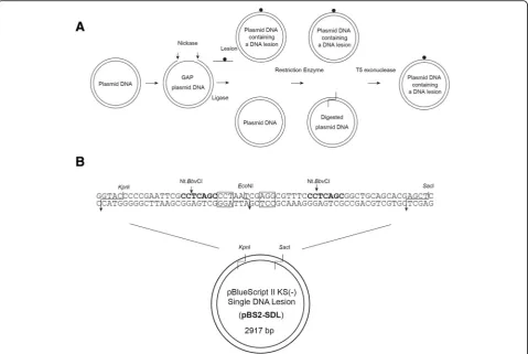
The pBlueScript II KS (−) Single DNA Lesion (pBS2-SDL) plasmid was constructed by replacing the
KpnI-SacI fragment of pBlueScript II KS (−) with the synthetic 64-bp DNA duplex containing the target site for nickase Nt.BbvCI, and the restriction enzymeEcoNI (Fig. 1b). Plasmid DNAs were purified by NucleoSpin Plasmid. The construct pBS2-SDL was checked by DNA sequencing.
Standard procedure for synthesis of in vitro DNA repair substrate
Nicking reaction mixture (20 μl) was comprised of pBS2-SDL (3 pmol) and Nt.BbvCI (5 units) in reaction buf-fer containing 50 mM Potassium acetate, 20 mM Tris-acetate (pH 7.9), 10 mM Magnesium acetate, and 100μg/ml bovine serum albumin. The reactions were incu-bated at 37 °C for 3 h and then at 80 °C for 20 min. They were then purified using PCR Clean-up kit (MACHEREY--NAGEL GmbH & Co.) to remove the 24-mer oligonucleo-tides digested by Nt.BbvCI. After purification, the DNA sample mixture (about 25μl) prepared in the same reaction buffer was incubated with chemically synthetized 24-mer DNA lesion oligonucleotide (15 pmol) at 95 °C for 5 min, 70 °C for 5 min and RT for 15 min for an annealing reac-tion. For DNA ligation reactions, the mixtures were incu-bated overnight at 16 °C in the presence of ATP (final concentration 1 mM) and T4 DNA ligase (800 units) in 30μl system, and the reaction was terminated by incubat-ing at 65 °C for 10 min. For restriction endonucleaseEcoNI reaction,EcoNI (5 units) was added to the reaction sample (30μl) followed by incubation of the mixtures at 37 °C for 1 h and at 65 °C for 20 min. For T5 exonuclease reaction, the mixtures were incubated with T5 exonuclease (5 units) at 37 °C for 1 h. For DNA purification step, T5 exonuclease treated mixtures were purified by PCR Clean-up kit (MACHEREY-NAGEL). For one-pot synthesis, first DNA purification step was omitted.
Agarose gel electrophoresis
The sample DNA substrates were subjected to 0.8% agar-ose gel, and visualized by ethidium bromide staining. DNA fragments in the agarose gel were analyzed using ImageJ software. % contamination = (Open circular DNA + liner DNA / (Open circular DNA + liner DNA + cova-lently closed circular DNA) × 100.
Results & discussion
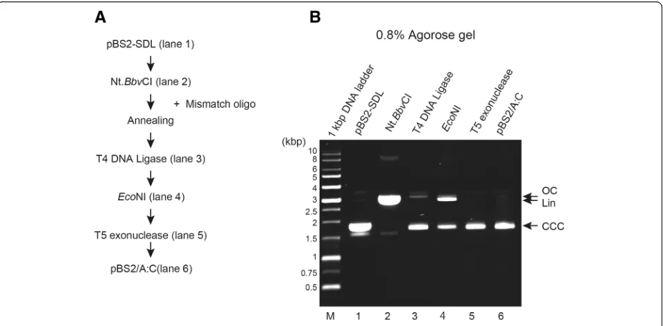
An experimental design has been shown in Fig. 1a. The plasmid DNA, pBluescript II KS(−) Single DNA lesion (pBS2-SDL), containing two nicking sites for endonucle-ase Nt.BbvCI and restriction endonuclease EcoNI, (Fig. 1b) was digested by Nt.BbvCI to produce 24-mer gap re-gion of ssDNA on the plasmid. The digested oligonucle-otides were then removed from the plasmid by a DNA purification step, followed by addition of the oligo-nucleotide containing DNA lesion. After ligation, the plasmid DNA was digested byEcoNI to remove plasmid containing residual non-lesion oligonucleotides. The
reaction mixtures were digested by T5 exonuclease that is capable of removing linear dsDNA and nicked plasmid DNA, but not supercoiled plasmid DNA [15, 16]. This method allows production of site-specific lesion-bearing DNA substrates.
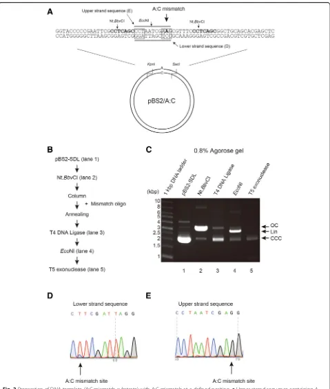
We tested the efficacy of this method in producing A:C mismatch substrates (Fig.2a) using mismatch oligo-nucleotide. Figure 2b shows experimental procedure for purification of pBS2/A:C. Purified pBS2-SDL (Fig. 2c, line 2) was digested by Nt.BbvCI to produce the 24-mer gap and was purified to remove Nt.BbvCI-digested oligo-nucleotides (Fig.2c, line 3). The mixture was incubated with T4 DNA ligase at 16 °C overnight (Fig. 2c, line 4), followed by EcoNI digestion (Fig. 2c, line 5). The mix-ture was finally treated with T5 exonuclease and purified by DNA purification step (Fig. 2c, line 6). Post purifica-tion, pBS2/A:C DNA substrate was observed as one sin-gle band on 0.8% agarose gel.
Fig. 2Preparation of DNA template (A:C mismatch substrate) with A:C mismatch at a defined position.aUpper strand sequence containing A base and lower strand substrate containing original C base in pBS2/A:C are shown diagrammatically.bExperimental procedure for purification of pBS2/A:C.cAliquots from various steps of the purification were analyzed on 0.8% agarose gel, and the DNA substrates were visualized by staining with EtBr. Lane 1, pBS2-SDL; lane 2, Nt.BbvCI-treatment; lane 3, T4 DNA ligase-treatment; lane 4,EcoNI-treatment; lane 5, T5 exonuclease-treatment. Lower panel shows final purified DNA products (%). Open circular DNA (OC), linear DNA (Lin), and covalently closed circular DNA (CCC) are indicated by arrow.dUpper strand sequence containing A base in pBS2/A:C was sequenced. An A:C mismatch site is indicated by the arrow.eLower strand sequence containing base A in pBS2/A:C was sequenced. The A:C mismatch site is indicated by the arrow
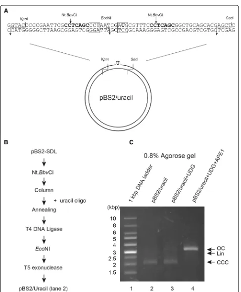
Fig. 4Preparation of DNA template (uracil substrate) with uracil at a defined position.aUpper strand sequence containing uracil base and lower strand substrate containing original C base in pBS2/Uracil are shown diagrammatically.bExperimental procedure for purification of pBS2/uracil using uracil oligo.c
sequenced. While the upper strand sequence showed mainly T and a little C at the mismatch position, the lower strand sequence showed only G, indicating that this method can produce A:C mismatch on the DNA substrate. We observed slight contamination with the original plasmid.
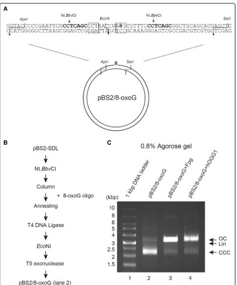
Next, we tried to introduce an oxidative DNA lesion 8-oxoG in the plasmid DNA using this method. 8-oxoG was located within/at theEcoNI recognition site of the oligonucleotide, causing inhibition of EcoNI mediated digestion (Fig. 3a). Figure 3b shows the experimental steps followed for the purification of pBS2/8-oxoG. To confirm if this in vitro DNA repair substrate contained 8-oxoG, the substrates purified by this method were treated with Fpg [17, 18] or hOGG1 [19]. The two DNA repair enzymes act as both an N-glycosylase and an AP-lyase. The N-glycosylase activity releases dam-aged purines from double stranded DNA, generating an AP site. The AP-lyase activity cleaves both at 3′and 5′ ends of the AP site, thereby removing it and leaving a single-base gap. When pBS2/8-oxoG DNA substrates were incubated with these enzymes, open circular con-formation of plasmid DNA substrate was observed on 0.8% agarose gel, whereas linear DNA was not seen (Fig.3c, lanes 3 and 4). In addition, most of the super-coiled DNA substrates converted to open circular form, indicating the presence of 8-oxoG lesion in this DNA substrate.
For further applications of this method, we introduced a deaminated uracil as the DNA lesion on the plasmid DNA. Uracil was positioned at the EcoNI recognition site of the oligonucleotide to inhibit EcoNI digestion (Fig. 4a). Figure 4b shows the experimental procedure for purification of pBS2/uracil. Again, to confirm whether this in vitro DNA repair substrate contained uracil, the purified substrate was treated with UDG [20] and then APE1 [21, 22]. UDG catalyzes the release of free uracil from uracil-containing DNA, producing the AP site. Thus, upon incubation with UDG, no change in migratory behavior of pBS2/uracil was observed on agar-ose gel (Fig.4c, lane 3). When UDG and APE1 were in-cubated with the uracil containing DNA substrates, most of the supercoiled DNA substrates transformed into open circular plasmids (Fig. 4c, lane 4), suggesting that this method is able to produce uracil lesion within the plasmid DNA.
We used this method by recruiting restriction endo-nuclease and T5 exoendo-nuclease to induce site-specific DNA lesion in the plasmid. To simplify this method fur-ther, we attempted to synthesize the DNA substrate in one 1.5 ml microcentrifugation tube. Figure 5a, when compared with Fig.2b, shows the experimental proced-ure for purification of pBS2/A:C omitting a DNA purifi-cation step. When this procedure was executed, population with incorporation of a mismatched oligo-nucleotide seemed to be better with respect to the
Fig. 5One-pot synthesis of DNA repair substrate.aExperimental procedure for purification of pBS2/A:C omitting a column purification step.b
procedure involving the additional DNA purification step (compare Fig.2c, lane 5 and Fig.5b, lane 4). More-over, in the method containing the DNA purification step, 81.1% of products were digested by EcoNI, indicat-ing 81.1% contamination, while in case of one-pot syn-thesis, only 53.6% of products were digested by EcoNI. Some reagents in the DNA purification step might inter-fere with the following reactions for the DNA synthesis. Thus, one-pot synthesis worked more efficiently under our experimental conditions. Therefore, we have suc-cessfully established the one-pot synthesis protocol for in vitro DNA repair substrate containing site-specific DNA lesion.
Conclusions
To analyze DNA repair mechanisms in vitro, DNA sub-strates having DNA lesions at particular sites are neces-sary. There are several procedures to produce the DNA lesion bearing substrate. In some cases, however, it is not easy to prepare the DNA substrate and proves to be expensive. Here we establish the procedure for preparing DNA substrate containing site-specific DNA lesion. This modified method using restriction enzyme and T5 exo-nuclease allows the preparation of the DNA substrate on which the DNA repair enzymes function. Moreover, one-pot synthesis method makes it more simple and convenient to prepare the DNA substrate. We believe that this method is useful to investigate DNA repair mechanisms in vitro.
Abbreviations
6–4 pp.:pyrimidine (6–4) pyrimidone photoproduct; oxoG: 8-oxoguanine; AP: Apurinic/apyrimidinic site; APE1: Apurinic/apyrimidinic endonuclease; BER: Base excision repair; CPD: Cis-syn cyclobutane pyrimidine dimer; dsDNA: Double strand DNA; EtBr: Ethidium bromide; Fpg: Formamidopyrimidine [fapy]-DNA glycosylase; hOGG1: human 8-oxoguanine DNA glycosylase; MMR: Mismatch repair; NER: Nucleotide excision repair; PCNA: Proliferating cell nuclear antigen; ssDNA: single strand DNA; UDG: Uracil-DNA Glycosylase
Acknowledgements
We would like to thank Editage (http://www.editage.jp) for English language editing.
Funding
This work was supported by a Grant-in-Aid for Scientific Research (B) 25281018 from the Ministry of Education, Culture, Sports, Science and Technology (MEXT) of Japan.
Availability of data and materials
Not applicable
Authors’contributions
YS and MH performed experimental work. SI designed the research. IK designed the research and wrote the manuscript. All authors have read and approved the final manuscript.
Ethics approval and consent to participate
Not applicable.
Consent for publication
Not applicable
Competing interests
The authors declare that they have no competing interests.
Publisher’s Note
Springer Nature remains neutral with regard to jurisdictional claims in published maps and institutional affiliations.
Received: 5 September 2018 Accepted: 12 October 2018
References
1. Lindahl T, Wood RD. Quality control by DNA repair. Science. 1999;286: 1897–905.
2. Friedberg EC, Walker GC, Siede W, Wood RD, Schultz RA, Ellenberger T. DNA repair and mutagenesis. Washington, DC: American Society for Microbiology Press; 2005.
3. Suzuki T, Kamiya H. Mutations induced by 8-hydroxyguanine (8-oxo-7,8-dihydroguanine), a representative oxidized base, in mammalian cells. Genes Environ. 2017;39:2.https://doi.org/10.1186/s41021-016-0051-y.
4. Kubota Y, Nash RA, Klungland A, Schär P, Barnes DE, Lindahl T. Reconstitution of DNA base excision-repair with purified human proteins: interaction between DNA polymerase beta and the XRCC1 protein. EMBO J Eur Mol Biol Organ. 1996;15:6662–70.
5. Krokan HE, Bjoras M. Base excision repair. Cold Spring Harb Perspect Biol 2013;5: a012583–a012583.https://doi.org/10.1101/cshperspect.a012583. 6. Huang JC, Svoboda DL, Reardon JT, Sancar A. Human nucleotide excision
nuclease removes thymine dimers from DNA by incising the 22nd phosphodiester bond 5″and the 6th phosphodiester bond 3″to the photodimer. Proc Natl Acad Sci. 1992;89:3664–8.
7. Moggs JG, Yarema KJ, Essigmann JM, Wood RD. Analysis of incision sites produced by human cell extracts and purified proteins during nucleotide excision repair of a 1,3-intrastrand d(GpTpG)-cisplatin adduct. J Biol Chem. 1996;271:7177–86.
8. Huang JC, Hsu DS, Kazantsev A, Sancar A. Substrate spectrum of human excinuclease: repair of abasic sites, methylated bases, mismatches, and bulky adducts. Proc Natl Acad Sci. 1994;91:12213–7.
https://doi.org/10.1073/pnas.91.25.12213.
9. Matsumoto Y, Bogenhagen DF. Repair of a synthetic abasic site involves concerted reactions of DNA synthesis followed by excision and ligation. Mol Cell Biol. 1991;11:4441–7.
10. Matsumoto Y, Kim K, Bogenhagen DF. Proliferating cell nuclear antigen-dependent abasic site repair in Xenopus laevis oocytes: an alternative pathway of base excision DNA repair. Mol Cell Biol. 1994;14:6187–97. 11. Varlet I, Radman M, Brooks P. DNA mismatch repair in Xenopus egg
extracts: repair efficiency and DNA repair synthesis for all single base-pair mismatches. Proc Natl Acad Sci. 1990;87:7883–7.
12. Wang H, Hays JB. Preparation of DNA substrates for in vitro mismatch repair. Mol Biotechnol. 2000;15:97–104.https://doi.org/10.1385/MB:15:2:97. 13. Wang H, Hays JB. Mismatch repair in human nuclear extracts. J Biol Chem.
2002;277:26136–42.https://doi.org/10.1074/jbc.M200357200.
14. Du W, Kinsella TJ. A rapid, simple DNA mismatch repair substrate construction method. Front Oncol. 2011;1:8.https://doi.org/10.3389/fonc.2011.00008. 15. Sayers JR, Eckstein F. Properties of overexpressed phage T5 D15
exonuclease. Similarities with Escherichia coli DNA polymerase I 5″-3″ exonuclease. J Biol Chem. 1990;265:18311–7.
16. Shivji MKK, Moggs JG, Kuraoka I, Wood RD. Dual-incision assays for nucleotide excision repair using DNA with a lesion at a specific site. In: DNA repair protocols. New Jersey: Humana Press; 1999. p. 373–92.
https://doi.org/10.1385/1-59259-675-4:373.
17. Boiteux S, O'Connor TR, Laval J. Formamidopyrimidine-DNA glycosylase of Escherichia coli: cloning and sequencing of the fpg structural gene and overproduction of the protein. EMBO J. 1987;6:3177–83.
18. Tchou J, Bodepudi V, Shibutani S, Antoshechkin I, Miller J, Grollman AP, et al. Substrate specificity of Fpg protein. Recognition and cleavage of oxidatively damaged DNA. J Biol Chem. 1994;269:15318–24.
19. Bjoras M, Luna L, Johnsen B, Hoff E, Haug T, Rognes T, et al. Opposite base-dependent reactions of a human base excision repair enzyme on DNA containing 7,8-dihydro-8-oxoguanine and abasic sites. EMBO J. 1997;16: 6314–22.https://doi.org/10.1093/emboj/16.20.6314.
20. Lindahl T, Ljungquist S, Siegert W, Nyberg B, Sperens B. DNA N-glycosidases: properties of uracil-DNA glycosidase from Escherichia coli. J Biol Chem. 1977;252:3286–94.
21. Demple B, Herman T, Chen DS. Cloning and expression of APE, the cDNA encoding the major human apurinic endonuclease: definition of a family of DNA repair enzymes. Proc Natl Acad Sci. 1991;88:11450–4.