Synthesis and Characterization of New Fe-Complex
and Its Nanoparticle Oxide Using the Novel
Photolysis Method
Wijdan Amer Ibrahim, Zaid Hamid Mahmoud
Department of Chemistry, College of Science, Diyala University, Diyala, Iraq.
ABSTRACT
New ferric complex of 4-(Dimethylamino) benzylidene-4-nitroaniline ligand and its nanoparticles oxide (alpha phase) were fabricated and characterized by elemental analysis, FTIR, 1HNMR, TGA, XRD and TEM. The ligand appeared during the reaction 4-Nitroaniline with 4-(Dimethylamino) benzaldehyde while the nanoparticles of oxide appeared utilizing the photolysis of the complex and when the salt is burnt at 400ºC. The obtained FTIR analysis proved the coordination between ferric ions with the nitrogen atom of (-C=N-) group, while the results of atomic flame adsorption and C.H.N.O analysis obtained the release of chlorine atom from coordination after the photolysis of the complex. The magnetic measurements converted the complex from Para magnetic state to Dia state during photolysis. The results that were yielded from both XRD and TEM explained that the particles prepared are in nano size.
Key Words: Hematite, Nanoparticles, Photolysis, Irradiation, Novel
eIJPPR 2018; 8(1):57-61
HOW TO CITE THIS ARTICLE: Wijdan Amer Ibrahim, Zaid Hamid Mahmoud. (2018). “Synthesis and Characterization of New Fe-Complex and Its Nanoparticle Oxide Using the Novel Photolysis Method” , International Journal of Pharmaceutical and Phytopharmacological Research, 8(1), pp.57-61.
INTRODUCTION
At present, a large group of researchers are aiming at preparing nanomaterials in a very simple way through improving their methods of production. These goals and interests include controlling the nanoscale size and increasing the yield with minimum side effects[1]. Iron oxides gained much importance because of their attractive properties such as their small size, low toxicity and high magnets[2]. A large type of methods have been communicated in the literature for the fabrication of iron
oxide NPs like sonochemical Synthesis[3],
microemulsion[4], electrochemical deposition, [5]
hydrothermal processes, [6] and photolysis methods [7]. Presently, the preparation of iron oxides using a simple method and at the same time gaining a high yield is important and economical due to the high activity and cost. Photolysis is a novel method to synthesize iron oxide from its complexes or salts utilizing the system of irradiation, and this method is found to be more economical compared
to other conventional methods. This method also offers many advantages such as small particle, high yield and no need for energy, etc.
MATERIALS AND METHODS
General
4-Nitroaniline, 4-(Dimethylamino) benzaldehyde and ferric chloride hexahydrate were used as starting materials to prepare nanoparticles. The structure of the fabricated intermediate (ligand and complex) and final product (Fe3O4 NPs) were characterized using different techniques.
The melting point of ligand and complex were measured using Electro thermal IA9100 digital). The analysis of element (C.H.N.O) for starting materials is determined via LECO CHNS-932 analyzer (LECO Corporatio USA). The spectral properties (FT-IR and UV-Vis) of the intermediate compound carried out with the help of (65-FT-IR, Perkin Elmer, USA) and (JASCO Asia Portal UV-visible model V-650), respectively. NMR spectra of ligand showed
Corresponding author: Wijdan Amer Ibrahim
Address: Department of Chemistry, College of Science, Diyala University, Diyala, Iraq
E-mail: wijdan.amer84 @ gmail.com
Relevant conflicts of interest/financial disclosures: The authors declare that the research was conducted in the absence of any commercial or financial relationships that could be construed as a potential conflict of interest .
58
utilizing JEOL ECA400 FT NMR while the structure, the size of particles and morphology of NPs were obtained
using (A Shimadzu-XRD-6000) with Cu K
radiation, Transmission Electron Microscope model (JEOL JEM -2100) and SEM, respectively.Preparation of the ligand, complex and magnetite NPs
Firstly, (0.01 mole) of 4-Nitroaniline, 4-(Dimethylamino) benzaldehyde were dissolved separately in 10ml of ethanol and refluxed after adding acetic acid drop wise for 4h\ hours utilizing water bath. Then the mixture was collected and dried in air until an orange precipitate has appeared (see scheme 1)[8].
Scheme 1
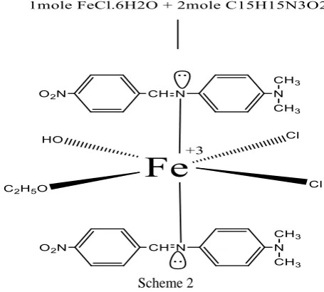
Then, 30ml of chloroform is used to dissolve ferric chloride hexa hydrate after which the ligand and the above element were mixed in a ratio 2 mole (ligand): 1 mole
(metal) accompanied with high stirring, until the red precipitate appeared (see scheme 2).
Scheme 2
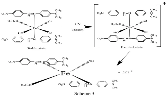
Finally, 100ml of ethanol/H2O (50ml:50ml) was added to
(0.5g) Fe-complex and sonicated until dissolved. Then, it was irradiated for 5 hours using 125W irradiation system (fig 1). Then the formed precipitate was isolated, washed,
dried and burned at 400°C. Brown powder, without magnetic properties, was formed according to the photolysis mechanism (scheme 3)
59
Scheme 3
RESULTS AND DISCUSSION
The physical and analytical measurements of the ligand, Fe-complex and its oxide were listed in table 1. The
percentage ratio of elements of legend and complex was found using (C.H.N.O) and the flame atomic absorption is in good agreement with the general molecular formula suggested for ferric complex.
Table 1. The physical properties and element analysis of the prepared legend and ferric complex (before and after irradiation)
Compound Color Magnetic properties
Molar
conductance
(-1cm2mol-1)
C%
clac.(found)
H%
clac.(found)
N%
clac.(found)
O%
clac.(found)
M%
clac.(found)
Ligand Orange / / 66.91 (66.57)
5.57
(4.70)
15.61
(14.74)
11.91
(12.06) /
Fe-complex
(Before irradiation) Red 1.57 4.24
53.11
(52.87)
4.84
(4.57)
11.60
(11.42)
13.2
(12.98)
7.33
(7.25)
Fe-complex
(After irradiation) Brown 0 3.31
58.80
(57.56)
5.36
(4.98)
12.8
(12.56)
14.7
(14.25)
8.12
7.89
During FTIR and 1H NMR analyses of ligand, amino and
aldehyde groups, the starting materials (PNA and DMAB) were disappeared whereas new strong peaks appeared. In
FTIR, two strong peaks, appeared at 1595 and 1575 cm-1
assigned to (C=N) and NO2 respectively (fig. 2) while the
analysis of 1H NMR is shown at (fig. 3). The singlet peak
observed at 8.26 ppm corresponds to the proton of imine group. Multiple peaks appeared at 8.22, 7.75, 7.22 and 6.70 ppm assigned to aromatic protons while strong peak back to methyl group showed up at 3.05ppm[7].
60 Fig. 3. 1HNMR spectrum of ligand
When comparing the FTIR spectrum analysis of complex with its ligand (fig 4, A and B), the imine group (C=N) got shifted to low frequency (1588cm-1) which indicates
coordinate metal (Fe) with nitrogen atom, and two strong bands appeared at 564 and 409 cm-1 assigned to (Fe-N) and
(Fe-O) respectively due to the process of making a
complex and entry of ethanol molecules in the complex structure. After irradiation of iron complex, there is a shift of (C=N) observed at high frequency due to the ferrous ion produced from irradiation which has low electronegativity when compared with ferric ions. This causes the decrease of attraction power between ions and (C=N) bond[8].
Fig 4. FTIR spectrum of Fe-complex (A) before irradiation and, (B) after irradiation
The figure 5 illustrates the XRD patterns of the hematite with a hexagonal structure fabricated using photolysis methods and all the results are in good agreement with the card (JCPDS 86-0550) and it explained that the complex
was prepared in nano size equal to (13.76nm) using Debye-Scherer equation:
D=Kλ/βcosθ……… (1)[9]
61
where D: the size of particles, k: constant equal (0.9), λ: is the wavelength equal (0.15418 nm), β: is the (FWHM) using (104) plane, and θ is the diffraction angle.The peaks obtained at 24.13°, 33.10°, 35.61°, 39. 25°, 40.60°, 43. 50°, 49.42°, 54.06°, 57.40°, 62.43°and 63.97° angles and they can refer to the 012, 104, 110, 006, 113, 202, 024, 116, 018, 214 and 300 miller index and they are corresponding to the pure hematite. In this method (photolysis), narrow peaks
appeared, and this indicates that the particles were prepared with high purity.
The size and shape of the particles were recorded utilizing TEM as shown in fig. 6. The analysis was conducted by taking aliquots of hematite NPs and placing on a grid of carbon-coated copper and drying under the ambient conditions. The analysis suggested that the particles are in spherical shape and the size is in agreement with XRD analysis.
Fig.6. TEM image of hematite NPs
CONCLUSIONS
The ferric complex, with DMAB-PNA (before and after) irradiation and its ligand fabricated through this method, have a monomer structure as proposed by FTIR, element
analysis, magnetic measurement and 1HNMR data.
Furthermore, the current research obtained that the complex of ferrous synthesized after irradiation of the ferric complex could be converted to nanoparticles of hematite by calcining it at 400C. The sample is calcined after the irradiation process gets NPs in the order of size 13 or larger as shown in the XRD and TEM results.
REFERENCES
[1] Leslie L., Nitin N. and Gang B., Mater. Today., 2005; 8: 32-38. DOI.org/10.1016/S1369-7021(05)00893-X.
[2] Daksha P., Je Y.M., Yong M.C., Tae J.K. and Gang
H., Colloid Surf. A., 2008, 313–314: 91-94. DOI.org/10.1016/j.colsurfa.2007.04.078.
[3] Kenneth S.S.,Science., 1990, 247: 1439-1445. DOI: 10.1126/science.247.4949.1439
[4] Solans C., Izquierdo P., Nolla J., Azemar N.
andGarciaCelma M., Curr. Opin. Colloid Interface
Sci.,2005, 10: 102.
DOI.org/10.1016/j.cocis.2005.06.004.
[5] Cabrera L., Gutierrez S., Menendezb N., Morales M.P.
andHerrasti P., Electrochim. Acta., 2008, 53: 3436.DOI.org/10.1016/j.electacta.2007.12.006. [6] Xiaohe L., Guanzhou Q., Aiguo Y., Zhong W. and
Xingguo L., J. Alloy Compd., 2007, 433: 216-220. DOI.org/10.1016/j.jallcom.2006.06.029.
[7] Umesh N. and RN R.,J. Fluoresc., 2017, 27:2263– 2277. DOI. org/10.1007/s10895-017-2168-1.
[8] Colthup N.B.; Daly L.H.; Wiberley S.E.,
“Introduction to Infrared and raman Spectroscopy” 3rd ed, Academic Press, New York and London, 1990.
[9] Mousa A.A., AbbasR., FardinS., SamiraB. and