Original Research Article.
440 |P a g e Int J Med Res Prof.2018 Jan; 4(1); 440-45. www.ijmrp.com
Clinical Profile and Risk Factor of Coronary Artery Disease in Young Indians
Keshavamurthy Ganapathy Bhat
1, Kamal Hasan Kandanuru
2, Bhupinder Kaur Anand
3*,
Marwaha Manvinder pal Singh
4, Anil Kumar
5, Mahesh Nalin Kumar
61Senior Adviser Medicine and Cardiologist, Air Force Central Medical Establishment, New Delhi, India. 2Assistant Professor, Dept. of Medicine, Shadan Institute of Medical Sciences, Hyderabad, Telangana, India. 3*Professor, Department of Community Medicine, SGT Medical University, Gurugram, Haryana, India. 4Classified Specialist Aerospace Medicine, Air Force Central Medical Establishment, New Delhi, India. 5Department of Cardiology, Command Hospital (Western Command), Chandimandir, Haryana, India. 6Head of Department of Cardiology, Base hospital, Delhi, India.
ABSTRACT
Objective: The aim was to study the clinical profile and risk factors in young Coronary Artery Disease (CAD) patients. Materials & Methods: We studied 132 consecutive young adults with CAD presenting to two tertiary care centres in India over a period of one and half years. We recruited our subjects from various wards of our hospitals where patients presented with acute coronary syndrome (ACS), CSA, Heart failure and Asymptomatic Electrocardiogram (ECG) abnormalities. Asymptomatic individuals who had incidentally detected ECG abnormality during routine medical examination or pre anesthetic checkup and subsequently diagnosed to have CAD were also included in the study.
Results: In this study, Hypertension, Diabetes, past and family history of CAD were similar in the two groups. Tobacco consumption among cases (37.88%) was significantly high (p= 0.0045) compared to controls. Dyslipidemia was very common in both the groups (82 and 87%). HDL- Cholesterol < 40 mg/dl was the commonest lipid abnormality (~ 50%) in both the groups. More than one lipoprotein abnormality was noted in 77 (58.33%) patients in the study group.
Conclusion: Typical angina is the commonest presenting symptom and Myocardial infarction is the commonest diagnosis at presentation in young. Anterior wall is the most commonly involved myocardial territory in young Indians.
Keywords: Coronary Artery Disease (CAD), Risk Factor, Hypertension, Diabetes.
*Correspondence to:
Dr. Bhupinder Kaur Anand,
Professor, Department of Community Medicine, SGT Medical University, Gurugram, Haryana, India. Article History:
Received: 01-11-2017, Revised: 14-12-2017, Accepted: 28-01-2018
Access this article online
Website:
www.ijmrp.com
Quick Response code
DOI:
10.21276/ijmrp.2018.4.1.091
INTRODUCTION
Coronary artery disease (CAD) is the leading cause of death worldwide.1 It is a devastating disease precisely because an otherwise healthy person in the prime of life dies or becomes disabled without warning. Age above 45 years is considered a risk factor for CAD. However, 5-10 % of CAD occurs in individuals < 45 years. CAD occurs 5-10 years earlier in Indians compared to western population.2,3 The risk of CAD in Indians is 3-4 times higher than White-Americans, 6-times higher than Chinese and 20 times higher than Japanese.4,5 Indians are prone as a community to CAD at a much younger age.6,7 In the Western population incidence of CAD in the young is up to 5% as compared to 12-16% in Indians.8,9 Various studies have noted that higher visceral adiposity, higher small dense LDL, higher lipoprotein (a)
[pronounced as “little a”] and metabolic syndrome in Asians could be contributing to CAD.10-12 The frequency of asymptomatic CAD, Chronic stable angina (CSA), unstable angina (UA) and Myocardial Infarction (MI) in younger population and their risk
factor profile has not been reported in India. Hence we studied the
CAD in Young Indians ≤ 45 years of age with respect to their risk
factor profile and clinical presentation with the aim to find out whether we are missing common risk factors.
MATERIALS AND METHODS
medical examination or pre anesthetic checkup and subsequently diagnosed to have CAD were also included in the study. Inclusion Criteria
(1) Age group 18-45 years
(2) Patients with diagnosis of CAD established unequivocally by one or more diagnostic tests – ECG, Cardiac enzymes, Echocardiography, Myocardial Perfusion Imaging (MPI) and Coronary Angiography (CAG).
Exclusion Criteria
Those who refused to give informed consent were excluded from
the study. During the same period, young adults ≤ 45 years, who
presented with chest pain or asymptomatic ECG abnormalities and had positive stress test but normal coronary angiogram (No CAD) were taken as controls for comparison and analysis of coronary risk factor profile.
Consent, Screening and Evaluation
All subjects were counseled in detail about the study and informed consent was obtained. Detailed history was obtained. All underwent a thorough physical examination including height, weight, body mass index (BMI), waist circumference and waist hip ratio (WHR) at the earliest available and appropriate opportunity. In patients with Acute Coronary Syndrome (ACS), detailed history and physical examination were deferred for obvious reasons. All relevant investigations were carried out.
All subjects underwent lipid profile, Lipoprotein (a) [Lp(a)], Serum homocysteine and blood glucose estimation. Total cholesterol was estimated by CHOD-POD method, triglycerides by GPO, high density lipoprotein cholesterol (HDL-C) and low density lipoprotein cholesterol (LDL-C) by direct estimation method. Lp(a) was estimated using Enzyme immune assay (EIA) and serum
homocysteine was estimated by HPCL method. Fasting samples were collected after a minimum of ten hours of fasting. All individuals underwent ECG and 2 D Echocardiography. Stress testing - either exercise ECG treadmill test(TMT) or Stress Myocardial Perfusion Imaging( Stress MPI) was carried out if
clinically indicated. LVEF ≥ 55% was taken as normal. Patients
were categorized into mild, moderate and severe LV dysfunction based on LVEF (mild LV Dysfunction: LVEF 45-54%; moderate LV Dysfunction: LVEF 31-44%; severe LV Dysfunction: LVEF
≤30%).13
All patients were subjected to CAG. The diagnosis of significant CAD was made in cases having ≥ 50% stenosis in coronary
lumen in coronary angiography. In cases where CAG was normal or had < 50% of lesion, to make a diagnosis of CAD, they required to have unequivocal evidence of ACS with symptoms, ECG changes, cardiac enzymes or Echocardiography. Such cases were labelled non obstructive CAD/ Recanalysed CAD. Incidental detection of minor coronary plaques was not considered as CAD. Statistical Analysis
Sample size was calculated by using STATCALC from epi info software using confidence level of 95%, power of 80%, ratio of controls to cases 0.25, odds ratio of 8.14 and proportion of exposed among cases 30% and among controls 5%. The calculated sample size was 118 and controls 30. We studied 132 cases and 33 controls. Frequency of risk factors was noted. Means with standard deviations (SD) of continuous variables and proportions of discrete variables were calculated. Proportions were compared using chi square test and means of continuous variables were compared using unpaired t-test (Student t test). A P value of < 0.05 was taken as significant. Risk ratio and 95% Confidence Interval were estimated wherever relevant.
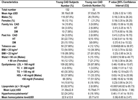
Table 1: Clinical characteristics and risk factors Characteristics Young CAD Subjects
Number (%)
Young non CAD Controls Number (%)
P value [95% Confidence Interval (CI)] Total number
Mean Age ±SD
132 38.19±5.58
33
37.03±5.79 0.29(-3.32 to 1.00)
Males (%) 116 (87.9%) 26 (78.8%) 0.18(-3.36 to 26.24)
Females (%) 16 (12.1%) 7 ( 21.2%) 0.18(-3.35 to 26.25)
Family h/o CAD 14 (10.6%) 3 (9.09%) 0.80(-13.56 to 10.26)
HT 04 (3.03%) 2 (6.06%) 0.41(-3.25 to 16.7) DM 10 (7.58%) 3 (9.09%) 0.77(-6.80 to 16.39)
Past h/o CAD 04 (3.03%) 2 (6.06%) 0.41(-3.25 to 16.70)
HT 30 (22.73%) 5(15.15%) 0.34(-9.41 to 19.16) DM 20 (15.15%) 3(9.09%) 0.37(-9.3 to 15.32)
Tobacco use 50 (37.88%) 4 (12.12%) 0.0049(8.66 to 36.97)
BMI > 25 kg/m2 72 (54.55%) 13 (39.39%) 0.12(-3.78 to 32.02)
BMI > 23 kg/m2
Waist > 90 cm (Males) > 80 cm (Females)
118 (89.39%) 70 (53.03%) 16 (12.12%)
27 (81.82%) 5 (15.15%) 7 (21.21%)
0.23(-3.95 to 24.3) 0.0001(19.98 to 49.77)
0.18(-3.38 to 26.24) Dyslipidemia LDL > 160 mg/dl
TC > 200 mg/dl TG > 150 mg /dl HDL < 40 mg/dl (Males) <50 mg/dl (Females)
109 (82.58%) 6 (4.55%) 40 (30.30%) 50 (37.88%) 66 (50%) 14 (10.61%)
29 (87.88%) 1 (3.03%) 8 (24.24%) 11 (33.33%) 17 (51.52%) 6 (18.18%)
0.46(-10.88 to 15.67) 0.69(-11.01 to 7.12) 0.49(-12.19 to20.17) 0.63(-14.22 to 20.58) 0.88(-16.82 to 19.58) 0.23(-3.95 to 24.31) Lipoprotein (a) > 30 mg/dl
Mean Lp(a) ±SD
83 (62.87%) 31.39±22.8
2 (6.06%) 15.79±6.71
442 |P a g e Int J Med Res Prof.2018 Jan; 4(1); 440-45. www.ijmrp.com
Figure 1: Sex wise distribution of subjects in Young CAD
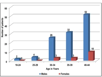
Figure 2: Age wise distribution of CAD subjects
Table 2: Clinical Spectrum of CAD in Young
Clinical syndrome Number ( Percentage)
STEMI 94 (71.21%)
NSTE MI 12 (9.09%)
Unstable Angina 14 (10.60%)
CSA 06 (4.54%)
Asymptomatic 06 (4.54%)
SCA NIL CAD: Coronary Artery Disease; STEMI: ST segment elevation Myocardial Infarction; NSTEMI: Non ST segment elevation Myocardial Infarction; MI – Myocardial Infarction; CSA – Chronic Stable Angina; SCA- Sudden Cardiac Arrest
Males 88%
Females
12%
Males Females
0 10 20 30 40 50 60
18-24 25-29 30-34 35-39 40-44
2 4
26
32
52
0 0 3 3
10
Nu
m
ber
o
f p
at
ien
ts
Age in Years
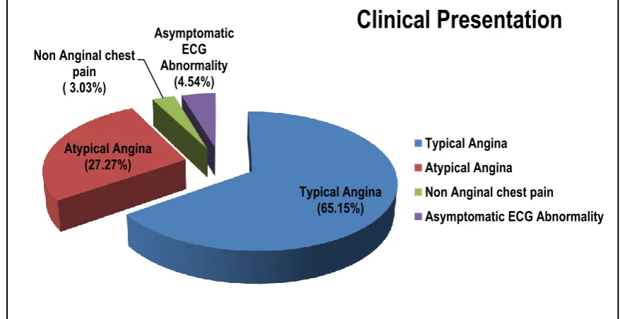
Figure 3: Symptoms at presentation (in percentage)
Table 3: Site of MI in Young Adults Site of MI Number (%) AWMI 60 (56.60%)
IWMI 26 (24.53%)
Lateral Wall MI 8 (7.55%) > One wall
Anterior and Inferior wall
Inferolateral wall 12 (11.32%)
8 (7.55%) 4 (3.77%) MI – Myocardial Infarction; AWMI – Anterior Wall MI; IWMI- Inferior Wall MI
Table 4: Left Ventricular Ejection Fraction (LVEF) in study subjects
LVEF Baseline Number (%)
NORMAL (≥55%) 76 (57.57%)
Mild LV Dysfunction (45-54%) 26 (19.69%) Moderate LV Dysfunction (31-44%) 26 (19.69%) Severe LV Dysfunction (≤30 %) 04 (3.03%)
OBSERVATIONS AND RESULTS
Over a period of one and a half years, 1432 CAD patients were treated out of which 132 patients (9.21%) were young (< 45 years). Out of 132 patients studied (Cases); 116 (87.87%) were males and 16 (12.13%) were females (Figure 1). During the same period, age and sex matched young individuals who presented with symptoms of possible CAD but CAD was ruled out after investigations were studied as controls for analysis of risk factors. The clinical characteristics and risk factors of both the groups are summarized in table 1. The youngest was a 20 year old male. Frequency of CAD increased with age initially linearly up to 30 years and thereafter exponentially with maximum number of patients (47%) being in 40 to 44 years age group. (Figure 2). In each age group males were more commonly affected.
Analysis of Conventional Risk Factors
Hypertension, Diabetes, past and family history of CAD were similar in the two groups. Tobacco consumption among cases (37.88%) was significantly high (p= 0.0045) compared to controls.
Dyslipidemia was very common in both the groups (82 and 87%). HDL- Cholesterol < 40 mg/dl was the commonest lipid abnormality (~ 50%) in both the groups. More than one lipoprotein abnormality was noted in 77 (58.33%) patients in the study group.
Eight patients were obese with body mass index (BMI) more than 29.99 kg/m2 (6.06%) among cases and 2 (6.06%) among controls. However, overweight was more common with 72 (52.55%) patients having BMI >25 kg/m2. Further, when cut off for normal BMI was reduced to 22.99 kg/m2 as recommended for Indians14, 118 patients (89.39%) had either overweight or obesity. There was no significant difference in BMI between the two groups. However, abdominal (Central) obesity with waist circumference > 90 cm (cutoff for Indians and Asians) 13 was more common in CAD group than controls (p= 0.0001).
Among cases, 62.87% had elevated Lipoprotein (a) [Lp(a) >30 mg/dl] compared to only 6.06% among controls (p<0.0001) with mean Lipoprotein (a) 31.39±22.8 mg/dl compared to 15.79±6.71 in controls (p=0.0002). There was no significant difference between the two groups in homocysteine level.
Presenting symptoms are shown in Figure 3. Chest pain was the presenting symptom in 92.42% of cases. Clinical spectrum of the CAD group is summarized in table 2. Myocardial Infarction was the commonest diagnosis (80.30%). Anterior wall was the commonest (56.6%) wall involved in MI (Table 3) followed by Inferior wall (24.53%).
Left ventricular Ejection Fraction of our study subjects if summarized in Table 4. In our study 76 patients (57.57%) were having normal LVEF, 26 patients (19.69%) had moderate LV dysfunction and same number (19.69%) had mild LV dysfunction. Four (3.1%) patients were in severe LV dysfunction category. We also analyzed the LVEF among the STEMI and NSTEMI patients separately. Among the STEMI patients there were 42(44.68%) subjects in normal LVEF, 24 subjects (25.53%) each in mild and moderate LV dysfunction category. There were 04 patients (3.03%) with severe LV dysfunction. Among the NSTEMI group, there was one patient each in mild and moderate LV dysfunction category. There was none having severe LV dysfunction with NSTEMI.
Typical Angina (65.15%) Atypical Angina
(27.27%) Non Anginal chest
pain ( 3.03%)
Asymptomatic ECG Abnormality
(4.54%)
Clinical Presentation
Typical Angina Atypical Angina Non Anginal chest pain
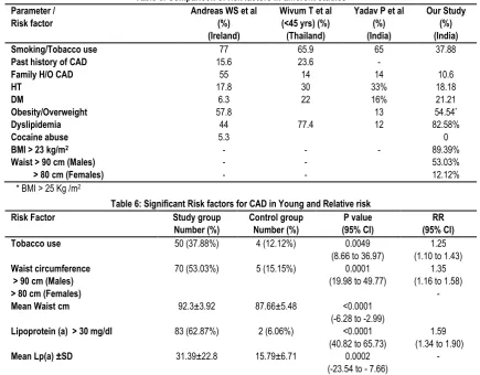
444 |P a g e Int J Med Res Prof.2018 Jan; 4(1); 440-45. www.ijmrp.com Table 5: Comparison of risk factors in different studies
Parameter /
Risk factor
Andreas WS et al (%) (Ireland)
Wivum T et al (<45 yrs) (%) (Thailand)
Yadav P et al (%) (India)
Our Study (%) (India)
Smoking/Tobacco use 77 65.9 65 37.88
Past history of CAD 15.6 23.6 -
Family H/O CAD 55 14 14 10.6
HT 17.8 30 33% 18.18
DM 6.3 22 16% 21.21
Obesity/Overweight 57.8 13 54.54*
Dyslipidemia 44 77.4 12 82.58%
Cocaine abuse 5.3 0
BMI > 23 kg/m2
Waist > 90 cm (Males) > 80 cm (Females)
- - -
- - -
- 89.39%
53.03% 12.12% * BMI > 25 Kg /m2
Table 6: Significant Risk factors for CAD in Young and Relative risk
Risk Factor Study group
Number (%)
Control group Number (%)
P value (95% CI)
RR (95% CI)
Tobacco use 50 (37.88%) 4 (12.12%) 0.0049
(8.66 to 36.97)
1.25 (1.10 to 1.43) Waist circumference
> 90 cm (Males) > 80 cm (Females) Mean Waist cm
70 (53.03%)
92.3±3.92
5 (15.15%)
87.66±5.48
0.0001 (19.98 to 49.77)
<0.0001 (-6.28 to -2.99)
1.35 (1.16 to 1.58)
-
Lipoprotein (a) > 30 mg/dl
Mean Lp(a) ±SD
83 (62.87%)
31.39±22.8
2 (6.06%)
15.79±6.71
<0.0001 (40.82 to 65.73)
0.0002 (-23.54 to - 7.66)
1.59 (1.34 to 1.90)
-
DISCUSSION
Young adults constituted 10.54 % of all CAD patients in our study with males constituting 87.9%. In an Indian study, authors studied 200 patients from age 31 yrs to 80 yrs15. In their study 8% of male patients and 2 % of female patients belonged to age group 31- 40 yrs and 17 % male patients and 4% female patients belonged to age group 41 -50 yrs. In other studies, males constituted about 85%16, 17. Our finding of majority of young CAD patients being males is consistent with other studies in the region. Wiwun T et all observed that typical angina was the presenting symptom in 86.3% and atypical angina in 9% of their study population under the age of 45 yrs17. Chest pain as the presenting symptom constituted 95.4% in the same study. Another study observed that 94% of study population (31-81 yrs) presented with chest pain15. In our study, 65.15% presented with typical angina and 95.54% presented with chest pain consistent with other studies.
Risk factors: Various risk factor profile in our study is compared with other studies in Table 5. In our study, 37.88% were tobacco consumers, significantly less compared to other studies possibly because of better health awareness and annual medical check-up of our study subjects who are largely serving soldiers or veterans. There were no morphine or cocaine abusers among the study group. Overweight and obesity were similar when cut off for BMI was taken as 25kg/m2. However, when Indian standards were applied, a vast majority of cases (89.39%) were overweight or obese with majority having abdominal obesity (65.15 % with waist > 90 cm). These are the unique findings of our study since these parameters were not included in other studies. Risk factors which were statistically significant with hazard ratio >1 are summarised in Table 6. In our study we found that 62.12% had elevated serum
homocysteine and 24.24% had elevated serum lipoprotein (a) levels. There was no significant difference between study and control groups for homocysteine level; however, CAD patients had significantly higher Lipoprotein (a) compared to controls. We could not find any previously reported studies in this regard in young Indian CAD population.
Clinical presentation of CAD: Wiwun T et al, noted that under the 45 yrs group with CAD- 13.4% belonged to UA category, 19.3% to Non STEMI (NSTEMI) and 67.3% belonged to STEMI17. Singh A et al observed UA as the commonest presentation when all age groups were taken into consideration18. In our study we analysed the complete spectrum of CAD (STEMI, NSTEMI, UA, CSA and asymptomatic ECG abnormality/silent MI). We observed that 80.30% patients presented with MI (71.21% - STEMI and 9.09% - NSTEMI), 10.6% presented with UA consistent with other studies in young patients. In our study, 1.52% presented with CSA and asymptomatic ECG abnormality (silent CAD) was noted in 4.54%. We could not find any other study where complete spectrum of CAD presentation including CSA and Silent CAD are analysed.
Site of infarction: Yadav P et al reported 54% Anterior Wall (AWMI); 41% Inferior Wall MI (IWMI) and 5% multiple wall involvement in their study15. We observed 56.6% AWMI, 24.53% IWMI, 7.55% lateral wall MI and 11.32% multiple wall involvement. AWMI was the most frequent presentation consistent with other studies.
LVEF. Mild and moderate LV dysfunctions were observed in 19.69% each. Only 3.1% patients had severe LV dysfunction. LV function findings of our study are largely consistent with other studies; inclusion of complete spectrum of CAD including CSA and asymptomatic ECG abnormality/silent MI in our study probably account for minor differences in proportions observed.
STRENGTHS OF OUR STUDY
It is an observational study conducted at two centers with where the complete spectrum of coronary artery disease including silent CAD has been analyzed. Indian standards for overweight and obesity studied. Study population was representative sample of whole of India since soldiers hailing from all parts of the country are positioned in a station.
LIMITATIONS
It is relatively a small, two centre study. The study population is mostly from the armed forces and veterans. Hence relatively healthy people are included in the study compared to general population.
CONCLUSIONS
Coronary Artery Disease in young Indians constitutes about 10% of all CAD. It is a different entity compared to older adults with different risk factor profile and clinical presentation. While tobacco use is decreasing as a risk factor, it still remains a significant contributing risk. Abdominal obesity and overweight are ubiquitously prevalent but commonly overlooked. In younger age group, males are more commonly affected than females (9:1) since CAD in females below 45 years is rare. Elevated Lipoprotein (a) is a significant risk factor for CAD. Young CAD commonly presents with typical angina. Typical angina is the commonest presenting symptom and Myocardial infarction is the commonest diagnosis at presentation in young. Anterior wall is the most commonly involved myocardial territory in young Indians. Primary and primordial preventive measures should focus on detection and reduction of abdominal obesity and tobacco use cessation. Lipoprotein (a) should be a part of routine coronary risk stratification in young.
REFERENCES
1. GBD 2015 Risk Factors Collaborators. Global, regional, and national comparative risk assessment of 79 behavioural, environmental and occupational, and metabolic risks or clusters of risks, 1990–2015: a systematic analysis for the global burden of disease study 2015. Lancet. 2016; 388(10053):1659–1724
2. Jaloweil DA, Hill JA. Myocardial infarction in young men and women. Cardiovasc Clin 1989; 20:197–206
3. Gupta R. Burden of coronary heart disease in India. Indian Heart J. 2005; 57: 632-38.
4.Enas EA, Garg A, Davidson MA et. al. Coronary heart disease and its risk factors in the first generation immigrant Asian Indians to the United States of America. Indian Heart J 1996; 48: 343-54.
5. Rissam HS, Kishore S, Trehan N. Malignant Coronary Artery Disease in Young Indians-A Challenge of the New Millennium. JK Science 1999; 1:197-202.
6. National Institutes of Health. National Heart, Lung, and Blood Institute. Morbidity & Mortality: 2012 Chart Book on Cardiovascular, Lung, and Blood Diseases. Bethesda, Susan B. Shurin, M.D: National Heart, Lung, and Blood Institute; 2012.
7 Reis SE et al. Coronary microvascular dysfunction is highly prevalent in women with chest pain in the absence of coronary artery disease: results from the NHLBI WISE study. Am Heart J 2001; 141:735-41
8. Han SH, Bae JH et al. Sex differences in atheroma burden and endothelial function in patients with early coronary atherosclerosis. Eur Heart J 2008; 29:1359-69.
9. Hemingway H, McCallum A et al. Incidence and prognostic implications of stable angina pectoris among women and men. JAMA 2006; 295:1404-11.
10. Sainani GS, Karatela RA. Association of Plasma Homocysteine and Insulin Resistance in Coronary Artery Disease. J Assoc Physicians India.2009; 57: 439-442
11. Yang WX, Yang Z, Wu YJ, Qiao SB, Yang YJ, Chen JL. Factors
associated with coronary artery disease in young population (age ≤
40): analysis with 217 cases. Chin Med Sci J. 2014; 29:38-42 12. Yusuf S, Hawken S, Ounpuu S et al. INTERHEART Study Investigators. Effect of potentially modifiable risk factors associated with myocardial infarction in 52 countries (the INTERHEART study): case-control study. Lancet. 2004;364(9438):937–952.
13. Lang RM, Bierig M et al: Recommendations for chamber quantification: A report from the American Society of
Echocardi-ography’s Guidelines and Standards Committee and the Chamber
Quantification Writing Group, developed in conjunction with the European Association of Echocardiography, a branch of the European
Society of Cardiology. Echocardiography’s Guidelines and Standards
Committee; European Association of Echocardiography. J Am Soc Echocardiogr 2005;18:1440-63
14. Misra A et al. Consensus Statement for Diagnosis of Obesity, Abdominal Obesity and the Metabolic Syndrome for Asian Indians and Recommendations for Physical Activity, Medical and Surgical Management. J Assoc Physicians India. 2009; 57:163-70.
15. Yadav P et al. Clinical Profile & risk factors in acute coronary syndrome. National Journal of Community Medicine 2010; 1: 150- 52 16. Andreas W S, Dragana R et al. Acute coronary syndromes in young patients: Presentation, treatment and outcome. International Journal of Cardiology. 2011; 148: 300–4
17. Wiwun T et al. Acute Coronary Syndrome in Young Adults: The Thai ACS Registry. J Med Assoc Thai 2007; 90 (Suppl 1): 81-90. 18. Singh A, Singh V, Ranjan R. Survey of Assessment and Management of Coronary Heart Disease Patients (SMART) in India. Journal of The Association of Physicians of India. 2017; 65: 22-26 19. Christus T, Shukkur AM et al. Coronary Artery Disease in Patients Aged 35 or less - A Different Beast? Heart Views. 2011;12:7–11. 20. Pahlajani DB , Chawla MH ,Kapashi KA . Coronary artery disease pattern in the young. The Journal of the Association of Physicians of India. 1989; 37(5):312-314.
Source of Support: Nil. Conflict of Interest: None Declared.
Copyright: © the author(s) and publisher. IJMRP is an official publication ofIbn Sina Academy of Medieval Medicine & Sciences, registered in 2001 under Indian Trusts Act, 1882. This is an open access article distributed under the terms of the Creative Commons Attribution Non-commercial License, which permits unrestricted non-commercial use, distribution, and reproduction in any medium, provided the original work is properly cited.