2 0 N O V E M B E R 2 0 1 0
Oral Communications
TRAUMATOLOGY 1
The role of Girdlestone resection arthroplasty in a case
of posterior hip dislocation associated to femoral neck
fracture, transverse acetabular fracture and sciatic
nerve injury
R. Giancola, F. Valli, G. Antonini, C. Crippa
Department of Orthopaedic and Trauma Surgery, San Carlo Borromeo Hospital (Milan, ITALY)
Dislocations of the hip have become a common injury in recent years and it can occur in conjunction with a fracture of the femoral shaft or neck or head. Posterior dislocation of the hip associated with femoral neck fracture along with fracture of the acetabulum but an intact femoral head is an unusual but not an unknown injury. This paper reports a patient who sustained posterior dislocation of the hip asso-ciated with fracture of the femoral neck of the ipsilateral femur, transverse with posterior wall acetabular fracture and sciatic nerve injury. Such injuries present a therapeutic dilemma. Controversy usually exists regarding primary arthroplastyversus open reduction and internal fixation of the femoral neck fracture, but in this case the authors have to be considered the acetabular fracture, where total hip replacement is rarely indicated in the acute phase. However the open reduction and internal fixation’s technique presents an high risk, 20–30%, of subsequent avascular necrosis and an high rate, 35%, of reprise; posttraumatic arthritis has been demonstrated in 12–67% of the patients with acetabular fracture, even when the acetabular bone stock could be restored with minimal deformity, while the posttrau-matic avascular necrosis of the femoral head following an acetabular fracture has been reported to range from 2 to 40%. Our patient was managed within five hours from injury by open reduction and internal fixation of acetabular fractures and proximal femoral resection arthroplasty (Girdlestone’s technique). After 6 months a total hip arthroplasty was performed. The patient at 4 years from trauma pre-sents a full range of motion, no pain, a good integration of arthroplasty, no osteolysis sign, but a paralysis of external sciatic nerve.
The purpose of this case report is to suggest a new surgical approach for the treatment of this association of lesions, introducing the
Girdlestone procedure like an intermediate option before definitive total hip replacement. To our knowledge in the English-language literature there is not a similar treatment solution.
Management of fractures ipsilateral femur and pelvis:
floating hip
R. Spagnolo1, G. Fioretta1, S. Angelini2, P.G. Davini2
1Department of Orthopaedics and Traumatology, Ospedale di Desio
(Milan, ITALY);
2Department of Orthopaedics and Traumatology, Ospedale di Empoli
(Florence, ITALY)
ObjectiveWe review 5 cases of ipsilateral femur and pelvis fractures and describe associated lesions, treatment, results, and complications.
Material and methods The lesion mechanism involved a street accident for all patients, car to car collision in 3 cases and running over in 2 cases. According to Liebergall et al., 2 patients were type A, 3 type B, no patient type C. The time of surgery was determined by the hemodynamic status of the patient. Results were evaluated according to the criteria of d’Aubigne` and Postel modified by Matta et al.
Results None of the patients died. According to the criteria of d’Aubigne` and Postel modified by Matta et al., results were excellent in 3 cases, good in 2.
DiscussionIpsilateral fractures of the femur and pelvis are complex lesions requiring specific management training, they are severe injury usually caused by high energy trauma. The acetabulum and pelvic ring are more commonly fractured together than either alone. The acetabular fractures were treated with open reduction and internal fixation (ORIF) in all cases. The patients with pelvic ring fractures were surgically stabilized with external fixation in emergency and subsequently with ORIF. In case of femur fractures, acetabular fractures were first treated, while in case of association of pelvis and femur fractures, femur was treated after pelvic stabilization.
Suggested readings
1. Burd TA, Hughes MS et al. (2008) The Floating Hip: Complica-tions and Outcomes. The journal of Trauma 64:442–448 2. Liebergall M et al. (1992) The floating hip. Ipsilateral pelvic and
TRAUMATOLOGY 2
Biomechanical properties of different pin types
and configurations for percutaneous fixation of 2-part
proximal humeral fractures
D. Bonasia1, F. Castoldi1, R. Rossi1, A. Sankey2, F. Dettoni1, D. Blonna1, A. Amis3
1Ospedale Mauriziano Umberto I, Universita` degli Studi di Torino
(Turin, ITALY);
2St. Mary Hospital (London, UK); 3Imperial College (London, UK)
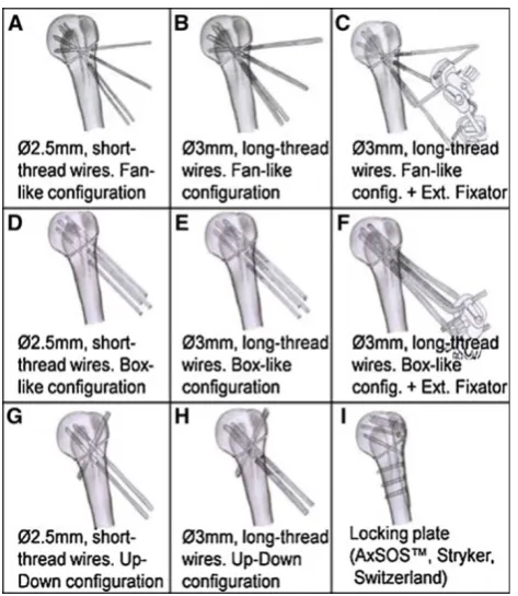
IntroductionIn 2005 a new percutaneous device has been developed for the treatment of proximal humeral fractures. This device has 2 main features: (1) long thread wires, and (2) an external fixator to link the wires together. The goal of this study was to investigate the biomechanical properties of different wire constructs (changing the size and type of the wires, and combining them in different config-urations) compared to the locking plate (Fig.1).
Material and methodsA two-part proximal humeral fracture was created in 72 fourth-generation Sawbones. There were 9 groups of 8 humeri each. Each group was fixed with one of 9 different methods (Fig.1). An Instron 8800 servohydraulic tension–torsion material testing machine was used for: (1) Cyclic torsion test for stiffness (Nm/ deg of rotation) and maximum rotation (deg); (2) cyclic axial com-pression-plus-varus (20 degrees) bending test for stiffness (N/mm) and maximum displacement (mm); (3) ultimate load to failure in the axial compression-plus-varus bending mode. Statistical analysis: ANOVA and post-hoc analysis with Bonferroni correction Signifi-cancep\0.05.
Results 1. Stiffness: the external fixator tended to increase the tor-sional stiffness, but significantly only for the Box group (*p=0.03).The locking plate group had the highest torsional stiff-ness, but not significantly different from group F (**p=1). Maximum rotation: data were similar to torsional stiffness.2. Stiff-ness: the only construct significantly weaker (p=0.03) was H when compared to C and F. No other significant differences were found. C and F were stiffer than the Plate (p=0.07). Maximum displacement: no differences were noted.3.With some constructs (8/8 of group I, in 5/8 of F, and in 4/8 of C), at 2000N, failure occurred at the shaft. The other constructs failed at fracture site at significantly lower loads.
DiscussionIn group F, the properties did not differ significantly from the locking plate. Group C was comparable to F and I in every test, except in the torsion tests, where it showed inferior properties.
TRAUMATOLOGY 3
The treatment of C2-3 distal humeral fractures
in elderly patients with osteosynthesis
(parallel-plate technique)
F. Stacca, L. Malagoli, A. Celli
Clinica Ortopedica e Traumatologica, Policlinico di Modena (Modena, ITALY)
Introduction The gold standard in the treatment of C2-3 distal humeral fracture is the ORIF procedure. The target is the recovery of the anatomical complexity of the elbow with a strong stabilization, so to allow a early mobilization. This target is difficult in the adult, and more difficult in the elderly patient, who has a poor bone quality, so that an important part of the international literature indicates the arthroplasty as the primary treatment. Elbow prosthesis allows normal daily activities, but it could be a limitation in case of high functional requests. The aim of this study is the evaluation of the elderly patient with C2-3 distal humeral fractures treated in our clinic with parallel plates ORIF technique.
Material and methods In the period 2003–2009 we treated 14 patients over 65 with complex distal humeral fracture (AO:C2-3). All the patients had high functional requests. All the patients were treated with parallel plates (Mayo Clinic) as described by O’Driscoll. The evaluation interested all the patients: F.U. 29.2 months, main age:71.7 years, 6 male and 8 female. In 9 cases the fracture interested the dominant arm. Standard RX scan were used. Clinical evaluation followed standardized test: MEPS one and DASH one.
ResultsAll the patients obtained a complete healing of the fracture. We observed good or excellent results in 13 patients, the main MEPS was 91.1 and main DASH was 17.5. R.O.M.: 18°–131°in flexion– extension, 74°–73° in prono-supination. Pain was absent in 10 patients. Only one patient did not have a satisfactory outcome because of articular stiffness: there was the presence of many comorbidities and he couldn’t attempt at a correct rehabilitation.
Discussion and conclusionsThe recovery of the anatomy is the first step in the treatment of C2-3 distal humeral fractures. A strong sta-bilization is also very important, so to allow the mosta-bilization and avoid articular stiffness. In our experience we observed that the use of parallel plates system helps the surgeon for a correct reduction of the fracture, and can give a great stability of the fragments also in elderly patients with a poor bone quality. In conclusion it can represent the first choice also in over 65 patients, especially with high functional requests.
Treatment of fractures of the humerus diaphysis
with Seidel endo-medullar pins
L. Nalbone, G. Alongi, N. Galvano, G. Milicia, M. D’Arienzo
Clinica Ortopedica e Traumatologica del Policlinico dell’Universita` degli Studi di Palermo (Palermo, ITALY)
The aim of this paper is to carry out a retrospective clinical and radiological study on our patients treated in the Orthopaedic Clinic of Palermo University.
Between 2005 and 2008 we operated on 51 patients with fractures of the diaphysis of the humerus, 29 males and 22 females with a mean age of 53, the youngest being 19 and the oldest 93. Forty fractures resulted from trauma, 11 were pathological of which 9 from meta-static tumours (1 from Paget and 2 from plasmocitoma).
In all patients the pin was blocked proximally with one or two screws and distally with a particular kind of anti-torsional blockage typical of the Seidal pins without screws.
Follow-up checks were carried out after 1, 3, 6 and 12 months. 46 patients were checked with an mean follow-up of 3 years (minimum 2–maximum 5). The fractures healed in 10–16 weeks; mobilization was generally early and the functional recovery was generally excellent. In the pathological fractures the ‘‘healing’’ time was gen-erally longer. Of the 35 of the post-traumatic fractures 2 had neurological complications (lack of sensitivity of the radial nerve) but both recovered without any treatment. The results were excellent in 35 cases, fair in 7, poor in 4.
In the pathological fractures we observed a downward movement of the pin in one case but it did not affect the elbow function. In all the cases there was an improvement in the pain.
The endomedullar pins have less morbidity. The Seidal pin has a single distal blockage with a screw that, when it withdraws proxi-mally, allows the opening of three distal flanges that prevent torsional movements.
In the cases of pathological metastatic fractures, it is possible to carry out a palliative operation blocking the fracture site without touching it, eliminating the pain and improving the quality of life.
Aseptic forearm nonunions treated by plate and fibular
opposite and intercalary autograft
C. Faldini, S. Pagkrati, M.T. Miscione, D. Leonetti, M. Nanni, F. Acri, S. Giannini
Istituto Ortopedico Rizzoli (Bologna, ITALY)
Objective Forearm nonunion frequently changes the relationship between the radius and ulna and may lead to impairment of forearm function. We propose a new surgical technique for aseptic forearm nonunions combining a fibular cortical autograft strut with a metal plate and a fibular intercalary autograft in cases with a segmental bone defect.
Material and methodsWe retrospectively reviewed 20 patients with a mean age of 31 years (range, 17–48 years) at the time of surgery, presenting in 2 cases an isolated nonunion of the radius, in 14 cases an isolated nonunion of the ulna and in 4 cases the non-union of both bones. Nonunions were atrophic in 18 cases and ipertrophic in 6 cases. Mini-mum follow-up was 12 years (mean, 14 years; range, 12–21 years).
ResultsThere were no intraoperative or postoperative complications. At last follow-up, all forearm bones had remodelled. The mean visual analog pain scale was 1 (range, 0–3). Forearm function improved; there were no radiographic signs of ankle arthritis at follow-up.
Conclusions Surgical treatment of aseptic forearm nonunions by combining a massive fibular cortical autograft strut with a plate and associating a fibular intercalary autograft in case of a segmental bone defect led to bone healing, improved forearm function, and a durable outcome with long-term follow-up.
TRAUMATOLOGY 4
Treatment of the persubtrochanteric fractures
of the femur with a new long pertrochanteric nail:
preliminary results of 22 cases treated with long
Veronail
N. Godi, G. Peruzzi, F. Cortese, S. Rigotti, C. Zorzi
Ospedale Sacro Cuore, Negrar (Verona, ITALY)
IntroductionThe incidence of femoral persubtrochanteric fractures continues to rise. The scope of this study was to give a first evaluation of this new method of intramedullary osteosynthesis, Veronail (Ortho-fix, Verona) in its long configuration that provides fixation through two converging screws locked into the femoral head and a dynamic or static distal locking. This type of nail allows the surgeon to realize a stable synthesis at proximal and distal level, reducing therefore the post-operative pain, blood loss but especially it allows an early weight bearing upon the patient’s operated leg with all the consequent benefits.
Material and methodsFrom 1st January 2008 until 31st December 2009 twenty-two patients were treated with this new system in our Department: the fractures were classified according to the AO clas-sification, all the fractures were treated with the long Veronail in its converging screws configuration locked into the femoral head and the distal locking configuration was decided on the basis of the fracture type. The functional recovery of the patients was assessed with the modified Harris Hip score.
ResultsAll patients healed and did not experience any early or later complications, they have a good functional recovery. We believe that the use of this method for these fractures reduces intraoperative complications (surgery duration, blood loss, anesthesiologic time), improves patient’s compliance during the postoperative period (immediate mobilization of the hip joint and knee joint, decrease of the hospitalization time) and the results (healing time, early weight bearing) in comparison with other methods.
ConclusionsThe exclusive configuration with the converging screws locked into the femoral head allows the surgeon to realize a stable osteosynthesis also in the persubtrochanteric and multifragmented fractures, and it represents a innovation among the intramedullary nails which are nowadays available in the market. This would be a pre-liminary study, and even if it is encouraging, due to the early results, more cases are required to properly evaluate the efficacy of this device.
LCP trochanteric plate to treat comminuted proximal
femoral fractures. Preliminary report
D. Lazzara, A. Aquino, F. Del Prete, G. Caruso, A. Petrini
Ortopedia, Ospedale San Giovanni di Dio (Florence, ITALY)
evolution of the LCP system specifically designed for the treatment of unstable fractures of the trochanteric region, such as the A3 fractures (according to AO classification), with reverse or subtrochanteric extension, as a possible alternative to intramedullary nailing. It con-sists of a diaphyseal plate made of stainless steel with holes for the diaphyseal region (4–10) and a proximal part (shaped to fit to the trochanteric region, for this reason there are plates for left and right femur) where it is possible to apply 3 angle locking screws (two of 7.3 and one of 5) to secure the femoral neck reduction.
In our Department from June 2009 to March 2010 we performed 23 operations using this system. In particular, 17 women and 6 men aged between 64 and 96 years with a mean age of 82 years. Except for two cases: a pathological fracture and a fracture nonunion with breakage of the previously implanted DCS, the remaining 21 procedures were performed on recent fractures. We used a 4 holes plate in one case, 6 holes in 13 cases, 8 holes in 7 and a 10 holes plate in the remaining 2 cases (with the option to use locking or traditional diaphiseal cortex screws).
In these patients usually we do not grant an immediate post-operative weight bearing, but that decision was not taken on the basis of the means of synthesis, but on the type of fracture (mainly based on the severity of comminution).
All the patients had a clinical and radiographical follow-up every 40–45 days until consolidation occurred. The follow-up ranging from 9 months to 40 days. In some cases, those with more complex frac-ture rhymes, at the first control has been granted only partial load. All the fractures healed in a mean time of 3 months. We recorded only one complication, the shift of the proximal screws, probably due to a technical error, but the fracture was stable.
In our opinion, the preliminary results look encouraging, even allowing for the fact that we are limiting the use (as specifically noted) to those with particularly complex fractures where other methods of synthesis do not seem to give a tight enough.
The percutaneous compression plate (PC.C.P)
in the treatment of pertrochanteric fractures:
functional recovery and mortality after 1 year and their
correlation with the ASA classification
R. Giancola1, G. Antonini1, D. Berruti2, E. Blanchietti3, V. Di Placido4, V. Francione5, P. Greco6, T.C. Russo7, L. Pietrogrande8
1A.O. San Carlo Borromeo (Milan, ITALY); 2A.O. San Gerardo (Monza-MI, ITALY); 3Ospedale Civico (Chivasso-TO, ITALY); 4Asur 2 (Urbino, ITALY);
5A.O. Mazzini (Pesaro, ITALY); 6A.O. Caserta (Caserta, ITALY);
7A.O. Busacca Maggiore (Modica, ITALY); 8A.O. San Paolo (Milan, ITALY)
IntroductionOutcome of surgical treatment is superior to that of conservative treatment for hip fractures, nevertheless for some patients operation is unfit due to the poor and unstable medical conditions. Literature shows a higher prevalence of patients classified as ASA I or II in comparison with the ASA III and IV for surgical treatments. The ASA classification is a good predictor of mortality and recovery of the mobility, issue that remains a problem for patients with this kind of fracture.
Material and methods Here we present the results of 8 Italian Centers concerning 187 cases of 31A1 and 31A2 fractures treated with the percutaneous compression plate (PC.P.C) between March
2008 and January 2009. The parameters evaluated were the intraop-erative details, mortality and function recovery (Harris hip score) at 1 year as well as their correlation with patient groups: ASA I-II (group A) and ASA III-IV (group B).
ResultsThere were 138 women and 49 men with an average age of 82 years (18–104). 75% of patients was ASA 3 and 4. The average duration of the operation was 43 min. Postoperative blood transfusion was necessary in 49% of patients. The mortality at 1 year was 21% (n=39). The mortality between the groups was significantly dif-ferent (group A: 4%, group B: 25%;p\0.05). At 1 year the average modified Harris Hip Score was 56 points, 87% of pre-trauma value. The average change of the final score in comparison with the pre-trauma value was not statistically significant (group A:-11 points; group B:-10 points;p=0.146).
DiscussionThe PC.C.P. fulfilled the concept of minimally invasive surgery and was efficacy used for the surgical treatment of fractures in fragile and very old patients. The ASA score has been confirmed as a predictor of mortality, but this plate permitted an equivalent func-tional recovery in both the groups.
TRAUMATOLOGY 5
Treatment of the tibial plateau fracture with high
energy by the hybrid external fixation
P. Barbato, F. Nicolosi, W. Leonardi
Aornas Garibaldi, U. O. Ortopedia e Traumatologia, Presidio Garibaldi Nesima (Catania, ITALY)
IntroductionThe tibial plateau fractures are an important chapter of the trauma, in particulary those with high energy have a damage to the structures and to the function of the knee joint. As a matter of fact, because of the biomechanics of the anatomical components, a surgical treatment that maximally respects the anatomical zone is required.
Material and methodsThe patients subjected to surgical treatment were 18 until 70 years old with a peak between 30 and 50 years old. The fractures were evaluated with Shatzker classification, particulary the fractures of the III, IV, V and VI type. The Hybrid External Fixator was used as tool. This allowed the treatment of the bone damage without further harm of the soft tissue that covers the knee joint and the leg. After a trauma with high energy, the soft tissues are damaged and the surgery treatment often is delayed until the improvement of the soft tissue.
Results The use of that tool allows the treatment of the fractures reducing the damages to soft tissues and improve the rehabilitation of the knee joint. No patient showed infection of soft tissues and oste-omyelitis. On average the patients had full loading after 45 days. The healing time was on average 150 days.
ConclusionsThe targets to be achieved in the treatment of these frac-tures are the anatomical reduction, the lasting synthesis and the improvement of rehabilitation. The hybrid external fixation proved itself as an efficient tool of synthesis for the achievement of these results.
The mineral bone substitute in tibial plateau fractures
E. Betti, F. Gazzarri, L. Magistrelli
Azienda Ospedaliera Universitaria Pisana (Pisa, ITALY)
collapse of the metaphyseal bone, located beneath the reconstructed articular surface, makes the osteosynthesis mechanically unsafe for long. Injecting and fast setting mineral bone substitute used to fill the bone void, with its initial mechanical strength allows to get optimal stability integrating minimal ostheosyntesis
Material and methodsPercutaneus indirect articular reduction was performed by raising articular fragments wedged in the metaphiseal bone collapse. Temporarily fragments were held with a Kirshner wire and definitely fixed with a cannulated screw. Mineral bone substitute (Norian SRS) was injected to fill the residual bone void. Seventy patients affected by tibial plateau fractures (type B2 and B3 according to AO classification) were treated between 1997 and 2008 using this minimal invasive technique. Injectable and fast setting bone substitute Norian SRS, with early compressive strength, allows the mobilization the day after the surgery and partial weigh-bearing after 2 weeks. The minimum follow-up was 1 year. We used the Hohl assessment form for clinical evaluation, and the criteria of Rasmussen X-rays for the radiographs.
ResultsThe final conclusions, resulting from integrated analysis of the clinical data and X-rays data, can be represented as follows: 62 cases excellent-good (88.5%), 7 fair (10%), and 1 poor (1.5%).
DiscussionIn these fractures, where the metaphiseal cancellous bone loss plays a leading role, the injectable and fast setting mineral bone substitute, with the complete filling of the metaphiseal bone void, provides an immediate mechanical stability that could not be other-wise safely ensured by any hardware.
ConclusionsThe treatment of unicondylar tibial plateau fractures (B2 and B3 AO/OTA classification) with percutaneous indirect reduction, minimal ostheosynthesis and mineral bone substitute let us reach the goal of the immediate restoration of joint function and the prevention of late post-posthraumatic osteoarthritis.
Suggested readings
1. Gui L (1973) Fratture e lussazioni. Gaggi Ed., Bologna 2. Rasmussen PS (1973) Tibial condylar fractures. impairment of
knee joint stabilitas an indication for surgical treatment. J Bone Joint Surg 55:1331–1350
3. Simpson D, Keating JF (2004) Outcome of tibial plateau fractures managed with calcium phosphate cement. Injury 35(9):913–918
Applicability of the Solomin method to internal
osteosynthesis
M. Massobrio, G. Antonietti, F. Necci, P. Albanese, C. Esposito, U. Bianco
(Rome, ITALY)
IntroductionThere is no universal, objective, and scientific method of surgical description, concerning internal synthesis. The employment of osteosynthesis devices is not associated to an adequate description method. This complicates the study and the transmission of data for practical and research scopes. In 2002 Solomin [1] suggested the application of an unified method to describe osteosynthesis related to external fixation (MUDEF), in order to encode all the different appli-cation methods of this technique. We evaluated the possibility to use the descriptive alfa-number Solomin system in internal osteosynthesis.
Material and methodsThe Solomin method is a formula made by: – Roman numbers from 0 to IX to show longitudinally the bone
level wherek-wire or the screw is inserted.
– Arab numbers from 1 to 12 to show, in a tomographic section, the sector of the circumference corresponding to the position of the
k-wire or the screw.
– Comma (,) and hyphen (–) to distinguish the position and the orientation of the screw ork-wire or their route.
– Degrees showing the insertion angle.
Starting from 2007 we studied a group of patients (n=40) affected by fractures and operated with internal osteosynthesis. We verified the applicability of the Solomin descriptive method to classify the performed treatment with this method. The method was applied to the 4 long segments: omerus, radius-ulna, femur, tibia-fibula. For each segment we randomly chose 10 patients affected by fractures of different types, classified with the A.O. method. They had been already treated with internal osteosynthesis. The method was applied on the post-operative X-rays, and we described the lesion location, the eventual exposition, the extension of the synthesis.
ResultsIt was possible to apply the method in all cases, indicating precisely the fracture and exposition site, and the synthesis extension. The method does not seem to be useful in the description of osteo-synthesis of the femoral proximal lateral and medial fractures.
Discussion and conclusionsThe system seems suitable to identify precisely the pathological levels, the osteosynthesis implant sites, and their extension. It is also effective to locate a neoplastic lesion or an altered morphologic bone lesion. Therefore it is applicable in radio-therapy, for example, for the identification and the mapping of the pathologic site. The system is compatible with the creation of a software [2] for the transmission and the storage of data. It could be possible the creation of a national archive of osteosynthesis.
References
1. Solomin L, Kornilov NV (2003) Importance of ‘‘Method of unified designation of external fixation’’. J Bone Joint Surg Br 86[Suppl 3]:300
2. Raguzzi PP, Mora R (2006) Future perspectives: computer-assisted surgery; Nonunion of the long bones. Springer-Verlag, Milan
NEOPLASTIC DISEASES 1
A new scoring system to evaluate the risk of pathologic
fractures in patients with bone metastases
of the extremities
R. Casadei, N. Fabbri, M. De Paolis, G. Bianchi, M. Mercuri
Istituto Ortopedico Rizzoli (Bologna, ITALY)
Objective We evaluated the effectiveness of Mirels score in bone metastases of the extremities.
Material and methodsThree hundred and thirty-three metastases of the femur and 135 of the humerus were considered. Mirels system [1], applied to our patients, was not effective to predict pathologic frac-tures. Therefore, we evaluated many risk factors to find those statistically significant to elaborate a more objective scoring system.
Age (\50 years, p=1; 50–65 years, p=2; [65 years, p=3), isotype (good prognosis,p=3; fair prognosis,p=2; severe prog-nosis, p=1), femur (proximal, p=3; diaphyseal, p=2; distal,
p=1), humerus (diaphyseal,p=3; proximal,p=2; distal,p=1), longitudinal cortical erosion (femur:\3 cm, p=1; 3–4 cm,p=2;
[4 cm,p=3; humerus:\2,5 cm,p=1; 2.5–4 cm,p=2;\4 cm,
p=3), circumferential cortical destruction (\50%,p=1; 50–75%,
p=2;[75%,p=3). Patients with a score[10 were at high risk of pathologic fracture.
DiscussionMirels system is based on four factors: site, pain, exten-sion, radiographic appearance and it is used in both lesions of femur and humerus, but the incidence of pathologic fractures is different in these sites. In fact, in the humerus pathologic fractures occur more frequently in the diaphysis whereas in the femur in the trochanter area. Moreover, Mirels considers pain as a significant factor, but it is a very subjective parameter, not statistically significant in our series. The radiographic appearance of the lesion is another not objective factor in Mirels system because radiographic evaluation is related to the quality of X-rays and the experience of the examiner.
ConclusionsOur scoring system is more adequate because it is based only on objective factors and it is more accurate because it is different for femoral and humeral lesions. However, a more careful evaluation on a greater number of cases is required to validate this scoring system.
Surgical treatment of the femur for metastases:
an analysis of oncologic outcome and survival
comparing nailing versus prosthetic replacement
T. Calabro`, G. Drago, A. F. Mavrogenis, M. Romantini, G. Ussia, P. Ruggieri, M. Mercuri
Department of Orthopaedics, University of Bologna, Istituto Ortopedico Rizzoli (Bologna, ITALY)
Objective Femoral involvement is frequently observed in bone metastases, generally complicated by pathologic fractures. Aim of this study was to review surgical treatment of femoral metastases, comparing nailing versus resection and prosthetic reconstruction.
Material and methodsBetween 1975 and 2008, 110 patients were surgically treated for metastatic disease of the femur. Prostheses were implanted in 57 cases (16 HMRS Stryker, 38 MRP Bioimpianti, 2 Osteobridge and 1 GMRS). In 53 patients femoral nailing was per-formed with different types of locked nails (32 Gamma, 14 Grosse-Kempft and 6 T2-Stryker). Sites of primary tumor were breast (33 cases), kidney (18), lung (17), undifferentiated carcinoma (14), g.i. (8), bladder and prostate (5 each), endometrium and thyroid (3 each), skin (2), pheochromocytoma and pancreas (1 each). Indications to nails were given in patients with femoral metastasis and poor prog-nosis: multiple metastases, short free interval, unfavourable histotype, poor general conditions. Resection and prosthesis was preferred for patients with solitary metastasis, long free interval, favourable his-totype, good general conditions or in whenever the extent of the lesion was not amenable to a durable internal fixation. Complications were analysed. Univariate analysis by Kaplan–Meier curves of implant and oncological survival was performed. Functional results were assessed with MSTS system.
ResultsOncologic results showed: 23 patients alive with disease at a mean of 52 months, 57 patients died with disease at a mean of 9 months and 30 patients were lost to follow-up. Survival in patients treated with femoral nailing was about 10% at 5 years versus 20% for patients treated with resection. Patients with resections had a better survival curve at 2 years. Complications were: infections (4/110,
3.7%), aseptic loosening (1/110, 1%). No complications were observed in patients treated with nailing.
Conclusions Internal fixation is indicated in patients with multiple metastases and poor prognosis. When expected survival is longer and there is a solitary metastasis, resection is preferable. Indications of resections progressively increased due to newer medical treatment giving longer survival.
Surgical treatment of cervical spine osteoblastomas
A. Di Martino1, L. Denaro2, R. Papalia1, A. Marinozzi1, V. Denaro1
1Ortopedia e Traumatologia, Universita` Campus Bio-Medico (Rome,
ITALY);
2Cattedra di Neurochirurgia, Universita` di Padova (Padua, ITALY)
ObjectiveMain aim of the current work is to report the clinical and radiological picture and the results of surgery in nine patients affected by osteoblastomas at the cervical spine treated by a single surgeon with a standardized clinical and therapeutic approach.
Material and methodsNine patients affected by osteoblastomas at the cervical spine (Axial cervical spine, n=2; subaxial, n=7; 6 male and 3 female patients) were treated at our Institution in the last 8 years. Average age was 19 years, with a range of 7–24 years. Before surgery symptoms lasted on average 16 months (range 3–23 months). Diagnosis was confirmed by standard radiographs and bone scan, more often preformed before CT scan or MRI. CT guided biopsy was diagnostic for osteoblastoma in only 3 out of 9 patients. In all the other cases, the suspected diagnosis of osteoblastoma was confirmed postoperatively by pathological analysis. In all patients a marginal tumor resection with bone gap reconstruction with iliac crest harvested autologous bone graft, plus adjunctive fusion if required was performed. Preoperative arterography and tumor embolization was performed routinely in all patients.
ResultsNocturnal neck pain responsive to Aspirin and NSAIDs was the common symptom. Only one patients presented with a C7 radiculopathy, while two patients showed symptoms related to spinal cord compression. Torticollitis was present in patients with upper cervical spine involvement, while a cervicothoracic scoliosis was a consistent finding in tumors arising a the lower cervical spine. At follow-up, an improvement of clinical and neurological findings was observed in all patients, without cases of local tumor relapse.
Discussion and conclusions Preoperative arterography and tumor embolization of hypervascularized lesions such as osteoblastoma was effective in our experience to decrease intraoperative blood loss and ease in finding a surgical cleavage during decompressive surgery.
NEOPLASTIC DISEASES 2
GMRS prostheses in lower limb reconstructions
in musculoskeletal oncology
E. Pala, A. Angelini, T. Calabro’, C. N. Abati, A. F. Mavrogenis, J. D. Valencia, P. Ruggieri, M. Mercuri
Department of Orthopaedics, University of Bologna, Istituto Ortopedico Rizzoli (Bologna, ITALY)
Material and methodsthe GMRS Stryker Howmedica endopros-thesis (Stryker-Howmedica Inc, Rutherford, NJ) was used to reconstruct large segmental defects resulting from resection of the tumor. This newer model used is a modular system with a rotating hinge mechanism for the knee, uncemented straight-fluted stems, in titanium and chromium-cobalt-molybdenum with hydroxyapatite coating. Moreover adaptors are available to revise old implants HMRS Stryker with the new GMRS components (obtaining ‘‘Hybrid’’ implants). From October 2003 to December 2007, 175 GMRS pros-theses were implanted, including 108 primary implants and 67 revisions after failures of 55 primary reconstructions after tumor resection and 12 implants for non oncologic reasons. Sites of recon-struction included: 118 distal femur, 35 proximal tibia, 21 proximal femur, 1 total femur. Histological diagnoses were: 102 osteosarco-mas, 13 Ewing sarcoosteosarco-mas, 13 chondrosarcoosteosarco-mas, 12 other sarcoosteosarco-mas, 3 metastases, 20 giant cell tumor. Retrospective analysis of complica-tions and functional results (MSTS system) was performed and Kaplan–Meier curves of implant survival were defined.
Results At a mean oncologic follow-up of 2.5 years (min 2, max 5 years) 113 patients are continuously NED, 28 NED after treatment of relapse, 8 AWD, 14 DWD. There were 13 major complications (7.4%): 10 infections (5.7%) at a mean time of 17 months and 3 aseptic loosening (1.7%, at a mean time of 22 months from surgery: 2 in primary and 1 in secondary implants). Comparative statistical analysis of primary and secondary implants survival to major com-plications showed no significant difference. No cases of breakage were observed. Functional results evaluated in 154 cases were good or excellent in 96.7%.
Conclusions Modular megaprosthesis replacement has become a universally accepted treatment option after bone tumors resection of the lower extremities. GMRS prosthesis is a good reconstructive option for large segmental defects in selected non-tumors conditions, such as complex revisions of prosthetic failures with massive bone loss and allograft non-unions or fractures. Middle term results with GMRS are promising with excellent functional results and low inci-dence of complications. Good implant survival needs to be confirmed at long term follow-up.
Modular porous tantalum implants for reconstructive
primary and revision tumor surgery
N. Fabbri, P. Ruggieri, E. Rustemi, F. Vommaro, A. Angelini, M. Mercuri
Istituto Ortopedico Rizzoli (Bologna, ITALY)
ObjectivePorous tantalum has been successfully used during the last decade for THA and TKA revision surgery associated with severe bone loss. Purpose of this study is to assess clinical results using porous tantalum implants for primary reconstruction after oncologic resection or revision surgery of a failed pre-existing tumor reconstruction.
Material and methodsRetrospective study of 12 patients (6 male and 6 female) that underwent revision surgery of a failed tumor implant (9 cases) in the hip-pelvis (5 cases) or knee region (4 cases), and primary reconstruction of hip-pelvis after surgical resection of a malignant bone tumor. Mean age at the time of surgery was 35 years (22–55 years). Cause of failure in revision surgery was aseptic loosening (6 cases) or deep infection (3 cases). Revision of infected cases was performed in a staged fashion. All patients had a severe segmental bone defect as a result of either the treatment of primary tumor and/or the recent cause of failure. Bone defect was managed in
all patients with modular porous tantalum implants, uncemented at the interface with host bone and cemented in the contact zone with augments, used in all cases, associated with morcellised bone grafts to fill residual cavitary defects and with a megaprosthesis in 7 cases (4 proximal femur, 2 distal femur, 1 proximal tibia). Minimum follow-up in all cases is longer than 2 years, mean follow-up is 3.8 years (2.5–6 years).
Results There was 1 recurrent infection requiring further surgical treatment. In all cases the tantalum implant is well fixed and func-tional at the most recent follow-up.
ConclusionsPorous tantalum has been clinically very satisfactory at short term follow-up, demonstrating very reliable capability to obtain biologic fixation when a severe segmental bone defect is present. Longer follow-up is required to appreciate longer term shortcomings.
Total femur megaprostheses for bone sarcomas:
the Rizzoli experience
P. Ruggieri1, E. Pala1, G. Bosco2, T. Calabro`1, M. Montalti1, A. Angelini1, M. Mercuri1
1Department of Orthopaedics, University of Bologna, Istituto
Ortopedico Rizzoli (Bologna, ITALY);
2Department of Orthopaedics, Ospedale Maggiore (Bologna, ITALY)
ObjectiveAim of this study was to analyze the risk of local recur-rence, survival and functional outcome in total femur resection and prosthetic reconstruction for bone tumors at the Rizzoli Institute.
Material and methodsWe retrospectively reviewed 23 patients with total femur megaprostheses implanted between 1987 and 2006 after resection of bone tumors; 22 patients had large sarcomas and one had metastatic osteosarcoma after previous distal femur resection. Two patients lost at follow-up were excluded; the remaining 21 included 15 male and six female with a mean age of 21 years. Istological diagnoses included: 12 osteosarcomas, 7 Ewing’s sarcomas, 1 angi-osarcoma, 1 fibrosarcoma. Oncologic results were evaluated and Kaplan–Meier survival curves were defined. Function was assessed according to the Musculoskeletal Tumor Society system.
Results At last follow-up, 7 of the 21 patients were continuously disease-free (CDF) at 148 months (all had preoperative chemother-apy), one patient had no evidence of disease (NED) after treatment of recurrence at 4.5 years and 13 patients dead with disease (DWD) at a mean time of 17 months (five osteosarcomas, six Ewing’s sarcomas, one angiosarcoma, and one fibrosarcoma). Major complications included one vascular complication requiring hip disarticulation, one infection, one detachment of a trochanteric reattachment device for the glutei. One patient had polyethylene components changed at 53 months. Functional results in 15 evaluated patients were excellent (over 76% function) in 4 patients (26.7%), good (from 56 to 75% function) in 8 patients (53.3%) and fair (from 26 to 50% function) in 3 (20%). Average functional score was 20/30 (66%). The overall con-tinuously disease free survival of patients (patients surviving without evidence of recurrence or metastases) was 38% at 16 years. The continuously disease free survival (patients surviving without evi-dence of recurrence or metastases) of 11 patients with osteosarcoma was 45% at 10 years.
NEOPLASTIC DISEASES 3
Primary bone lymphomas in adults
G. Scoccianti1, D. A. Campanacci1, L. Rigacci2, B. Puccini2, M. Ippolito1, G. Beltrami1, R. Capanna1
1SOD Ortopedia Oncologica and2SOD Ematologia, AOU Careggi
(Florence, ITALY)
From 1999 to 2009 at the Hematology Department of the University of Firenze, in collaboration with the Orthopaedic Oncology Unit, 18 cases of primary bone lymphoma in adults were treated.
Most of the patients presented a histotype of diffuse large B cell lym-phoma (15 cases; 83.3%). In two patients a follicular lymlym-phoma was detected; in one patient a small cell linfocitic lymphoma was diagnosed. Age of the patients ranged from 19 to 82 years (mean 53.7). At presentation, 11 cases were classified as stage I and 7 as stage IV. In all cases no diffuse bone marrow involvement was detected by bone marrow biopsy. All skeleton sites were included in our series. 10 patients underwent a combination of chemotherapy and radiation therapy, 7 patients only chemotherapy, 1 patient only radiation ther-apy. Three patients received autologous stem cell transplant. Only three patients underwent surgical treatment: one patient was affected by pathological fracture of the proximal humerus (resection and megaprosthesis), one patient by a pathological fracture of the tibia (fixation with plate), one patient by a vertebral fracture. Five patients died of disease at a mean time of 12.2 months from diagnosis (range 3–36). One patient was lost at follow-up at 56 months from diagnosis and was at that time in disease remission. The remaining 12 cases are alive at a follow-up ranging from 4 to 125 months (mean 51.7). A complete remission was achieved in 12 cases; one of this patients presented a subsequent recurrence of dis-ease. According to Kaplan–Meier analysis, survival at 5 years was 69.3% (±0.11).
Prognosis of patients affected by primary bone lymphoma remains inconstant with cases presenting progression to death in a few months and cases with long survival, due to the efficacy of a combination treatment of chemotherapy and radiation therapy. Surgical treatment has few indications (16.7% of cases in our series underwent surgery) and should be limited to the patients affected by pathological fractures.
Tumors of the hand and wrist
A. Angelini1, M. Montalti1, G. Guerra1, E. Pala1, G. Ussia1, E. Rimondi2, M. Alberghini3, P. Ruggieri1
1Department of Orthopaedics, University of Bologna, Istituto
Ortopedico Rizzoli (Bologna, ITALY);
2Department of Radiology and3Department of Pathology, Istituto
Ortopedico Rizzoli (Bologna, ITALY)
ObjectiveTumors of the hand and wrist are rare. Although most of these are benign, a failure to appreciate their presence may delay diagnosis and treatment. A thorough understanding of the differential diagnosis and a comprehensive strategy for evaluation are central for effective treatment. Aim of this study was to evaluate the incidence, histologic features, treatment strategy of the most common tumors involving hand and wrist.
Material and methodsBetween 1900 and 2007, 1,491 tumors of the hand or the wrist were retrospectively analyzed. Imaging included radiographs in all patients, and CT or MRI when feasible. Diagnosis
was established in all cases with biopsy and histologic slides were reviewed. The lesions arose from soft tissue in 12 cases, and from bone in the remaining cases. Benign or pseudotumoral lesions were 1,372 (92%): multiple chondromas (499), enchondroma (362), giant cell tumor (141), osteoid osteoma (71), solitary osteochondroma (57), ‘‘brown tumors’’ in primary hyperparathyroidism (54), aneurysmal bone cyst (50), reparative giant cell granuloma (26), periosteal chondroma (21), and other entities with less than 15 cases each. Malignant lesions were 119 (8%): metastatic carcinoma (28), osteo-sarcoma (29), chondroosteo-sarcoma (26), Ewing’s osteo-sarcoma (17), and other more rare entities. Localizations were phalanges (756; 50%), meta-carpal region (369; 25%), and the wrist (366; 25%).
ResultsBenign lesions were generally treated with curettage±bone grafting. Giant cell tumors recurred frequently after curettage. For malignant lesions, chemotherapy is required pre- and postoperatively, and especially when the tumor involved the phalanges, partial or total amputation may be required.
ConclusionsMalignant tumors are relatively rare, but we re-emphasize the need to maintain a high level of attention on imaging, even when diagnosis seems straightforward. Whenever there is a doubt on the lesion, a biopsy should be done before proceeding with the definitive surgical procedure. In spite of the complex anatomy, adherence to proper onco-logic principles most often will lead to a satisfactory outcome.
Bone metastases of the foot: report of three cases
M. Marinelli, N. Orlando, C. Chillemi, G. Zanoli, L. de Palma
Cattedra di Ortopedia e Traumatologia, Universita` Politecnica delle Marche, Azienda Ospedaliero-Universitaria Ospedali R (Ancona, ITALY)
Bone metastases of the foot are rare and often misdiagnosed. Only tarsal localizations are relatively frequent. Primary tumors are mostly adenocarcinomas (colon, kidneys, lungs). Three cases of acrometas-tases, localized, respectively, on the ungul phalanx of the hallux, the first metatarsal and the first cuneiform, are reported. Primitive tumor sites differed in the three case (renal, pulmonary and mammary ori-gin). Adequate therapy must be preceded by an accurate staging. Surgery should aim to excise the tumor and restore, when possible, a good function of the foot without pain. Amputation should only be resorted to if it is not possible to perform the excision of the tumor in patients whose life expectancy is not too short. Wide local and sys-temic diffusion of the tumor and a short life expectancy are contraindications for any surgical approach. Treatment, in these cases, must try to achieve a satisfactory pain control and decrease the local expansion of the tumor.
INFECTIOUS DISEASES 1
Role of frozen section histology for diagnosis
of infection in hip and knee arthroplasty revision
surgery
P. Di Benedetto, V. Cainero, A. Beltrame, R. Gisonni, A. Causero
Clinica Ortopedica, Azienda Ospedaliero-Universitaria (Udine, ITALY)
surgery, it is not easy to assess whether the prosthesis is infected or not even though this may be important for the choice of surgical procedure. Intraoperative frozen section analysis is used in evaluating possible infections in cases of hip and knee revision arthroplasty, serving as an adjunct to preoperative and intraoperative studies [1].
Patients and methodsBetween November 2008 and January 2010 we examined sections and cultured periprosthetic tissues in prosthetic revision in 41 cases, including 23 hips and 18 knees. For preoperative diagnosis, we used the history (pain, drainage), laboratory analysis [erythrocyte sedimentation rate (ESR), protein C, blood count], radiology, nuclear medicine and synovial fluid analysis. Nevertheless these tests showed low confidence levels [2]. We assessed sensitivity (SE), specificity (SP), positive predictive value (PPV), negative pre-dictive value (NPV). Samples of tissues were taken to be analyzed immediately from frozen sections, to be processed on a routine basis later, and to be referred for bacteriological cultures. A finding of 5 or more polymorphonuclear leukocytes per field at a magnification of 4009was considered positive for infection.
ResultsThe analysis of frozen sections for infections was compared with the results of routine cultures. Concordance index between fro-zen sections and cultures was 86.9% and the Chi-Square Test was not significative. Comparison with the results of cultures showed a sen-sitivity of 71.4%, a specificity of 93.7%, a PPV of 83.3%, an NPV of 88.2%.
ConclusionsIn hip and knee prosthetic revision surgery frozen sec-tion analysis has a good specificity and sensibility, but the absence of polymorphonuclear leukocytes does not exclude periprosthetic infections. It is a quick and inexpensive test and we believe that frozen sections are of value in revision surgery when infections cannot be ruled out.
References
1. Kanner WA, Saleh KJ, Frierson HF Jr (2008) Reassessment of the usefulness of frozen section analysis for hip and knee joint revi-sions. Am J Clin Pathol 130(3):363–368
2. Levitsky KA, Hozack WJ, Balderston RA, Rothman RH, Gluck-man SJ, Maslack MM, Booth RE Jr (1991) Evaluation of the painful prosthetic joint. Relative value of bone scan, sedimentation rate, and joint aspiration. J Arthroplasty 6(3):237–244
Two-stage reimplantation for infected total knee
arthroplasty: diagnostic challenges and timing
of reimplantation
M. Lo Presti, F. Iacono, D. Bruni, A. Bondi, M. P. Neri, G. Raspugli, M. Marcacci
Istituto Ortopedico Rizzoli (Bologna, ITALY)
Diagnosis and treatment of infection after TKA is very demanding because there is no single test with proven absolute accuracy for the diagnosis of PJI. The purpose of this study was to evaluate the ability of two-stage procedure to eradicate infection and to determine the timing of reimplantation after resection arthroplasty. Our cohort consisted in 87 patients operated between January 1998 and December 2006. In all cases the Total Condylar version III (TCIII) was implanted. The most frequently isolated bacteria was the
Staphylococcus epidermidis (23%), followed by Staphylococcus Aureus(8%). At the present time, diagnosis remains dependent on serologic tests, analysis of aspirated joint fluid, 99mTc-labeled granulocyte scintigraphy and interpretation of intraoperative tissue and fluid results. Our data show that two stage exchange arthroplasty controlled infection in 77 out of 87 patients (89%). Serologic tests (ESR, CRP, Plasma Fibrinogen) may be used as screening exams,
while intraoperative tissue biopsy allows us to individuate microor-ganism in 58% of cases. At the same time granulocyte scintigraphy may be useful before reimplantation because of its high negative predictive value .
Primary arthroplasty with antibiotic loaded spacer
in septic arthritis as first step of hip replacement
M. Portigliatti Barbos, S. Marino, A. Barberis, B. Mognetti
Department of Clinical and Biological Sciences, University of Turin (Turin, ITALY)
The use of interim antibiotic loaded prosthesis-spacer in the treatment of infected hip arthroplasty has become a wide spread strategy in orthopaedic surgery. However there is poor experience of primary arthroplasty with antibiotic loaded spacer in septic arthritis as first step of definitive hip replacement. The benefits of this method are: direct treatment of source of infection and maintenance of joint mobility avoiding tissue contraction.
In a 57 y.o. woman contracting septicemia following lymphnode exeresis, S. aureus was isolated and antibiotic therapy adopted. Despite arthrocenthesis and drainage (under TC control) evidence of septic necrosis of the left hip drove to open debridement. In the same surgical time we implanted an antibiotated prosthesis spacer with the aim to save length of limbs and articularity waiting sepsis healing. Five months later spacer was removed and definitive arthroplasty was performed.
Observations: two luxations of implant (described complication) were observed in post-op of antibiotated spacer, probably due to muscle atrophy and non perfect congruence of prostheses spacer (only three dimensions of spacer/G Tecres are available).
Final result of arthroplasty was very good at 15 months after removing spacer and implant of total hip prosthesis.
In conclusion, the use of antibiotated hip prosthesis spacer is very uncommon as primary implant because of risk of instability and surgery difficulties of reimplant, but in our experience it was an effective method for primary treatment of severe hip infections as first step of definitive arthroplasty, avoiding scar formation, leg shorten-ing, osteoporosis from disuse, distorted anatomy with loss of articularity, and permitting identification of infecting organism, determination of antibiotic sensitivity, appropriate adjustment of antibiotic therapy and local release of antibiotic from antibiotated prosthesis spacer.
Surface oxidation of prosthetic UHMWPE
and bacterial adhesion
E. Brach Del Prever1, A. Bistolfi1, M. Boffano1, P. Bracco2,
L. Costa2, G. Banche3, V. Allizond3, A. M. Cuffini3
1Dipartimento di Traumatologia, Ortopedia e Medicina del Lavoro,
AO CTO/Maria Adelaide, Universita` degli Studi di Torino (Turin, ITALY);
2Dipartimento di Chimica IFM, Universita` degli Studi di Torino
(Turin, ITALY);
3Dipartimento di Sanita` Pubblica e Microbiologia, Universita` degli
Studi di Torino (Turin, ITALY)
Objective Peri-prosthetic infections are rare but, when they happen, imply very invasive and high-costly treatments. Some bacteria produce a biofilm over a biomaterial surface. The biofilm strongly adheres to the surface and obstructs antibiotics and umoral immunity intervention. The key-point is the initial bacterial adhesion. In this study we hypothesized that different kinds of Ultra High Molecular Weight Polyethylene (UHMWPE) with different surface properties could modify bacterial adhesive behaviour. The adhesion of biofilm-producing bacteria on standard UHMWPE and vitamin E-added UHMWPE was studied.
Material and methods Cylinders of standard UHMWPE and UHMWPE GUR 1050 annealed with vitamin E 0.5 and 0.1% were prepared. Biofilm-producing bacterial strains of Staphylococcus epi-dermidis ATCC 35984 were cultured on Tryptic Soy Agar (Merck KGaA, Darmstadt, Germany). Bacterial suspension was inoculated in tubes with cylindric samples. After centrifugation, bacterial adhesion was assayed at different incubation times (3, 7, 24 h). The results were analyzed by descriptive statistics and tested by unpaired T-Student test, in order to highlight significant differences (p\0.05) between the various biomaterials.
ResultsAt 3 and 7 h of in vitro incubation no significant differences between the two materials could be observed. On the contrary, at 24 h the bacterial adhesion on the vitamin E charged UHMWPE (0.1 and 0.5%) was significantly lower than on standard UHMWPE. No dif-ference was detected between the samples with the two different concentrations of vitamin E.
Discussion and conclusions The results suggest that the bacterial adhesion is lower on UHMWPE + vitamin E, probably due to the anti-oxidative properties of vitamin E. They also suggest that a minimal amount of the antioxidant is effective in reducing the bacterial adhesion. Further studies are needed to investigate the possible role of the oxidation and of the surface properties in bacterial adhesion. These studies can have relevant clinical implications and can con-tribute to the study and the comprehension of prosthetic infections. If a reduced bacterial adhesion is demonstrated for the vitamin E charged UHMWPE, this can make the material more attractive for a potentially higher resistance to infections.
Disposable Antibiotic Coating (DAC) for infection
prophylaxis of orthopaedic implants. An experimental
study
C. Romano`1, E. Meani2, R. Giardino3, G. Giammona4
1Istituto Ortopedico I.R.C.C.S. Galeazzi (Milan, ITALY); 2Istituto Ortopedico G. Pini (Milan, ITALY);
3Istituto Ortopedico I.R.C.C.S. Rizzoli (Bologna, ITALY); 4Universita` di Palermo (Palermo, ITALY)
ObjectiveVarious antibiotic coatings have been proposed to prevent bacteria colonization and infection of orthopaedic implants. While most of the available technologies seem to provide an effective implant protection from infection, unknown long-term effects of antibiotic coatings raise some concerns for extensive application. Aim of the present study was to develop and test a new fast-resorbable antibacterial carrier (Disposable Antibiotic Coating, DAC), to be used as a temporary coating to prevent early bacteria colonization of metallic implants.
Material and methodsThe patented tested hydrogel (Fig.1) is a co-polimer comprising hyaluronic acid (HA) and a biocompatible polyester (poly-lactic acid) with or without polyethylene glycol chains to further modulate hydrophilicity and anti-fouling character-istics of the compound. The HA derivative is then added to water and mixed, just before its use, with the chosen antibacterial agent. For the purpose of this study, different HA-PLA derivatives have been tested, with two vancomycin and tobramycin concentrations and manually spread to uniformly cover the surface of a titanium specimen. To evaluate the release of vancomycin or tobramycin, high performance chromatographic analysis (HPLC) was carried out.
ResultsAntibacterial hydrogels provided vancomycin release ranging from 47 to 80% in 2 hours to 100% (complete release) in 24–72 h, with antibiotic concentrations up to 400 times the minimum inhibiting concentration. The combined release of the two antibiotics (1% w/v) showed 26.8% release of vancomycin and 35.8% of tobramycin at 2 h and complete release at 72 h. Doubling antibiotic concentration (2% w/v), yielded 56.6% and 76.6% antibiotic release, respectively for vancomycin and tobramycin at 2 h and complete release at 48 h.
Discussion The Disposable Antibiotic Coating (DAC) has been showed in this study to completely release in vitro different antibi-otics in 48–72 h at high local concentrations.
ConclusionsHA chemical derivatization with polyesters leads to the formation of copolymers which can be used to produce antibacterial hydrogels with promising applications in the orthopedic field. These antibacterial hydrogels are in fact easily prepared and spread over a surface, showing the ability of releasing high concentrations of anti-biotics for a desired, limited, period of time. Adding antianti-biotics to the hydrogel just before its use, allows customized antibiotic choice and dosing, avoiding shelf-life problems.
INFECTIOUS DISEASES 2
Pott’s diseases: medical and surgical treatment
in different type of spinal tuberculosis
C. Zorzi, S. Rigotti, C.A. Luzi, N. Godi, A. Campacci, P. Avanzi
Ospedale Sacro Cuore, Negrar (Verona, ITALY)
ObjectiveThe aim of the study is to integrate with minimal invasive treatment the surgical approach of spinal tuberculosis following Oguz’s criteria [1].
Material and methodsBetween November 2009 and February 2010 we treated 3 patients with spinal tuberculosis, two women and one man, age 34 (26±41) with 2 dorsal and 1 dorso-lumbar location; nobody presented neurological deficit. We have studied patients with X-ray, CT, MRI with contrast, biopsy and classified them into three different types IA/B, II and III according to Oguz. Medical therapy was used with Ethambutol (15 mg/kg), Isoniazid (5 mg/kg), Pyra-zinamide (25 mg/kg) and Rifampicin (10 mg/kg) according to Bhojraj [2]. Indication for surgery was based on McLain’s criteria [3] and we used Oguz’s guide for surgical treatment: 2 patients of type IB were treated with drugs and abscess drainage; one of them became type II and was subjected to percutaneous posterior spinal stabiliza-tion; one type III was treated with drugs and standard posterior internal fixation.
Results Three months after abscess drainage and medical therapy with bracing, type IB patients were evaluated with CT. One patient with dorsal location showed an abscess resolution. One patient with dorso-lumbar location, had a cold abscess and destruction of bone and disc material with worsening of sagital index despite bed rest and bracing. We had treated this patient with posterior percutaneous stabilization. After 3 months CT evaluation showed normal sagital index and abscess solution. One type III patient with dorsal location was evaluated with CT, 3 months after posterior internal fixation and medical therapy, and showed bone graft, correction deformity and no abscess.
Discussion We agree with Oguz classification guide for surgical treatment, although it doesn’t consider the anatomic localization considered by Bhojraj. The different locations between dorsal, dorso-lumbar and dorso-lumbar need a different criterion for surgical approach.
ConclusionsType II patients with pathological fracture, kyphosis and stable deformity without neurological deficit can be treated with mini-invasive posterior percutaneous stabilization, instead of more mini-invasive anterior debridement and fusion.
References
1. Oguz E, Sehirlioglu A, Altinmakas M, Ozturk C, Komurcu M, Solakoglu C, Vaccaro AR (2008) A new classification and guide surgical treatment of spinal tuberculosis: original paper. Int Or-thopaed (SICOT) 32:127–133
2. Bhojraj S, Nene A (2002) Lumbar and lumbosacral tuberculous spondylodiscitis in adults: Redefining the indications for surgery. JBJS [Br] 84-B:530–534
3. McLain R, Isada C (2004) Spinal tuberculosis deserves a place on radar screen: review. Cleveland Clinic Journal of Medicine 71(7):537–548
ARTHROSCOPY 1
Does the nanohydroxyapatite-based bone graft
substitute improve bone-graft integration after ACL
reconstruction with hamstrings? An MRI study
A. Vadala`, R. Iorio, F. Conteduca, L. Basiglini, J. Conteduca, G. Argento, A. Ferretti
Orthopaedic and Traumatology Department, University La Sapienza (Rome, ITALY)
ObjectiveSeveral experimental and in vivo studies have shown that there is a tendon-bone fixation around 6–12 weeks after ACL
reconstructions with hamstrings. The purpose of our study is to evaluate the effectiveness of the use of nanohydroxyapatite on improving bone-tendon integration after ACL reconstruction with hamstring tendons.
Material and methodsTwenty consecutive male patients operated for ACL reconstruction were enrolled in this prospective study and they were randomly assigned to group A (10 patients, nanohydroxi-apatite group) and group B (10 patients, control group). All patients underwent an MRI exam at 30, 90 and 180 days after surgery with the aim to evaluate the following parameters as signs of bone-graft integration: (1) bone edema; (2) signal intensity in the central, in the peripheral part of the graft, and around the bone tunnel; (3) interface bone-graft; (4) amount of tunnel enlargement.
ResultsClinical evaluation did not show any difference between the two groups. Radiologically, regarding bone edema, in both groups edema tended to decrease gradually and then disappear by the 180-day follow-up. Regarding signal strength, at 180 180-days in both groups there was a recovery of basic hypointensity from the periphery toward the center. Regarding graft-bone interface, at 90 days, in Group A the interface thickness was 0.5 mm (0.4–0.8), while in Group B it was 0.8 (0.7–1.1); at 180 days, no hyperintensity bands were detected in any patients of both groups. Mean tibial tunnel diameter increased sig-nificantly from 9.04±0.06 mm to 9.68±0.13 mm in group A and from 9.03±0.26 mm to 10±0.43 mm in group B (p\0.05). No differences were detected on the femoral side (p[0.05).
Conclusions Our study demonstrates that patients treated with nanohydroxyapatite show improved results in terms of resolution of cancellous bone edema, graft signal intensity, tendon-bone interface gap, and bone tunnel widening; moreover the use of nanohydroxy-apatite did not expose patients of group A to higher incidence of any type of complications compared to patients of group B.
The treatment by shock waves of the stiff knee
after anterior cruciate ligament surgery
C. Baiano1, M. Chierchia2, A. Romeo1, A. Tuzzi3, D. Ronca2
1Physical Medicine and Rehabilitation Division, Hospital A. Rizza,
ASP (Syracuse, ITALY);
2Department of Orthopaedics and Traumatology, 2nd University
(Naples, ITALY);
3Recovery and Rehabilitation Division of Campiglia Marittima, ASL
6 (Livorno, ITALY)
ObjectiveThe arthrofibrosis after ACL surgery is due to a formation of reactive connective tissue in and around the joint which can lead to fibrosis and ossification of the periarticular structures. Shelbourne defined arthrofibrosis any symptomatic reduction of joint movement and classified the joint stiffness into four stages according to severity of the process. Several authors suggest a surgical treatment. The option of using extracorporeal shock waves (ESWT) arises from therapeutic success obtained by this method in various calcified degenerative diseases of capsular, ligament and tendon structure.
evaluation was performed at the beginning of the treatment, at the end of the treatment and 2 months after the end of the treatment. We evaluated subjective pain during motion (VAS), joint range of motion (ROM) and disability (FIM). Analysis of results was performed in two ways: (1) intra group, with the Wilcoxon test; (2) inter group with the Mann– Whitney test, with significance ofp\0.05.
ResultsThe results indicate that in the treatment of knee stiff after ACL surgery, the association ESWT-kinesitherapy has a greater effect on reducing pain and restoring the range of motion than others therapy.
DiscussionMany factors can cause stiffness of the knee after ACL surgery. Several authors reported an increased incidence of arthrofi-brosis following open surgery and in patients operated too early after the trauma. The loss of the extension is the most important joint damage which reduces notably the daily activities. In this study, we considered particularly the restoring of extension by combining ESWT and kinesitherapy.
Conclusions There are numerous limitations in our study: small number of patients, different operators, different sort of transplant in ACL surgery, but in our experience the use of ESWT represented a valid therapeutic option in a conservative rehabilitation program to supplement kinesitherapy.
ARTHROSCOPY 2
Does chronic MCL laxity in the setting of ACL
reconstruction influence the clinical results?
Prospective evaluation from surgery at a minimum
3-year follow-up
S. Zaffagnini, T. Bonanzinga, G.M. Marcheggiani Muccioli, S. Bignozzi, G. Giordano, M. Nitri, N. Lopomo, M. Marcacci
Laboratorio di Biomeccanica, Istituto Ortopedico Rizzoli (Bologna, ITALY)
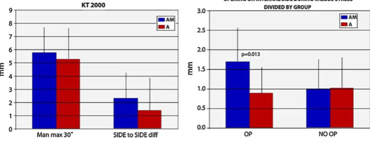
ObjectiveCombined ACL+MCL lesions are challenging to treat. A time zero evaluation with a navigation system showed that patients with preoperative Anteromedial (AM) instability had similar Anter-oposterior (AP) laxity at 30°but greater valgus laxity at 30°after the ACL reconstruction. This study investigates the same cohorts of patients at 3 years follow-up to see if the residual valgus laxity led to different results.
Material and methods In the previous study 57 patients were included, 20 with ACL+MCL lesion (group AM) and 37 with pure ACL lesions (group A). All patients underwent arthroscopic
double-bundle ACL reconstruction with STG. In group AM the MCL lesion was treated non-operatively. Fifty-one patients were available at follow-up: 19 for group AM and 32 for group A. AP laxity was measured using KT-2000 with Manual Maximum Test at 30°; valgus laxity was evaluated with a Telos X-Ray test with a 10 Nm valgus force. We evaluated clinical scores (IKDC, Lysholm, Tegner and Womac), muscle girth and time to return to activities.
Results We did not find any statistically significant difference con-cerning to clinical evaluation. A statistically significant difference was found in the medial joint opening during the valgus stress radiographs on the reconstructed knee (1.7±0.9 mm group AM, 0.9±0.7 mm group A;p=0.013). Finally there was no statistically significant difference respect to the AP laxity at 30° considering neither the AP displacement on the index side (5.8±1.9 mm group AM, 5.3±2.4 mm group A) or the side to side difference (2.4±2.5 mm group AM, 1.3±2.2 mm group A) (Fig.1).
ConclusionsThe results showed that the residual valgus laxity found at time zero in the group AM still exist but it did not affect the AP laxity significantly at a minimum follow up of 3 years. A trend was seen for AM group to have higher AP laxity value but this did not affect functional results. These findings suggest that no additional surgical procedure on the MCL lesion is needed in combined lesions.
Intra-operative analysis of knee dynamic stability
during ACL reconstruction: anatomic double-bundle
versus single-bundle plus lateral plasty
S. Zaffagnini1, N. Lopomo1, S. Bignozzi1, T. Bonanzinga1, C. Signorelli1,2, M. Marcacci1
1Istituto Ortopedico Rizzoli (Bologna, ITALY); 2Politecnico di Milano (Milan, ITALY)
IntroductionPivot-shift test is commonly used to qualitative assess the dynamic stability of the knee. Different ACL reconstructions are available: with a rationale, a specific reconstruction wanted to com-bine intra and extra-articular procedures using a single-blundle (SB) with an additional extra-articular lateral plasty-LP; with a different rationale the anatomical double-bundle (ADB) reconstruction was proposed trying to mimic the native ACL. The objective of this study was to quantify intra-operatively the recovery given both by an ADB reconstruction and by a SB+LP one, by means of PS test.
Material and methods 15 patients that underwent ADB ACL reconstruction and 10 patients that underwent SB+LP ACL recon-struction were involved in the study. A commercial navigation system (Orthokey, USA) was used for kinematic acquisitions. The surgeon
performed PS test before and after ACL reconstruction. We analysed the maximal anterior translation of the tibial lateral compartment during subluxation and the acceleration reached by the lateral com-partment during tibial reduction.
Results All the dynamic instabilities (translation, acceleration) were significantly reduced both by the ADB and SB+LP reconstruction (p\0.0005). There were not statistical significance if we considered the recovery in anterior translation due to both the techniques (9.5±7.7 mm and 10.3±2.1 mm respectively, p=0.7656). SB+LP patients pre-sented more acceleration after the reconstruction (457±110 mm/s2
versus661±208 mm/s2, p=0.0060), but there were no statistical differences in the recovery due to both the reconstructions (p=0.420).
ConclusionsThe presented intra-operative analysis of knee stability allowed to highlight possible different dynamic behaviours due to the use of two different ACL surgical reconstructions. All ACL-deficient knees showed positive pivot-shift test before the surgery revealing huge values in the tibial subluxation and in the acceleration reached by the tibia, that is linked to tibial reduction during PS phenomenon. However, even with some difference due to the execution of the test, both ADB and SB+LP reconstruction seemed to well control pivot-shift phenomenon reducing tibial translation and thus controlling the dynamic instability of the joint.
Results of isolated postero-lateral corner reconstruction
L. Camarda1, V. Condello2, V. Madonna2, F. Cortese2,
G. Caradonna1, M. D’arienzo1, C. Zorzi2
1Department of Orthopaedic and Traumatology, University of
Palermo (Palermo, ITALY);
2Department of Knee Surgery, Ospedale Sacro Cuore Don Calabria di
Negrar (Verona, ITALY)
BackgroundIsolated postero-lateral corner (PLC) tears are relatively rare events. Various surgical techniques to treat postero-lateral knee instability have been described; because surgical results are linked to cruciate reconstructions it has been difficult to date to define whether one surgical procedure has better prognosis than another. The goal of this study is to determine the clinical outcome of PLC reconstruction following fibular-based technique.
Material and methodsWe retrospectively evaluated a case series of patients who received isolated PLC reconstruction between March 2005 and January 2007. Ten patients were surgically treated for isolated injuries and were available for follow-up; mean patient age was 27.4 years (range 16–47 years). All patients were treated following the fibular-based technique: double femoral tunnel was performed in six patients, while in the remaining four patients, the reconstruction of the PLC was performed with a single femoral tunnel. Six patients had semitendinosus allograft and four had semitendinosus autograft. All patients had the same evaluation and the same rehabilitation protocol.
ResultsMean follow-up was 27.5 months (range 18–40 months). Mean range of motion (ROM) was 143.5 degrees for flexion (range 135–150°) and 0.5 degrees for extension (range 0–3°). Three patients showed 1+ on varus stress test, while on Dial test another three patients showed 10°
reduction of external rotation compared with contra-lateral knee. The mean Lysholm score was 94 points (range 83–100), and the mean International Knee Documentation Committee (IKDC) subjective result was 88.48 (range 74–96.5). Based on Lysholm score, the results were excellent in eight knees and good in two knees. On IKDC evaluation, two patients were grade A and eight were grade B. No significant difference in clinical results was observed between single and double femoral tunnel.
ConclusionsFibular-based technique showed good results in terms of clinical outcome, restoring varus and rotation stability of knees in treatment of chronic isolated PLC injury.
ACL reconstruction and development of knee arthritis:
long term clinical and radiographic follow-up
and analysis of correlations
M. Surace, P. Bulgheroni, D. Marcolli, A. Fagetti, P. Cherubino
Dipartimento di Scienze Chirurgiche Ricostruttive e Tecnologie Avanzate (Varese, ITALY)
Anterior cruciate ligament (ACL) lesion is a frequent pathology in athletes.
Aim of our s