Extra-articular hip impingement:
a narrative review of the literature
Scott W. Cheatham, PT, DPT, PhD(c), OCS, ATC, CSCS
11 Assistant Professor, Director Pre-Physical Therapy Program, Division of Kinesiology and Recreation, California State University Dominguez Hills
Corresponding Author: Scott W. Cheatham
Division of Kinesiology, California State University Dominguez Hills, 1000 E. Victoria St. Carson, CA 90747 Tel: (310) 243-3794
Email: [email protected] © JCCA 2015
Conflicts of interest and source of funding:
The author declares that there is no conflict of interest or source funding for this manuscript.
There is growing subgroup of patients with poor outcomes after hip arthroscopy for intra-articular pathology suggesting unrecognized cause(s) of
impingement may exist. Extra-articular hip impingement (EHI) is an emerging group of conditions that have been associated with intra-articular causes of impingement and may be an unrecognized source of pain. EHI is caused by abnormal contact between the extra-articular regions of the proximal femur and pelvis. This review discusses the most common forms for EHI including: central iliopsoas impingement, subspine impingement, ischiofemoral impingement, and greater trochanteric-pelvic impingement. The clinical presentation of each pathology will be discussed since EHI conditions share similar clinical features as the intra-articular pathology but also contain some unique characteristics.
(JCCA. 2016;60(1):47-56)
k e y w o r d s: hip pain, extra-articular, impingement,
diagnosis, review
Un nombre croissant de patients ont des résultats négatifs après l’arthroscopie de la hanche pour la pathologie intra-articulaire, ce qui indique l’existence de causes non reconnues de pincement. Le pincement extra-articulaire de la hanche est un groupe émergent d’états qui ont été associés à des causes intra-articulaires de pincement et peuvent constituer une source méconnue de douleurs. Le pincement extra-articulaire de la hanche est causé par un contact anormal entre les régions extra-articulaires du fémur proximal et le bassin. Cette étude examine les formes les plus courantes du pincement extra-articulaire de la hanche, y compris : le pincement central psoas-iliaque, le pincement sous-vertébral, le pincement ischio-fémoral et un pincement trochantérien-pelvien plus important.La présentation clinique de chaque pathologie sera discutée puisque les conditions du pincement extra-articulaire de la hanche ont des caractéristiques cliniques similaires à celles de la pathologie intra-articulaire, tout en contenant des caractéristiques uniques.
(JCCA. 2016;60(1):47-56)
m o t s c l é s : douleurs de hanche, extra-articulaire,
Introduction
Intra-articular causes of impingement such as femoral acetabular impingement (FAI) and acetabular labral tears have become well known causes of hip pain and impinge-ment in younger non-arthritic individuals. Arthroscopic and open surgical procedures are often indicated and have shown good outcomes with returning individuals back to pre-injury levels of function and sports activity.1-3 How-ever, there is growing subgroup of patients with poor out-comes after surgery suggesting unrecognized cause(s) of impingement may exist.4-6
More recently, an emerging body of literature has identified extra-articular causes of hip impingement that are associated with patients who have poor outcomes to intra-articular surgical procedures.5,6 Extra-articular hip impingement (EHI) is caused by abnormal contact be-tween the extra-articular regions of the proximal femur and pelvis and may coexist with intra-articular FAI.7 Re-gions of abnormal contact may exist between the greater trochanter, lesser trochanter, extracapsular femoral neck and the ilium or ischium.4,5 The causes of EHI have been further classified into specific conditions: central iliop-soas impingement, subspine impingement, ischiofemoral impingement, and greater trochanteric-pelvic impinge-ment.3,8
The research on these specific conditions is still emer-ging. A recent systematic review by de Sa et al5 appraised the literature on surgical interventions for these condi-tions. The authors found only 14 qualifying studies that varied in methodology and overall quality. The authors concluded that a small amount of evidence does exist sup-porting the surgical interventions for these conditions and that further research is necessary. The lack of evidence leaves a gap in our understanding of the pathophysiology of these conditions and how they relate to intra-articular pathology. Moreover, there is a need to determine the best diagnostic criteria for identifying these conditions and de-termining which interventions influence recovery. Extra-articular conditions share similar clinical fea-tures as the intra-articular pathologies but also contain some unique characteristics. Clinicians must have a work-ing knowledge of the clinical presentation of these condi-tions in order to enhance accuracy during the examination and differential diagnosis process. This manuscript will review common extra-articular conditions with a focus on clinical presentation.
Prevalence
The epidemiological data on EHI is still emerging and the available research has revealed some preliminary trends in patient demographics. Riccardi et al4 conducted a retrospective review of 1765 patients (2075 hips) who underwent hip preservation surgery (hip arthroscopy, periacetabular osteotomy, femoral osteotomy, and sur-gical hip dislocation) between 2010 and 2013. The au-thors analyzed two cohort groups: (1) EHI group and (2) intra-articular FAI group. The diagnosis of EHI was made preoperatively based on history, clinical examination, and radiographic studies. Seventy-five patients (86 hips) met the criteria for the EHI group and 1690 (1989 hips) pa-tients for the FAI group. Papa-tients in the EHI group were younger than the FAI group (24 ±7 years versus 30±11 years). The EHI group had an increased proportion of fe-males than the FAI group (85% to 49%). The right hip was the most commonly affected side in both groups (57% in each group). EHI patients were more likely to have under-gone prior hip surgery than the FAI group (44% to 10%) which consisted of hip arthroscopy (N = 24) and pelvic osteotomy (N = 6). The EHI group had lower preoperative outcome scores for the modified Harris hip score (mHHS) and Hip Outcome Scores activities of daily living (HOS ADL) (55 ± 15 versus 63 ± 15) after adjustments for age, sex, and type of revision surgery. Sixteen percent of the EHI patients were diagnosed with previous hip pathology which included: Leg Legg-Calve´-Perthes (N = 7), de-velopmental dysplasia of the hip (N = 2), slipped capital femoral epiphysis (N = 1), Ehlers-Danlos (N = 1), and postinfectious deformity (N = 1).4
The research by Riccardi et al4 suggested that EHI pa-tients tend to be younger, female, and have undergone previous FAI surgery. Also, the presence of EHI was about 4% (75 of 1765) which is infrequent compared to the intra-articular pathology. The authors suspect that the diagnosis of EHI pathology may have been missed during the initial diagnosis. This hypothesis has been supported by other clinical trials investigating the outcomes of FAI revision surgery. 9-11
Central Iliopsoas Impingement
extend into the anterosuperior portion of the labrum (e.g. 1 to 2 o’clock position). The damage often occurs direct-ly adjacent to the iliopsoas tendon at the 2 to 3 o’clock position of the anterior labrum and is often confirmed via magnetic resonance arthrography (MRA) (Figure 1).5,12,13 It is postulated that the impingement is caused by a repeti-tive traction injury by the iliopsoas tendon that is scarred and adherent to the capsule-labrum complex of the hip or by a tight or inflamed iliopsoas tendon that causes im-pingement during hip extension.5,8 Iliopsoas impingement has also been reported after total hip resurfacing and total hip arthroplasty when a larger femoral head component is used.14-17
The strongest available evidence on CII consists of retrospective case series from the United States with no randomized controlled trials.5 The current research sug-gests that this condition may be more common in younger females (pooled age range 19 to 35 years) and individuals involved in regular sports activities.5,12,18 Patients often re-port anterior hip pain with active flexion and may rere-port a
snapping sensation. The clinical examination may reveal non-specific focal tenderness over the iliopsoas tendon at the anterior joint line, positive hip impingement test (e.g. Flexion, Adduction, Internal Rotation (FADIR) test) (Fig-ure 2), and pain or apprehension with resisted straight leg raise (Table 1).12,18 Patients may report little or no relief after intraarticular injection of a local anesthetic. MRA is often ordered to further diagnose the condition. MRA has shown good diagnostic properties with strong intra-ob-server agreement.13 A labral tear at the 3-o’clock position (immediately below the iliopsoas tendon) suggests the diagnosis of iliopsoas impingement; especially if it does not extend above the 2-o’clock position.13
Non-surgical intervention such as activity modifica-tion, rehabilitamodifica-tion, and therapeutic injections may be pre-scribed first. However; the efficacy of these interventions have not been investigated.5 If conservative measures fail, then surgery may be an option. Often, patients will have concomitant labral injury with the iliopsoas pathology requiring arthroscopic resection or repair of the
acetabu-Figure 1.
a) Region of subspine impingement, b) Region of central psoas impingement
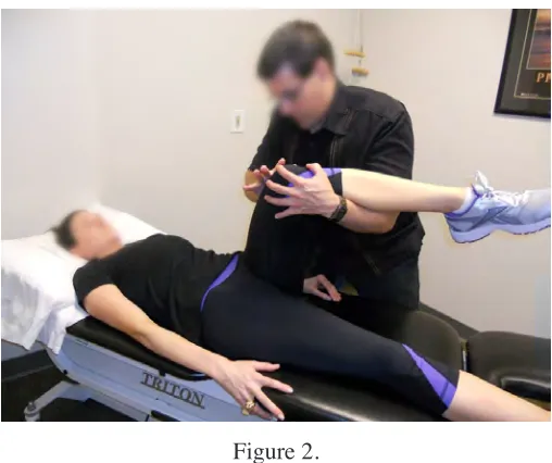
Figure 2.
Flexion-Adduction-Internal Rotation Test. The patient is lying supine. The affected hip is passively moved into 90 degrees of hip and knee flexion. The hip is then passively adducted with internal rotation and overpressure in both directions. A positive test is reproduction of the patient’s
lar labrum and iliopsoas tenotomy at the level of the la-brum.12,18 Studies have shown good short-term outcomes at 1 year post-operative for return to sports activity, re-stored range of motion (ROM), decreased symptoms, and scores on the mHHS and HOS ADL and sport HOS (Table 2).5,12,18,19 Further studies are needed to validate these find-ings and further develop diagnostic criteria for this path-ology. To date, the clinical trials have primarily reported post-surgical outcomes and briefly described post-opera-tive rehabilitation or did not mention if it was prescribed for these patients.5,18,20 Lindner et al.21 briefly outlined a post-surgical program. After surgery, the patient is partial weight bearing (e.g. 9.7kg (20lbs) flat-foot weight bear-ing) with crutches and a hip brace locked at 0 to 90 de-grees for the first 2-weeks. Two weeks post-surgical, the brace and crutches are discontinued and the patient con-tinues rehabilitation with an emphasis on regaining joint ROM, strengthening the gluteus medius and core mus-cles. The available details regarding the role of post-sur-gical rehabilitation is under reported. Further studies are needed to objectively assess the effects of post-operative rehabilitation for these individuals.
Figure 3.
The Subspine Impingement Test. The patient is lying supine. The affected hip is passively moved into maximum hip flexion (neutral adduction and internal rotation). A positive test is reproduction of the patient’s
concordant anterior hip pain.
Subspine Impingement
Subspine impingement (SSI) is caused by a prominent anterior inferior iliac spine (AIIS) abnormally contacting the distal femoral neck during hip flexion (Figure 1).5 SSI is thought to be caused by excessive muscular activity of the rectus femoris during repetitive knee flexion with hip extension resulting in an avulsion injury of the AIIS. Upon healing, the apophysis may be inferiorly displaced leading to a malunion which often results in an enlarged bony protrusion at the AIIS that abnormally abuts the femoral neck.8,22 Avulsion injuries are common in ado-lescent athletes. This repetitive traction injury is common in running sports and sports involving rapid high energy kicking such as soccer.5,22 Avulsion injuries to the AIIS are reported to be the second most common with ischial avulsions being the most common.10 SSI has been related to CAM-type FAI and may be corrected with surgery.8 The strongest available evidence on SSI consists of case reports and series from the United States, United Kingdom, and China with no randomized controlled trials.5 The current research suggests that this condition is more common in younger active males (age range 14 to 30 years).5,8 Patients often report anterior hip or groin pain that is aggravated by active hip flexion and activities such as running or kicking. The clinical exam may reveal palpable AIIS pain and limited passive hip flexion with end range anterior hip pain. The patient may or may not have a positive hip impingement test (Table 1).22 Poult-sides et al23 described the subspine impingement test that includes passively flexing the hip into maximum flexion (neutral adduction and internal rotation) (Figure 3). Re-production of the patient’s anterior pain is considered a positive test.23 Currently, there are no studies that have as-sessed the clinimetric properties of this test. Patients may report little or no relief with hip flexion after intraarticular injection of a local anesthetic. Radiographs may reveal a prominent AIIS deformity that extends distally to the level of the anterior-superior acetabular rim.22 The radiographs may also reveal sclerosis at the AIIS (inferior) and dis-tal femoral neck junction. Computed tomographic (CT) scans have also been used with a classification system to categorize the type of SSI.24 Researchers have also asso-ciated the ROM limits with each SSI classification (Table 3).24 To date, the diagnostic properties of this imaging has not been reported in the literature.
modifica-tion, rehabilitamodifica-tion, and therapeutic injections may be prescribed first but their efficacy have not been investi-gated.5 Some patients may be recalcitrant to conservative treatment and require surgical interventions. An open AIIS decompression is commonly performed through the
standard anterolateral and mid-anterior hip arthroscopy portals. At times, a concomitant arthroscopic procedure is conducted to address any intra-articular pathology.22 Studies have shown good short-term outcomes at up to 2 years post-surgical follow-up for return to sports activity, Table 1.
Common types of extra-articular hip impingement Extra-Articular
Condition Patient Demographics Pathological Characteristics Clinical Presentation
Central iliopsoas
impingement Pooled age range: 19–35 years
Gender: Females more than males
The pathology may be caused by: (1) a repetitive traction injury by the iliopsoas tendon that is scarred and adherent to the capsule-labrum complex of the hip, (2) a tight or inflamed iliopsoas tendon that causes impingement during hip extension
Patients often report anterior hip pain with active hip flexion and may report a snapping sensation. Clinical findings include non-specific focal tenderness over the iliopsoas tendon at the anterior joint line, positive hip impingement test (e.g. FADIR test), and pain or apprehension with resisted straight leg raise. MRA is often used to further diagnose the condition.
Subspine
impingement Pooled age range: 14–30 years
Gender: Males more than females
The pathology is caused by a prominent AIIS abnormally contacting the distal femoral neck during hip flexion. This may be due to an avulsion injury to the AIIS due to excessive muscular activity of the rectus femoris during repetitive knee flexion and hip extension.
Patients typically report anterior hip or groin pain that is aggravated by active hip flexion and activities such as running or kicking. Clinical findings include palpable AIIS pain and limited passive hip flexion with end range anterior hip pain. The patient may or may not have a positive subspine hip impingement test. Plain radiographs and computed tomography are commonly used to further diagnose the condition.
Ischiofemoral
impingement Pooled age range: 14–30 years
Gender: Females more than males
The pathology is caused by a narrowed space between the ischial tuberosity and the lesser trochanter resulting in repetitive impingement of the quadratus femoris muscle.
Patients often report non-specific pain in the hip, groin, and buttocks with active adduction and external rotation. Pain is often increased with sports related activity such as gymnastics or dance or activities of daily living such as long-stride walking. Referral pain may occur down the lower extremity due to possible irritation of the adjacent sciatic nerve. Clinical findings include pain with active or passive hip extension, external rotation, and adduction. In some cases, snapping may occur during hip flexion or extension during weight bearing activities. Magnetic resonance imaging and plain radiographs are commonly used to further diagnose the condition.
Greater trochanteric-pelvic impingement
Pooled age range: 5 to 41 years
Gender: No predilection
The pathology is caused by a painful and pathological contact between the greater trochanter and ilium when the hip is actively or passively moved into abduction and extension.
Patients typically report both lateral hip and groin pain that is reproduced with active hip abduction and extension. A blocking of the joint may be felt at the end range of these combined motions. Clinical findings include limited and painful active or passive hip abduction and extension, a shortening of the involved leg, and a Trendelenburg gait pattern. Plain radiographs are commonly used to further diagnose the condition. Abbreviations: FADIR: flexion-adduction-internal rotation; AIIS: anterior inferior iliac spine; MRA: magnetic resonance
increased ROM, scores on the HHS, visual analog scale, and short form 12.9,22,25 The post-surgical rehabilitation for SSI is poorly reported among the published surgical in-vestigations. Hestroni et al12 do recommend 2 to 4 weeks of protected weight bearing with crutches and ROM exercises until basic muscle strength is regained. Further strengthening and proprioception exercise should be pre-scribed as tolerated. Anti-inflammatory medications are also recommend for the first 3 to 4 weeks after surgery to help decrease the risk of heterotrophic ossification. 22 To date, post-surgical rehabilitation has not been objectively studied and its role in the post-operative period is poorly detailed in the literature.5
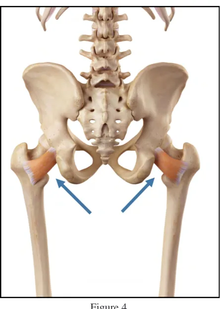
Ischiofemoral Impingement
Ischiofemoral impingement (IFI) is characterized by a narrowed space between the ischial tuberosity and the lesser trochanter resulting in repetitive impingement of the quadratus femoris muscle (Figure 4).5,7 The condition has been reported as primarily congenital but may also be acquired from a hip fracture, superior medial migration of the hip joint with osteoarthritis, or total hip arthroplasty when offset is not fully restored.7,8
The strongest available evidence consists of case re-ports and series outside the United States with no ran-domized controlled trials.5 The research suggests that ischiofemoral impingement is more prevalent in females Table 2.
Common non-arthritic patient reported outcome measures for the hip Patient Related Outcome
Measure Type of Questions Description
Hip Outcome Score 24 questions measuring activities of daily living and physical function during sports activity.
Each subscale is scored separately. The highest potential score for the ADL scale is 68 and 38 for the sports subscale. The scores are converted to a percentage. A higher score represents a higher level of physical function. modified Harris Hip Score 2 rating scales and 8 items.
The domains covered are pain, function, and functional activity.
Each item has a unique numerical scale. There are 100 total points. A higher score represents a higher level of physical function.
Abbreviation: ADL: activities of daily living
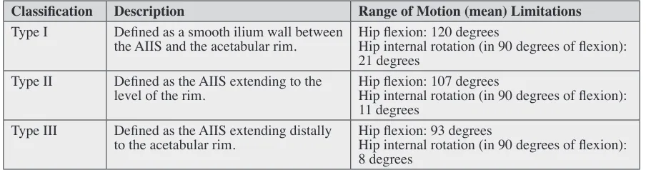
Table 3.
Classification of subspine impingement
Classification Description Range of Motion (mean) Limitations
Type I Defined as a smooth ilium wall between
the AIIS and the acetabular rim. Hip flexion: 120 degreesHip internal rotation (in 90 degrees of flexion): 21 degrees
Type II Defined as the AIIS extending to the
level of the rim. Hip flexion: 107 degreesHip internal rotation (in 90 degrees of flexion): 11 degrees
Type III Defined as the AIIS extending distally
to the acetabular rim. Hip flexion: 93 degreesHip internal rotation (in 90 degrees of flexion): 8 degrees
versus males and older individuals (mean age of 51-53 years, pooled age range 14-77 years).5,7,8 Bilateral IFI is believed to occur in approximately 15 to 30% of cases of individuals diagnosed with IFI. There is an increased risk of IFI in patients who have suffered prior proximal hamstring avulsion fractures or multiple hereditary exos-toses.7 Patients often report non-specific pain in the hip, groin, and buttocks with active adduction and external ro-tation. Pain is often increased with sports related activity such as gymnastics or dance or activities of daily living such as long-stride walking.7 Referral pain may occur down the lower extremity due to possible irritation of the adjacent sciatic nerve.7,8
The clinical examination may reveal pain with active or passive hip extension, external rotation, and adduction. In some cases, snapping may occur during hip flexion or ex-tension during weight bearing activities (Table 1).7 There are no specific special tests for IFI which is often mistaken
for intra-articular pathology and is largely dependent on magnetic resonance imaging (MRI).8 On MRI, decreased space between the ischium and lesser trochanter is often identified as a risk for IFI. Singer et al26 conducted a me-ta-analysis of MRI studies (2005 to 2014) and determined that a cut-off threshold of ≤ 15 mm (ischio-femoral space) showed a sensitivity of 77%, specificity of 81%, and over-all accuracy of 78.3% for diagnosing IFI (defined as the presence of quadratus femoris edema and/or atrophy, and ipsilateral pain). Edema of the quadratus femoris muscle may be visible in patients with IFI and some patients may present with fatty infiltration of the quadratus femoris muscle which is sometimes combined with muscle atro-phy.7,8
Plain radiographs are often negative but may reveal sclerosis or cystic changes within the lesser trochanter or ischium, decreased femoral offset, or bony prominences from ischial avulsion injury or multiple hereditary exos-Figure 4.
toses.7 Patients may report relief after intraarticular injec-tion of a local anesthetic.27
Non-surgical management is often prescribed first with a focus on avoiding activities that involve combined hip adduction and external rotation. Rehabilitation should be directed towards strengthening the hip external rotators and abdominal core. Stretching of the hip adductors and external rotators should also be done in the presence of decreased muscle length.7 Nonsteroidal anti-inflamma-tory (NSAID’s) medication and therapeutic corticosteroid injections may also be beneficial. Investigations have re-ported good outcomes with the combination of activity modification, rehabilitation, NSAIDS, and therapeutic in-jections.7,28,29
If non-surgical interventions fail, then surgical management may be suggested. The objective of surgery is to widen the space by resecting the bone from the lesser trochanter or ischium or releasing the quadratus femoris muscle. The surgery is commonly done through the stan-dard anterolateral arthroscopy portal. Psoas weakness is a potential complication. Resection of the lesser trochanter and release of the quadratus femoris creates a risk for dis-rupting the lateral circumflex artery and avascular necro-sis of the femoral head.7 The studies reporting outcomes from this surgery have mainly been case reports which make intra-study comparisons difficult due to the low level of evidence.5 However; the published case reports have shown good outcomes up to a 3.5 year follow-up for decreased symptoms, increased ROM, and return to func-tion.5 To date, post-surgical rehabilitation for IFI has not been objectively studied and its role in the post-operative period is poorly detailed in the literature.
Greater Trochanteric-Pelvic Impingement
Greater trochanteric-pelvic impingement (GTPI) is de-scribed as painful and pathological contact between the greater trochanter and ilium when the hip is actively or passively moved into abduction and extension (Figure 5).7 During development an elongation of the great-er trochantgreat-er occurs due to partial or complete arrest of the proximal femoral physis. With a complete arrest of the proximal femoral physis, the femoral neck becomes shortened, thickened, and develops a varus deformity. The trochanteric epiphysis may also be elongated further predisposing individuals to GTPI. GTPI is commonly as-sociated with Legg–Calvé–Perthes disease but also may
be related to the ischemia that occurs with the treatment of congenital hip dislocation, hip infection, traumatic in-jury, and infantile coxa vara.5,7
in-creased strength, and improved gait. No osteonecrosis events were reported.5 Despite the favorable results, the weakness in the evidence must be considered when inter-preting these findings. To date, post-surgical rehabilita-tion for GTPI has not been objectively studied and its role in the post-operative period is also poorly detailed in the case reports.
Conclusion
Understanding the clinical presentation of common EHI conditions is vital to the hip differential diagnosis process. Currently, the evidence on EHI is weak but does present some preliminary insight into the clinical presentation of these conditions. The one related characteristic among all EHI pathologies is that they may co-exist with intra-articu-lar causes of impingement and may be overlooked during the examination process. This must be considered during the examination and differential diagnosis process in order to accurately diagnosis all causes of hip impingement.
References:
1. Kemp JL, Collins NJ, Makdissi M, et al. Hip arthroscopy for intra-articular pathology: a systematic review of outcomes with and without femoral osteoplasty. Br J Sports Med. 2012;46(9):632-643.
2. Papalia R, Del Buono A, Franceschi F, et al.
Femoroacetabular impingement syndrome management: arthroscopy or open surgery? Int Orthop. 2012;36(5):903-914.
3. Clohisy JC, St John LC, Schutz AL. Surgical treatment of femoroacetabular impingement: a systematic review of the literature. Clin Orthop Relat Res. 2010;468(2):555-564. 4. Ricciardi BF, Fabricant PD, Fields KG, et al. What are
the demographic and radiographic characteristics of patients with symptomatic extraarticular femoroacetabular impingement? Clin Orthop Relat Res. 2015;473(4):1299-1308.
5. de Sa D, Alradwan H, Cargnelli S, et al. Extra-articular hip impingement: a systematic review examining
operative treatment of psoas, subspine, ischiofemoral, and greater trochanteric/pelvic impingement. Arthroscopy. 2014;30(8):1026-1041.
Figure 6.
The “Gear Stick” Sign. The patient is sidelying with the affected side up. The hip
is passively moved into abduction and extension without excessive lumbopelvic movement. A positive test is limited range of motion and reproduction of the
6. Cvetanovich GL, Harris JD, Erickson BJ, et al. Revision hip arthroscopy: a systematic review of diagnoses, operative findings, and outcomes. Arthroscopy. 2015;31(7):1382-1390.
7. Beckmann JT, Safran MR, Abrams GD.
Extra-articular impingement: ischiofemoral impingement and trochanteric-pelvic. Oper Techniques in Sports Med.2015. [Published ahead of print]
8. Sutter R, Pfirrmann CWA. Atypical hip impingement. Am J Roentgenol. 2013;201(3):W437-W442.
9. Larson CM, Kelly BT, Stone RM. Making a case for anterior inferior iliac spine/subspine hip impingement: three representative case reports and proposed concept. Arthroscopy. 2011;27(12):1732-1737.
10. Ross JR, Stone RM, Larson CM. Subspine Impingement. Operative Techniques in Sports Medicine.
11. Sardana V, Philippon MJ, de Sa D, et al. Revision hip arthroscopy indications and outcomes: a systematic review. Arthroscopy. 2015.[Published ahead of print] 12. Domb BG, Shindle MK, McArthur B, et al. Iliopsoas
impingement: a newly identified cause of labral pathology in the hip. HSS J. 2011;7(2):145-150.
13. Blankenbaker DG, Tuite MJ, Keene JS, et al. Labral injuries due to iliopsoas impingement: can they be diagnosed on MR arthrography? Am J Roentgenol. 2012;199(4):894-900.
14. Cobb JP, Davda K, Ahmad A, et al. Why large-head metal-on-metal hip replacements are painful: the anatomical basis of psoas impingement on the femoral head-neck junction. J Bone Joint Surg Br. 2011;93(7):881-885. 15. Browne JA, Polga DJ, Sierra RJ, et al. Failure of
larger-diameter metal-on-metal total hip arthroplasty resulting from anterior iliopsoas impingement. J Arthroplasty. 2011;26(6):978.e975-978.
16. Piggott RP, Doody O, Quinlan JF. Iliopsoas tendon rupture: a new differential for atraumatic groin pain post-total hip arthroplasty. BMJ Case Rep. 2015. [Published ahead of print].
17. Jerosch J, Neuhauser C, Sokkar SM. Arthroscopic treatment of iliopsoas impingement (IPI) after total hip replacement. Arch Orthop Trauma Surg. 2013;133(10):1447-1454.
18. Cascio BM, King D, Yen YM. Psoas impingement causing labrum tear: a series from three tertiary hip arthroscopy centers. J La State Med Soc. 2013;165(2):88-93.
19. Nelson IR, Keene JS. Results of labral-level arthroscopic iliopsoas tenotomies for the treatment of labral
impingement. Arthroscopy. 2014;30(6):688-694.
20. Jerosch J, Neuhäuser C, Sokkar S. Arthroscopic treatment of iliopsoas impingement (IPI) after total hip replacement. Arch Orthop and Trauma Surg. 2013;133(10):1447-1454. 21. Lindner D, Stake CE, El Bitar YF, et al. Endoscopic
iliopsoas tenotomy for iliopsoas impingement on a collared femoral prosthesis. Arthrosc Tech. 2013;2(3):e205-208. 22. Hetsroni I, Larson CM, Dela Torre K, et al. Anterior
inferior iliac spine deformity as an extra-articular source for hip impingement: a series of 10 patients treated with arthroscopic decompression. Arthroscopy. 2012;28(11):1644-1653.
23. Poultsides LA, Bedi A, Kelly BT. An algorithmic approach to mechanical hip pain. HSS J. 2012;8(3):213-224.
24. Hetsroni I, Poultsides L, Bedi A, et al. Anterior inferior iliac spine morphology correlates with hip range of motion: a classification system and dynamic model. Clin Orthop Rel Res. 2013;471(8):2497-2503.
25. Hapa O, Bedi A, Gursan O, et al. Anatomic footprint of the direct head of the rectus femoris origin: cadaveric study and clinical series of hips after arthroscopic anterior inferior iliac spine/subspine decompression. Arthroscopy. 2013;29(12):1932-1940.
26. Singer A, Subhawong T, Jose J, et al. Ischiofemoral impingement syndrome: a meta-analysis. Skel Radiol. 2015;44(6):831-837.
27. Backer MW, Lee KS, Blankenbaker DG, et al. Correlation of ultrasound-guided corticosteroid injection of the quadratus femoris with MRI findings of ischiofemoral impingement. Am J Roentgenol. 2014;203(3):589-593. 28. Lee S, Kim I, Lee SM, et al. Ischiofemoral impingement
syndrome. Ann Rehabil Med. 2013;37(1):143-146. 29. Safran M, Ryu J. Ischiofemoral impingement of the hip: a
novel approach to treatment. Knee Surg Sports Traumatol Arthrosc. 2014;22(4):781-785.
30. Stevens P, Anderson L, Gililland J, et al. Guided growth of the trochanteric apophysis combined with soft tissue release for Legg–Calve–Perthes disease. Strat Trauma Limb Reconstr. 2014;9(1):37-43.