THE GENERATION AND ANALYSIS OF MICE BEARING A HUMAN ALPHA-1-ANTITRYPSIN GENE CONSTRUCT
CONTAINING THE Z MUTATION.
ROBIN R. ALI
A th e s is s u b m itte d In p a rtia l fu lfilm e n t for the degree of Doctor of Philosophy
of th e U n iversity of London.
MRC Human B iochem ical G enetics Unit, The G alton Laboratory,
ProQuest Number: 10106839
All rights reserved
INFO RM A TIO N TO ALL U SER S
The quality of this reproduction is dependent upon the quality of the copy submitted.
In the unlikely event that the author did not send a complete manuscript
and there are missing pages, these will be noted. Also, if material had to be removed, a note will indicate the deletion.
uest.
ProQuest 10106839
Published by ProQuest LLC(2016). Copyright of the Dissertation is held by the Author.
All rights reserved.
This work is protected against unauthorized copying under Title 17, United States Code. Microform Edition © ProQuest LLC.
ProQuest LLC
789 East Eisenhower Parkway P.O. Box 1346
A B S T R A C T
alpha-1-antitrypsin (AAT) is a serum protease inhibitor whose major physiological target is neutrophil elastase in the lung. The AAT Z variant is not properly secreted into the circulation, but accumulates in the endoplasmic reticulum of the hepatocytes that synthesise it. Individuals that are homozygous for the Z allele usually develop chronic emphysema and/or liver disease as adults, but a proportion (10%) develop neonatal liver disease. There is no heterogeneity in the Z allele. However, it is known that the risk of neonatal liver disease to a homozygous ZZ infant varies between families indicating that additional environmental or genetic factors may play a role in its etiology.
An attempt was made to reproduce aspects of the AAT related liver disease in mice in order to investigate which additional factors may contribute to the development of the childhood disease. An AAT Z construct was introduced into mice in order to test whether this would result in liver disease despite the presence of normal endogenous mouse AAT.
First the AAT Z gene construct was produced by a novel technique involving homologous recombination in E. Coli which was between a cosmid containing a normal human AAT gene and a plasmid containing an exon of the gene with the Z mutation. Secondly a facility for generating transgenic mice was established and two
lines of mice carrying the AAT Z construct were produced. One of these lines expressed the transgene. The expressing Z line was compared to other lines bearing the same construct and to lines bearing the normal human AAT gene.
ACKNOWLEDGEMENTS
I am grateful for the MRC studentship which allowed me to work for three years at the Galton Laboratory. My thanks to EMBO for providing a short term fellowship which allowed me to spend one month in Marco Tripodi's laboratory in "La Sapinza", Rome, where I carried out the homologous recombination experiment. A further trip to this laboratory, when I carried out Northern analysis of transgenic mice, was funded by the MRC.
I thank those who contributed to the work presented in this thesis: Marco Tripodi, Jenny Farrington, Margaret Fox, Carlo della Rocca, Paul Me Cullagh, Harry M illward-Sadler and Phil Johnson. I gratefully acknowledge them where their work is described.
The supportive friendship of many members of the Galton (past as well as present) has been very important to me. In this repect Kay Taylor and Godfrey Gillett deserve a special mention. I would also like to thank Tom Kirkwood and Bob Rosenberger for their much appreciated support during the last few months of preparing the thesis at the NIMR.
In addition, I would like to thank those whose general help and advice were invaluable: Yvonne Edwards who shared the struggle to make transgenic mice. David Whitehouse and Jenny Lovegrove who introduced me to the analysis of proteins and Paul Burgoyne and Steve Palmer who taught me much about mice.
I would like to thank Sue Povey and Cathy Abbott. I am grateful for the help, wise advise and encouragement that they have provided along the way. Moreover, I thank them for having faith in me when the chips were down. Cathy has provided more support than I could ever have expected from a friendship.
CONTENTS
ABSTRACT 2
ACKNOWLEDGMENTS. 4
CONTENTS 5
ABBREVIATIONS 17
CHAPTER 1 INTRODUCTION. 2 0
PART ONE: ALPHA-1-ANTITRYPSIN.
1.1 1 Structure and function of the AAT protein. 21
1.1.2 Expression of the AAT gene. 2 6
1.1.3 AAT gene structure. 3 0
1.1.4 Control of expression of the AAT gene. 3 3
1.1.5 AAT variants. 3 9
1.1.6 Evolution of the AAT variants. 4 4 1.1.7 Association of AAT deficiency with disease. 51
PART TWO: PiZ AND LIVER DISEASE.
1.2.2 The Z mutation. 5 3
1.2.2 Liver disease associated with PiZ. 5 9
1.2.3 Hepatic pathology. 61
1.2.4 Heat shock proteins and PiZ related liver
1.2.5 Incidence of neonatal liver disease in 6 5 PiZZ individuals.
1.2.6 Prognostic factors in neonatal liver 6 6 disease associated with PiZ.
1.2.7 Familial risk of neonatal liver disease 6 7 associated with PiZ.
1.2.8 Environmental factors and neonatal liver 6 7 disease associated with PiZ.
1.2.9 Genetic variation in the severity of 6 9 neonatal liver disease associated with PiZ.
1.2.9.1 Heterogeneity of the PiZ allele. 6 9
1.2.9.2 Genes linked to Pi. 6 9
1.2.9.3 Genes not linked to Pi. 7 2
PART THREE: A MOUSE MODEL OF PiZ RELATED LIVER DISEASE?
1.3.1 Animal models of human disease. 7 4
1.3.1.1 Experimental models. 7 4
1.3.1.2 Genetic models - the mouse. 7 5 1.3.2 Transgenic mouse models of human disease. 8 0 1.3.2.1 Introduction of additional protein- 81
encoding genes.
1.3.2.2 Targeted destruction of specific cell 8 4 types - toxigenics.
1.3.2.3 Inactivation of endogenous mouse 8 5 genes.
1.2.3.5 Systems for studying early onset 91 lethal diseases.
1.3.3 Transgenic mouse models of PiZ related 9 2 liver disease.
1.3.3.1 Murine AAT genes. 9 2
1.3.3.2 Aims of project. 9 4
CHAPTER 2 THE PiZ GENE CONSTRUCT.
2.1 Introduction. 9 6
2.2 M aterials. 9 8
2.2.1 Standard media and buffers. 9 8
2.2.2 Vectors and bacterial strains. 1 0 0
2.2.3 Autoradiography. 1 0 0
2.3 Methods. 101
2.3.1 Quantitation of DNA. 101
2.3.2 Double stranded DNA sequencing. 101 2.3.3 Preparation of competent cells. 1 0 4 2.3.4 Transformation of competent cells with
plasmid. 1 0 4
2.3.5 Transfection of competent cells with 1 0 5 packaged cosmid.
2.3.6 Large scale cosmid DNA preparations. 1 0 5 2.3.7 Small scale cosmid DNA preparations. 1 0 7
2.3.8 Packaging of cosmid DNA. 1 0 7
2.3 .1 0 Agarose gel electrophoresis. 1 0 7
2.3.11 Dry Southern blotting. 1 0 8
2 .3 .1 2 Kinase reaction. 109
2.3 .1 3 Oligo hybridisation. 110
2.4 General Strategy. 1 1 2
2.5 Results. 1 1 5
2.5.1 Recombinant donor plasmid and acceptor 1 1 5 cosmid.
2.5.2 Homologous recombination. 1 1 8
2.6 Discussion. 1 3 7
CHAPTER 3 GENERATION OF MICE BEARING THE PiZ CONSTRUCT.
3.1 Introduction. 139
3.2 Materials and methods. 1 4 5
3.2.1 Preparation of DNA. 1 4 5
3.2.2 M2 and M IS media for culturing mouse 1 4 7 embryos.
3.2.3 Requirements for animal husbandry and 150 m anipulations.
3.2.4 Animal manipulations. 1 5 4
3.2.4.1 Killing mice. 1 5 4
3.2.4.2 Intraperitoneal injection. 1 5 4
3.2.4.3 Anaesthetising mice. 1 5 4
3 .2 .4 .6 Collection of fertilised single cell eggs. 1 59
3 .2 .4 .7 Oviduct transfer. 1 62
3 .2 .4 .8 Uterine transfer. 169
3.2.4.9 Caesarian section and fostering. 171 3.2.5 Microinjection of fertilised single cell 1 7 2
embryos.
3.2.6 Testing offspring for presence of 1 8 3 transgene.
3.2.6.1 DNA preparation from tail tips. 1 8 3 3 .2 .6 .2 PCR of tail tip DNA. 1 8 5
3.3 Results. 1 8 6
3.4 Discussion. 1 9 0
3.5 Appendix. 191
3.5.1 Names and addresses of some specialised suppliers. 3 .5 .2 List of surgical/dissection materials and
equipment.
3 .5 .3 List of equipment for the microinjection of single cell embryos.
3.5.4 List of materials for the microinjection of single cell embryos.
CHAPTER 4 ANALYSIS OF MICE BEARING HUMAN AAT GENES.
4.1 Introduction. 1 9 7
4 .2 Materials and methods. 2 0 0
4.2.1 Venisection of mice. 2 0 0
4 .2 .2 Preparation of plasma. 2 0 0
4 .2 .4 Isoelectric focusing (lEF). 2 0 2
4.2.4.1 Treatment of samples. 2 0 2
4 .2 .4 .2 Preparation of acrylamide lEF gel. 2 0 3
4 .2.4.3 Electrophoresis. 2 0 5
4 .2 .5 Western blotting. 2 0 5
4 .2 .6 Fluorescence in situ hybridisation. 2 0 7
4.2.7 RNA Preparation. 2 1 5
4 .2 .8 Northern blot analysis. 2 1 7
4 .2 .9 Hybridisation of Northern blot. 2 1 9
4.2.10 Histological analysis. 221
4.3 Results. 2 2 2
4.3.1 Analysis of expression of P iZ I. 2 2 2 4 .3 .2 Cytogenetic analysis of PiZI 2 2 7 4.3.2.1 Number of sites of insertion of transgene 2 2 7
in P iZ I.
4 .3 .2 .2 Can the number of sites be determined 2 3 4 reliably with interphase nuclei?
4.3.2.3 Detection of homozygotes. 2 3 7
4 .3 .2 .4 Identification of mouse chromosome 2 4 3 carrying the expressed transgene.
4 .3 .2 .5 Discussion. 2 4 6
4.3.3 Characterisation of PiM and PiZ lines. 2 4 8
4.3.3.1 Transcriptional analysis. 2 4 8
4 .3 .3 .4
Histological analysis of transgenic mice.
25 6
4.4
Discussion.
2 68
4.4.1
The transgenic mouse model of PiZ related 2 6 8
liver disease.
4 .4.2
Other transgenic mouse models of PiZ
2 7 3
related liver disease.
4.4.3
Comparison of all the transgenic mouse
2 7 8
models of PiZ related liver disease.
CHAPTER 5 DISCUSSION.
5.1
Accuracy of mouse models of human
2 8 2
genetic disease.
5.2
Accuracy of mouse models of PiZ
2 8 5
related liver disease.
5.3
Toxicity of accumulated PiZ protein.
2 8 7
5.4
Factors which may affect liver disease in 2 8 9
neonates.
5.5
‘ Work in progress and future work.
29 2
LIST OF FIGURES AND TABLES.
Figure 1.1 Figure 1.2
Figure 1.3 Figure 1.4 Figure 1.5
Figure 1.6 Figure 1.7
Carbohydrate side chains of AAT. 2 2 Relative AAT mRNA content (units/cell) 2 8 of liver, blood monocytes and alveolar
macrophages (taken from Brantly et al., 1988).
Structure of the AAT gene. 3 2 Control of hepatic expression of AAT. 3 6 Polyacrylamide gel lEF (pH 4.0 to pH 5.0) 4 0 of human serum showing the major isoforms for a number of AAT variants.
Possible evolution of some AAT variants. 4 7 The CSIRO ribozyme hammerhead structure. 8 7
Table 1.1 Table showing amino acid differences between the oldest AAT variant (Pi M l
ALA 213) and other AAT variants with their corresponding DNA sequence changes.
4 8
Figure 2.1
Figure 2.2
Figure 2.3
Figure 2.4
Figure 2.5
Scheme for introducing the Z mutation 1 1 4 into an AAT gene construct by
homologous recombination in E. Coli.
Diagram of acceptor cosmid 1 1 7 pCOS RBP-CAT/A1AT.
Ego RI restriction map of 123
pCOS RBP-CAT/A1 AT in kilobases.
Integration of pUCQZ, via homologous 1 2 4 recombionation, into pCOS RBP-CAT/A1 AT
Figure 2.6 Eco RI restriction digests of cosmid DNA 1 28 from integration clone 16
Figure 2.7 Eco RI and Bam HI restriction digests of 130 cosmid DNA from 4 excision clones.
Figure 2.8 Eco RI restriction digests of cosmid DNA 1 34 from 7 excision clones.
Figure 2.9 Autoradiographs of M and Z specific oligo 1 3 5 hybridisations to Eco RI digested cosmid
DNA from 7 excision clones
Figure 2.10 DGGE of 218 bp DNA fragments produced by 1 3 6 PCR using primers flanking the region of
the AAT gene containing the Z mutation.
Figure 3.1 Diagram outlining three different strategies 1 4 2 that have been used to produce transgenic
mice.
Figure 3.2 Outline of method for producing transgenic 1 4 4 mice by microinjection of fertilised eggs.
Figure 3.3 Method for restraining mouse in order 1 5 5 to perform intraperitoneal injection.
Figure 3.4 Position of inscision for vasectomy. 1 57
Figure 3.5 Detail of vasectomy. 1 5 8
Figure 3.6 Collection of eggs from the oviduct. 161 Figure 3.7 Position of inscision for oviduct and 1 64
uterine transfers.
Figure 3.8 Detail of oviduct transfer. 1 6 5
Figure 3.9 Oviduct transfer pipette. 1 6 8
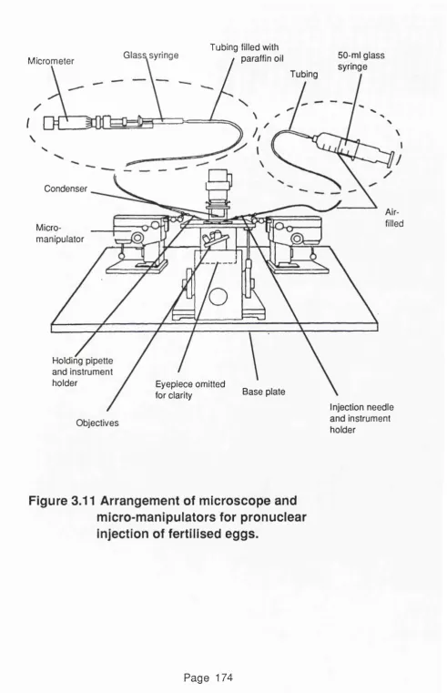
Figure 3.11 Arrangement of microscope and 1 7 4 micromanipulators for pronuclear
injection of fertilised eggs.
Figure 3.12 Diagram showing: A) The holding pipette 175 B) The microinjection needle.
Figure 3.13 Chamber for microinjecting fertilised 1 7 7 eggs.
Figure 3.14 Microinjection into the pronucleus. 181 Figure 3.15 Restrainer used for tail bleeding mice. 1 8 4 Figure 3.16 PGR analysis of tail DNA from 4 potential 189
transgenic mice
Figure 4.1 PGR analysis of the first litter from founder 2 2 3 P iZ I.
Figure 4.2 "Rocket" immunoelectrophoresis, using goat 2 2 4 anti-human AAT antibody, of plasma from
founder PiZI and its first litter.
Figure 4.3 Diagram showing the results of PGR and 2 2 6 "rocket" immunoelectrophoretic analysis of
the PiZI line.
Figure 4.4 FISH with FI cosmid to human metaphase 2 2 9 chromosomes.
Figure 4.5 FISH with FI cosmid to a metaphase 2 3 3 chromosome spread from mouse 1.6 (an
expressing transgenic mouse derived from P iZ I), showing the two sites of integration.
Figure 4.6 FISH with FI cosmid to a metaphase 2 3 4 chromosome spread from mouse 1.6.7
(expressing offspring of mouse 1.6
Figure 4.7 FISH with cosmid F1 to interphase cells 2 3 6 from a transgenic mouse known to have
only 1 chromosome carrying the PiZ transgene.
Figure 4.8 Results of in situ hybridisation analysis of 2 4 2 PiZ1 intercross offspring which are
potentially homozygous for the chromosome carrying the expressed integrated piZ construct.
Figure 4.9 FISH with cosmids F1 and 21/6 to a 2 4 5 metaphase spread from mouse 1.6.
Figure 4.10 Northern blot analysis of the relative 2 4 9 level of human AAT mRNA in five transgenic
mouse lines
Figure 4.11 Rocket" immunoelectrophoresis, using goat 2 5 2 anti-human AAT antibody, of plasma from
4 transgenic PiZ lines.
Figure 4.12 Rocket" immunoelectrophoresis, using goat 2 5 3 anti-human AAT antibody, of plasma from 5
transgenic PiM lines.
Figure 4.13 Immunodetection of AAT, using goat anti- 2 5 5 human AAT antibody, after lEF of plasma
from transgenic PiM and PiZ lines.
Figure 4.14 Section of liver from a non transgenic 2 6 2 C57BL/6 mouse aged 4 months.
Immunoperoxidase staining for human AAT is negative. The antibody had been pre-incubated with mouse serum.
(Magnification x 250).
Figure 4.15 Section of liver from a PiZI mouse aged 2 6 3 4 months showing periportal and
perivenular accentuation of cytoplasmic human AAT -stained brown. (Anti- human AAT immunoperoxidase staining,
magnification x 100).
Figure 4.17
Figure 4.18
Figure 4.19
changes as well as dark brown cytoplasmic globules of human AAT in hepatocytes around the terminal hepatic vein. (Anti-human AAT
immunoperoxidase staining, magnification X 250).
Section of liver from a PiM mouse 2 6 5 (line A T I 6) aged 15 months containing a
large nuclear inclusion of human AAT and showing fatty change. (Anti-human AAT immunoperoxidase staining, magnification X 100).
Section of a liver from a PiZI mouse aged 2 6 6 8.5 months. A large area of focal necrosis
- small dark blue mononuclear
inflammatory cells surrounding dead or dying hepatocytes- can be seen towards the right of the section. (Haemotoxylin and eosin,
magnification x 250).
Liver section from a human PiZZ individual 2 7 2 aged 27 years, showing cirrhosis with a band of abnormal fibrosis through the centre. The hepatocytes on each side contain globules of AAT. (Anti-human AAT immunoperoxidase staining, magnification x 100).
Table 4.1
Table 4.2
Table 4.3
Table showing results of FISH to interphase 2 3 5 cells from mice with a known number
of chromosomes carrying the PiZ transgene.
Results of histopathological analysis 2 5 8 by P. McCullagh and H. Millward-Sadler.
A B B R E V IA T IO N S
A AT a lp h a -1 -a n tltry p s in
AACT alpha-1 -antichym otrypsin AMG alpha-2-m acroglobulin
BARF beta-amyloid precursor protein
bp base pair
BRDU bromodeoxyuridine
CAT chloramphenicol acetyl transferase OF cystic fibrosis
CFTR cystic fibrosis transmembrane regulator
DAB diaminobenzidine
CIH2O distilled w ater
ddHaO distilled, de-ionised water DARI diaminophenolindole
DOGE denaturing gradient gel electrophoresis dl d e c ilitre
DTT d ith io th re ito l
EIG elastase inhibitory capacity EDTA ethelenediam inotetra-acetic acid
m endoplasmic reticulum
ES embryonic stem cell
FITC fluorescein isothiocyanate
g gram
HBV hepatitis B virus
HPRT hypoxanthine phosphoribosyl transferase HSV-1 Herpes simplex virus 1
HSV-1 tk Herpes simplex virus 1 thymidine kinase lAA Isoamyl alchohol
IE immediate early lEF isoelectric focusing FCS fetal calf serum IL6 interleukin 6
kb kilobase
kD kilodalton
I lit r e
LB Lauria broth
LM Lauria broth with maltose LPL low density lipid
LPS lipopolysaccharide
MHC major histocompatability complex MHV mouse hepatitis virus
NOD non obese diabetic NON non obese non-diabetic
PCR polymerase chain reaction Pi protease inhibitor
PI propidium iodide
PIL protease inhibitor-like gene psi pounds per square inch
RA rheumatoid arthritis
RBR rough endoplasmic reticulum hnRNA heteronuclear RNA
mRNA messenger RNA
CHAPTER 1
INTRODUCTION
PART ONE: ALPHA-1-ANTITRYPSIN.
1.1.1 Structure and function of the AAT protein.
AAT is a medium sized glycosylated protein of approximately 51 kD with an overall negative charge; it consists of a single polypeptide chain of 394 amino acids with one free cysteine residue and three oligosaccharide side chains (Carrell et al., 1982). There is a single reactive site - centered around methionine at position 358 (Brantly
at a!., 1988a). Of the amino acids, 30% are arranged in a -h e lic e s and 40% into p-pleated sheets. The first 24 N-terminal amino acids do not participate in any ordered structure (Loebermann et a!.,
Man-GIcNAc-Gal-Sia
-Asn-GlcNAc-GlcNAc-Man I I
I Man-GlcNAC’Gal-Sia
\
Man-GlcNAc-Gal-Sia
1
I
Asn = A sp arag in e
GlcNac = N -acetylg lu co sam in e Man = M a n n o s e
Gal = G a la c to s e
Sia = N -ac etyln e u ra m in ic acid
Figure 1.1 Carbohydrate side chains of AAT.
Analysis of a single AAT genotype using polyacrylamide gel electrophoresis at pH 4.9 5 , normally reveals eight bands or isoforms. The three major ones (2,4 and 6) have different numbers of sialic acid residues with 8, 7 and 6 respectively. In PIMM individuals band 6 accounts for about 50% of the total AAT, band 4 about 40% and band 2 about 8% (Mowat, 1984).
AAT is the major serum protease inhibitor. It accounts for 90% of the protease inhibitory capacity of the serum (Janoff, 1972). The small size and polarity of the molecule allows it to move readily from the circulation into tissue fluids. In normal subjects about 40% of the secreted AAT remains in the serum, with the remaining 6 0 % passing into the extravascular space (Laurell, 1975). Concentrations of 30-40% of that of the serum are found in breast milk with very much lower concentrations in the bile, jejunum, ileal washings and bronchial secretions (LaMontagne, et a/., 1981). AAT is a mild acute phase reactant (Vaughan at a/., 1982); the normal serum concentration is 150-350 mg/dl (Brantly at a/., 1988a ), but this can rise to four times the level during inflammation (Carrell a t
have an important role in protecting tissues from proteolytic enzymes released from the lysosomes of dying cells or from bacteria.
The only other protease inhibitor present at a relatively high concentration is alpha-2-macroglobulin (AMG) (Carrell, et al., 1982). This larger tetrameric molecule is found in the serum at a molar concentration which is ten-fold lower than that of AAT. However, where most protease/antiprotease interaction occurs, the molar ratio in favour of AAT is even higher (Laurell and Jeppsson, 1975). The major physiological target for AAT is thought to be neutrophil elastase in the lung. The evidence for this is circumstantial. Firstly, deficiency of AAT leads to chronic obstructive pulmonary em physem a, which can be mimicked in anim al models by administration of elastase (Janoff et a!., 1977). Secondly, the association rate constant for AAT with neutophil elastase is higher than with other proteases tested. They show decreasing order with: neutrophil elastase > chymotrypsin > cathepsin G > anionic trypsin > cationic trypsin > plasmin > thrombin (Carrell, et al., 1982).
Protease inhibition is accompanied by the formation of a 1:1 complex between AAT and its target enzym e. Under normal conditions, this is a suicide interaction for both molecules and although the elastase is not modified, it remains bound to the AAT. The reaction mimics that between the enzyme and its substrate, with the reactive centre of the inhibitor functioning as bait for the active site of the protease. Evidence suggests that cleavage is blocked and does not occur under physiological conditions (Carrell,
al., 1978), with protease/inhibitor complexes being cleared by the reticulo-endothelial system. Approximately 34 mg of AAT is produced per day per kg body weight (Jones et a!., 1978). Although methionine forms the reactive centre, substitution of a valine or alanine in the Pro-Met sequence would provide a more specific point for 'cleavage' by elastase. In addition, methionine has the apparent disadvantage of being readily oxidized so that it does not provide a favourable proteolytic 'cleavage' site for elastase. Consequently, AAT is readily inactivated by oxidizing agents such as oxygen radicals released by phagocytes. It has been suggested (Carrell a t a t, 1 982) that this lability may provide a self-reg ulatory mechanism to help control the process of inflammation. Discharges of bactericidal oxygen free radicals from neutrophils may allow the inhibition of AAT at the focus of an inflammation, where proteases, also released from neutrophils, need to act. However, AAT would continue to provide protection to outlying tissue from proteases, which have diffused from the centre of activity, because the released free radicals exist only over a short range. Since AAT is distributed throughout the extra-cellular compartment of the body, it has been assumed to have a general role in the regulation of proteases. The apparent restriction of most of the deleterious consequences of AAT deficiency to the lung might indicate that other organs possess additional anti-protease activity, or that the protease/antiprotease balance in the lung is more suceptible to disturbance.
lymphocyte mitogens with selective stimulation of B cells (Mowat, 1984). AAT may also influence inflammation by its action on the com plem ent system and in antigen-an tib ody form ation and clearan ce. Proteases released from granulocytes following com plem ent activation by the alternative pathw ay have an important role in the release and destruction of complement component factors which promote chemotaxis and secretion. Thus AAT may have an important role in regulating the inflammatory response by its inhibitory action on proteases (Venge, 1978). Elastase from human leucocytes splits IgG molecules leaving Fab-like fragments. AAT, by its inhibition of elastase may have a controlling role in both the formation and clearance of antigen-antibody complexes and also on antigen-antigen-antibody complex activation of complement (Folds et al., 1978). Aggregated, activated platelets may be taken up by macrophages including Kupffer cells. The AAT content of the leucocytes may inhibit the action of the proteases released.
1.1.2 Expression of the AAT gene.
120
0
U IC O
(/)
C
0
-4—>
c
o
o
DC E
<
<
8 0
6 0
4 0
20 0
Liver Blood monocyte Alveolar macrophage
Figure 1.2
Relative AAT mRNA content
A succession of increasingly sophisticated techniques have revealed expression of the AAT gene in a wide range of tissues. Macrophages and their precursors, mononuclear phagocytes, but not lymphocytes have been shown to contain AAT mRNA (Perlmutter et a/., 1985a). Neutrophils have also been shown to express the AAT gene (Mornex
et al., 1986). RNase protection assays are able to distinguish between macrophage and liver AAT mRNA transcripts. Such assays have been used to show the presence of AAT mRNA (without the possibility of blood contamination) in kidney, lung, small intestine and fetal small intestine (Kelsey et a!., 1987). The generation of transgenic mice containing the human AAT gene complete with S' and S' sequences (Kelsey et a/., 1987) and the use of RNase protection assays have revealed that the human pattern of expression is maintained.
(mostly in bronchiolar cartilage) and liver. AAT may have different local roles at the minor sites of expression.
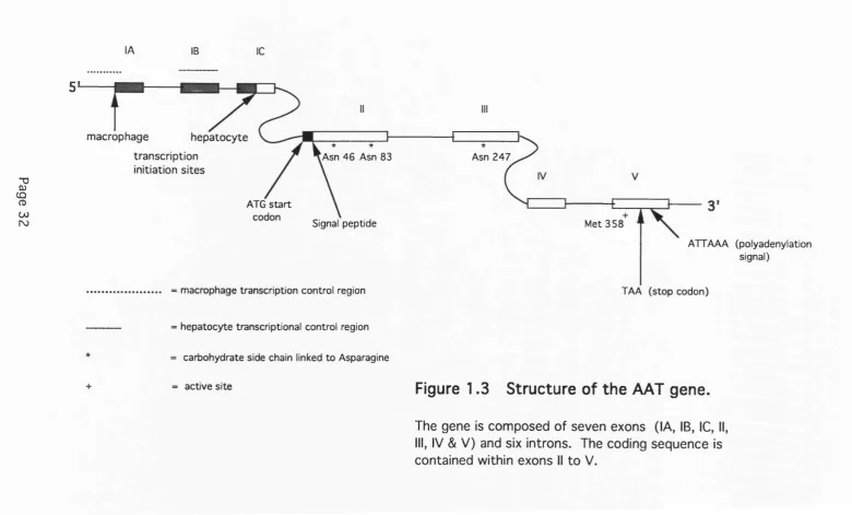
1.1.3 AAT gene structure.
Human AAT is coded for by a single gene, designated Pi, which is located on chromosome 14 at q31-32.3 (Darlington et al., 1982; Schroeder et al., 1985). Sequencing of the AAT gene has revealed it to be a member of the 'serpin' gene family (from serine protease inhibitor). Among other human members of this family are: alpha-1-antichymotrypsin (AACT) (the inhibitor of cathepsin G) which is approxim ately 250 kb from the AAT gene; antithrombin 111 (throm bin). C l-e s te ra s e inhibitor (C l-e s te ra s e and kallikrein), alph a-2-antiplasm in (plasm in), heparin cofactor II (throm bin), protein C inhibitor (activated protein C), plasminogen activator inhibitor (plasminogen activator) and angiotensinogen (substrate not known) (Brantly et al., 1988a). Approximately 10 kb downstream of the AAT gene is a sequence-related, non-expressed pseudogene, PiL (Kelsey et al., 1988a). All serpins are glycoproteins formed by a single polypeptide chain. They have common features of tertiary structure, functional domains and reactive sites. In general, they are suicide inhibitors that form a one-to-one complex with the target protease. The serpin gene family is thought to have evolved by duplications of an ancestral serine protease inhibitor gene prior to divergence (Carrell and Boswell, 1986).
lA IB 1C
T 3
0)
CÛ
CD 00
ro
5
macrophage hepatocyte transcription
initiation sites
Asn 4 6 Asn 83 Asn 2 4 7
ATG s ta rt codon
Signal peptide M et 3 5 8
ATTAAA (polyadenylation signal)
macrophage transcription control region TA A (stop codon)
hepatocyte transcriptional control region
carbohydrate side chain linked to Asparagine
active site
Figure 1.3
Structure of the AAT gene.
1.4 Control of expression of the AAT gene.
The human AAT gene has two promoters. A macrophage-specific promoter is located about 2 kb upstream of the hepatocyte-specific promoter (Perlino, 1987). They showed that transcription from the two promoters is mutually exclusive. AAT gene expression in hepatoma cells is controlled by a combination of cis-acting 5' flanking sequences and trans -acting DNA binding proteins (see fig. 1.4). There are three cis-acting regulatory elements which are contained in the region 500 bp upstream of the TATA box.
The most important regulatory element is the proximal element found between -137 and -37. It is essential for high level tissue specific expression and its presence alone is sufficient to activate heterologous promoters in hepatoma cells (Ciliberto et al., 1985; De Simone et al., 1987). Mutation analysis of this element has revealed two domains - "A" and "B" - both of which are necessary for efficient transcription (De Simone et al., 1987). Partial deletion or mutation of "B" when placed upstream of the SV40 promoter results in an increase in transcriptional efficiency in HeLa cells to levels approaching those seen in hepatoma cells. Thus it seems that this domain also acts as a tissue specific transcriptional repressor in
non hepatic cells (De Simone and Cortese, 1989).
was not tissue specific when tested using an SV40 promoter construct in hepatoma and HeLa cells. Despite this, the domain contains sequences common to other plasma protein gene regulatory regions. The centre of the domain is composed of an octamer similar to the canonical core enhancer. Upstream of this is an eleven base pair stretch homologous to that flanking the haptoglobin gene. Downstream are ten bases that are identical with the transcription factor AP-1 binding site found in the metallothionein and retinol binding protein genes.
The third regulatory element is located between -488 and -356 and gives a three- to four-fold increase in level of transcription (Shen
et a/., 1987).
A number of trans-acting DNA binding proteins that modulate transcriptional activity by binding to the cis regulatory elements have been identified. Those identified to bind to the "B" domain of the proximal element, so far, are: B1 (also known as HNF- 1), LF-B2, LF-B2 'like' protein and LF-B3 (also known as vHNF-1). LF-B1 and LF-B3 have the same consensus binding site and can form heterodimers (De Simone et al., 1991). This binding site is found in the promoter regions of a diverse number of genes expressed in the liver. These include rat a and p fibrinogen and the serum albumin, transthyretin and a-fetoprotein genes from human, rat and mouse as well as the human AAT gene (Courtois e ta !., 1987, 1988). Only one trans- acting factor, LF-A1 (also known as HNF-2), has been shown to bind to the "A" domain of the proximal element (Harden et a!.,
spleen (Monaci et al., 1988). Binding of LF-B1 seems essential for any expression of AAT, whereas the binding of LF-A1 seems to be important only for maximal expression of AAT in fetal liver and yolk sac and non-hepatic adult tissues which express AAT (Tripodi at a!.,
" O
û) (Q
(D ÜO (T)
scale indicates no. of bp from transcription start site
5'
-250distal enhancer element
-200 -150 -100 -50 +1
C/EBP HNF-3 NF-IL6 AP-1
3'
tissue specific proximal element
1 ^ 'A'
'B'-g
LF-Al (HNF-2) LF-B1 (HNF-1)
hepatocyte transcription start site
LF-B2 LF-B2 'like'
LF-B3
Figure 1 .4 Control of hepatic expression of AAT.
The AAT gene promoter elements and the
Macrophages also increase their production of AAT in response to IL6 and may respond to other mediators such as tumor necrosis factor and LPS. It has been shown that, after stimulation with IL6, the myelomonocytic cell line, U937, produces only one of the two mRNA transcripts normally seen in macrophages (Kalsheker et a/., 1990). This is the smaller transcript in which exon IB has been spliced out. It has been suggested that the most likely mechanism to explain the increased levels of AAT protein after IL6 stimulation is increased translational efficiency. Exon IB contains a potential initiation codon at 255-257 in an open reading frame with the recognised initiation codon at 522-524. There is also a stop codon in exon IB at 345-347. If translation is initiated in exon IB there may be competition with the recognised site and this could influence the rate of synthesis of the mature protein. This situation is seen in other eukaryotic genes with short potential coding regions found upstream of the main gene product (Mueller et al.,
1986). Levels of AAT in macrophages can also be modulated by neutrophil elastase (Perlmutter et a/., 1987). It was suggested that part of the mechanism controlling AAT concentration at the epithelial surface of the lung involves the protease/inhibitor complex or some peptide fragment derived from it.
monocytes/macrophages. Much less is known about the regulation of expression of the AAT gene in the many tissues that express AAT at even lower levels. However, it is known that they use the same promoter as the liver (Kelsey et a/., 1987) and that there is low level expression of some of the "liver enriched" trans-acting factors (e.g. LF-B1) in these tissues (De Simone and Cortese (1991).
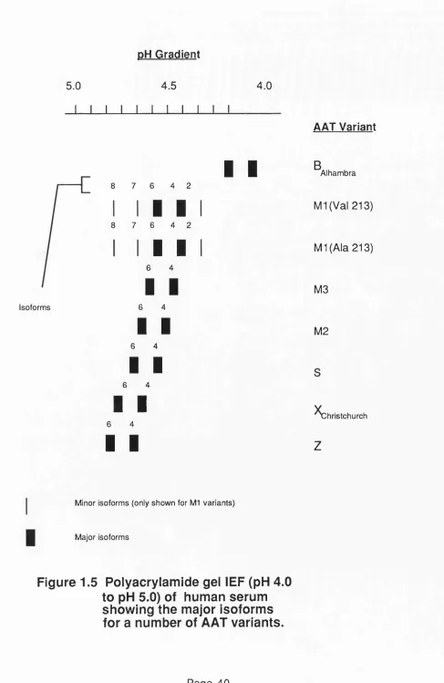
1.1.5 AAT variants.
AAT is a highly polymorphic protein with more than 75 known variants. Extensive lists are given in Brantley et al., 1988a. Variants have mostly been identified by thin-layer polyacrylamide gel isoelectric focusing (lEF) of serum at pH 4 to 5. They are named alphabetically according to their proximity to the cathode after lEF. lEF of serum from an individual homozygous for a normal M-type allele results in a pattern of five bands - two major and three minor (see fig. 1.5). This m icroheterogeneity is a result of the differences in the carbohydrate side chains and in the length of the polypeptide chain (Vaughan e ta !., 1982; Mercs, 1985). AAT in each of the major bands have polypeptide chains of normal length, but proteins in the "4" band have three bi-antennary carbohydrate side chains whereas those in the "6" band have two bi-antennary and one tri-antennary chain. The minor "2" band represents full length AAT polypeptide chains with two tri-antennary and one bi-antennary side chain attached. The minor bands "7" and "8" represent polypeptide chains that have lost five N- terminal amino acids, but have the same carbohydrate side chains as bands "4" and "6" respectively. More recently, a greater number of variants have been identified by analysis of the gene itself. This has been achieved, sometimes in conjunction with the use of the polymerase chain reaction (PCR), by restriction fragm ent length polym orphism s (R F L P ) analysis, oligonucleotide hybridization and direct sequencing of the DNA.
pH Gradient
Isoforms
5.0 4.5
. 1- 1 1 1 1 I I I 1
4.0
8 7 6 4 2
8 7 6 4 2
I I
) 4 :
I
I I
6 4
I I
6 4
I I
6 4
I I
6 4
I I
6 4
I I
I I
AAT Variant
B
Alhambra
M1(Val 213)
Ml (Ala 213)
M3
M2
C h ristch u rch
z
Minor isoforms (only shown for M l variants)
Major isoforms
Only one completely dysfunctional AAT variant, Pi Pittsburgh, has been
identified. Although it is found in the serum at levels in the normal range, this rare variant has been associated with a fatal bleeding condition and a reduced anti-elastase capacity. These conditions probably result from a mutation in the active inhibitory site of AAT (normal M et358 to Arg358). This causes the AAT to act as an inhibitor of thrombin (it now has the sam e active site as antithrombin III - another serpin) instead of elastase (Owen et a/., 1983). The AAT allele M mineral springs (normal Gly67 to Glu67) causes
a severe deficiency state primarily because the AAT protein functions poorly as an inhibitor of elastase (Curiel of a/., 1990).
The null alleles are a rare group which produce no detectable AAT in the serum. Null hong kong results from a two base pair deletion from
codon 318 that causes a frame shift with a stop codon at position 334. There is evidence of an accumulation of the truncated protein in AAT synthesising cells (Sifers at a/., 1988). Null isola di procida
results from the deletion of a 17kb fragment that includes exons II to V of the AAT gene (Takahashi and Crystal, 1990). Most of the other known null AAT alleles result from the formation of stop codons in the exons of the gene. In the case of Null granite fails and
Null beiiingham this leads to absence even of AAT mRNA in
AAT-synthesising cells (Nukiwa at al., 1987b; Garver at al., 1986).
refs). Although the the PiS allele results in reduced AAT serum levels (an individual homozygous for PiS has levels of 100 to 200 mg/dl) the PiS protein functions normally. The PiS allele is identical to the PiM1(Val213) except for a point mutation in exon III (Glu264 to Val264). This affects the Glu264 to Lys387 salt bridge and modifies the structure of the protein. Normal amounts of PiS mRNA are transcribed but, some of the newly synthesised PiS protein is degraded and probably shunted from the PER into lysosomes (Curiel et al., 1989a).
The PiZ allele is the most clinically important AAT variant and might be regarded as the classic AAT deficiency variant. It was this allele that was found in a homozygous form in the first cases of AAT deficiency to be described (Laurell and Eriksson, 1963). In U.S. Caucasians it is found with a frequency of 0.01 to 0.02, but is very rare in black or Asian individuals (see Brantley at al., 1988a for refs.). In Northern Europe the allelic frequency is even higher, reaching 0 .0 2 6 in Scandanavia (S veger, 1 9 7 6 ). Individuals homozygous for PiZ have serum AAT levels of between 15 and 50 mg/dl (Wewers at al., 1987). In addition to being present at reduced amounts the PiZ protein does not function normally as an inhibitor of neutrophil elastase. It has a lower association rate constant for neutrophil elastase. Thus it has been calculated that the time it would take PiZ to inhibit an equal amount of neutrophil elastase is 2.4 seconds compared with 0.2 seconds for normal PiM (Ogushi at al.,
1 9 8 8 ).
(Yoshida é ta l., 1976). Normal amounts of PiZ mRNA are transcribed and translated but deficiency of AAT in the serum arises because the mutant protein is not properly secreted and accumulates within the cell. The mutation and its effects are described in more detail la te r.
Other, much rarer, deficiency variants include M procida and M h eerien .
These result from single point mutations which lead to amino acid substitutions that affect the stability of the AAT molecule such that the protein is degraded in the cell before secretion (Brantly e t al., 1988a). M maiton results from a three base pair deletion in exon II that leads to loss of an amino acid (Phe52)(Curiel et al., 1989b ). Like the Z mutation it leads to defective secretion and intracellular accumulation of the protein in hepatocytes (Cox, 1976; Frazier e t al., 1989). Another rare deficiency allele, M d u a rte , has also been
shown to result in protein accumulation in the liver (Leiberman e t al., 1976).
1.1.6 Evolution of the AAT variants.
Haplotype analysis, using three different R.E. polymorphisms, showed that 51 out of 52 PiZ alleles were identical (Cox, 1985). It was suggested that all of the PiZ alleles studied had probably arisen from a single mutation which was estimated to have occurred about 6000 years ago. Once the M l alleles had been shown to be of two types, M1(Ala213) and M1(Val213), it was clear that the Z mutation had occurred on the M1(Ala213) background. The common origin of the majority of PiZ alleles has been confirmed by Meisen et a/., 1988 and Povey, 1990. The latter study characterized 129 PiZ alleles and showed that all but three of the PiZ alleles possessed alanine instead of valine at position 213 implying an M1(Ala213) background. Investigation of the proteins from two of the three exceptional PiZ alleles revealed no differences from the usual PiZ protein and it was not certain whether the origin of these PiZ alleles was by recombination or new mutation. Analysis of the third allele indicated that it contained the point mutation characteristic of PiZ but that this had occurred on an M2 background. This allele was designated PiZ tun (Whitehouse et a/., 1989a) and is also known
as PiZ a u g sb erg (Faber et a/., 1990). Its protein is slightly more basic
than the usual PiZ protein.
Deficient
Normal
Null
Single bp deletion results in S' shift and stop codon at Tyr 160 Glu 342
Lys 342
Pro 369 Leu 369
Lys 217 to Sto| Glu 376 to Asp376
Glu 264 to Val 264
Leu.41 to Pro 41
Arg 101 to His 101
M2 M3
Z tun M procida M heerien
Null beiiingham M1(Ala 213)
M l (Val 213) Null granite falls
Figure 1.6 Possible evolution of some AAT
variants.
Table 1.1
Table show ing am ino acid d ifferences
between the oldest A AT varian t (PI M l
ALA 213) and other A A T varian ts with
th e ir co rres p o n d in g DNA seq u en c e
changes.
Background changes are shown in italics. An asterisk (*) indicates separate possible C-T su b stitu tio n events at the CpG dinucleotide either on the sense or antisense strand.
AAT VARIANT P ro tein chang e DNA sequence ch an g e
F re q u e n c y of allele in U.S C a u c as ian s
Pi M l ALA 213 — — 0 .2 - 0 .2 3
PI M l VAL 213 Ala 213 to Val GCG to GTG * 0 .4 4 - 0 .4 9
Pi M2 Ala 2 1 3 to Val GCG to GTG 0 .1 -0 .1 1
(M3 background) Glu 3 76 to Asp GAA to GAC
Arg 101 to His G O T to C AT *
Pi M3 Ala 2 1 3 to Val GCG to GTG 0 .1 4 - 0 .1 9
(M l VAL 213 Glu 376 to Asp GAA to GAC
background)
Pi M heerien Pro 369 to Leu CCC to CTC ra re
P> M maiton Phe 52 deleted TTC deleted ra re
Pi M mineral springs Gly 67 to Glu GGG to GAG rare (Black)
Pi M nichinan Ala 2 1 3 to Val GCG to GTG ra re
(M l VAL 213 Phe 52 deleted TTC deleted
Pi M procida
(M1 VAL 213
background)
Pi F
Pi I
Pi Null beiiingham
Pi Null bolton
Pi Null devon
(M 3 background)
Pi Null granite falls
Pi Null hong kong
Pi Null ludwigshafen
Pi Null procida
Pi Null riedenberg
Pi Pittsburgh
Pi P lowell
(M l VAL 213
b ackground)
Pi P St. albans
Ala 2 1 3 to Val
Leu 41 to Pro
Arg 223 to Cys
Arg 39 to Cys
Lys 217 to TER
Frameshift at
Pro 3 62 with
1E R 373
Ala 2 1 3 to Val
Glu 376 to Asp
Gly 115 to Ser
Tyr 160 to TER
Leu 318 to
TER 334
lie 92 to Asn
Met 358 to Arg
Ala 2 1 3 to Val
Asp 256 to Val
Asp 341 to Asn
Asp 256 silent
GCG to GTG
GTG to COG
C G T to T G T
CGC to TGC
AAG to TAG
CCC to C-C
deletion
GCG to GTG
GAA to GAC
C CGC to C AGC
TAG to TA
fra m e s h ift
CTC to - C
deletion
ATC to AAC
17 kb deletion
(incl. exons 2-5)
complete deletion
of gene
ATG to AGG
GCG to GTG
GAT to GTT
C GAC to C A AC
GAT to GAC
Pi S Ala 2 1 3 to Va! GCG to GTG 0 .0 2
(M l VAL 213 Glu 264 to Val GAA to GTA
background)
Pi V munich Asp 2 to Ala GAT to GCT ra re
Pi W bethesda Ala 336 to Thr GCT to ACT ra re
P iZ Glu 342 to Lys 0 GAG to 0 AAG * 0.0 1
P iZ Aia 2 1 3 to Va! GCG to GTG ra re
(M l VAL 213 Glu 213 to Lys 0 GAG to C AAG *
background)
Pi Z augsberg Ala 2 1 3 to Val GCG to GTG ra re
(M 2 background) Glu 37 6 to Asp GAA to GAC
Arg 101 to His C G T to CAT
Glu 342 to Lys C GAG to 0 AAG *
Pi Z w rexham Glu 342 to Lys 0 GAG to 0 AAG * ra re
1.1.7 Association of AAT deficiency with disease.
AAT deficiency is associated with pulmonary emphysema which results from a reduced capacity to protect the lower respiratory tract from neutrophil elastase (Bell, 1970). The lower the levels of AAT, the higher the risk of disease. Thus, in PiNull-Null individuals em physem a always develops (see Brantley et al., 1988a for references) and in PiZZ individuals there is a 5 0 -6 0 % risk (Aagenaes, of a/., 1972). PiSZ heterozygotes carry a slight risk of developing emphysema, but the degree of risk for PiMZ and PiSS individuals is not much higher than that of the general population. The development of emphysema in all cases is thought to be markedly accelerated by the effects of cigarette smoking. This is due to the increase of oxidants in the lower respiratory tract which probably results in the oxidation of M et358 in the AAT protein (Carrell at a/., 1982). The variability in severity of disease is, however, is still unaccounted for, even in individuals matched for age, AAT serum levels and smoking history. One possibility is that there is genetic variation in the expression of the neutrophil elastase gene.
heterozygous for PiZ and one of the other three alleles associated with the condition. The low number of such reports reflect the extremely rare nature of these other alleles, rather than a reduced risk. Several cases of PiSZ and PiZNull patients with neonatal liver disease have been reported. However, the reliability of the data has been questioned (P ovey,1990). There have been no reported instances of neonatal liver disease in PiMZ heterozygotes or PiNull-Null homozygotes. In adults there is also an association between chronic liver disease (including hepatocellular carcinoma) and PiZZ homozygotes, but the frequency of the association is less well defined (see Mowat, 1984 for references). Although there appears to be no association between chronic liver disease and PiMZ heterozygotes, an increased prevalence of PiMZ has been reported in adult patients with relatively mild liver disease (Hodges et a/.,
1981). An association of the PiZ allele with several autoimmune disorders and with chronic inflammation has been also reported in a number of single patients (Fagerhol and Cox, 1981).
In conclusion, the only "common" AAT genotype that has a high risk of disease is the PiZZ homozygote.
PART TWO: PiZ AND LIVER DISEASE.
1.2.1 The Z mutation.
As previously stated, the Z mutation is a single-base substitution in exon V of the normal M1 allele that results in a Glu342 to Lys342 substitution in the molecule. It is this specific substitution which affects secretion of the m olecule, since the AAT protein accumulates whether the mutation occurs on an M1 (Ala213) or M1 (Val213) background and appears to accumulate when it occurs on an M2 background where it is known as PiZ tun (Whitehouse et a/.,
1989a). The levels of AAT mRNA are comparable in PiZZ and PiMM individuals and are translated with equal efficiency, but the PiZ protein is found to be secreted at only 10-15% of the level of the PiM protein (Mornex, 1986) the remainder accumulating inside the cell. The PiZ protein has been shown to accumulate within the monocytes of PiZZ individuals (Perlm utter et al., 1985b). Its secretory defect is maintained in X e n o p u s oocytes injected with the mRNA from the liver of PiZZ individuals (Foreman et a/., 1984) and in a variety of cell types transfected with PiZ cDNA constructs (Brantly et a/., 1988b; McCracken et a/., 1989; Sifers et a/., 1989a). Cell free translation studies of liver RNA using dog pancreas microsomes have shown that the PiZ protein is translocated across the E.R. membrane as efficiently as the normal PiM protein (Bathhurst et a/., 1983). Electronmicroscopy and studies on the endoglycosidase H sensitivity of intracellular PiZ protein indicate that it accumulates in the lumen of the E.R.. Whilst it is accepted that the reason the PiZ protein accum ulates within AAT-synthesising cells is the failure of transfer from E.R. to Golgi, it is not clear why the Glu342 to Lys342 substitution causes this.
effect of the Z mutation, that is more significant (Wu and Foreman, 1 9 9 1 ).
The Z mutation may create or expose a binding site for some protein normally resident in the E.R.. So far only one such protein, BiP (immunoglobulin heavy chain binding protein), has been implicated in the retention of proteins within this compartment. It is also, somewhat confusingly, known as GPR78 (glucose regulated protein 78) and hsc70 (cognate heat shock protein 70). BiP has been shown to be involved in regulating the transport of a number of membrane and secretory proteins from the E.R. to the Golgi. It was first shown to be involved in the post-translational processing of immunoglobulin heavy chains before their assembly with light chains. Unassembled heavy chains remain associated with BiP and are not transported to the Golgi apparatus (Bole et al., 1986). Synthesis of BiP in cell culture is increased by glucose starvation (Shiu et a/., 1977) and by treatment with inhibitors of N-linked glycosylation (Olden et a/., 1979). When protein folding or normal N-linked glycosylation is blocked BiP apparently binds glycoproteins to a greater degree (Gething et a/., 1986; Bole et al., 1986).
in the degree of complexity of their intracellular processing and N-linked glycosylation patterns. Dorner et al., ( 1987) demonstrated that, in the absence of normal N-linked glycosylation, the unglycosylated protein displays an increased association with BiP and efficient secretion is impaired. Increased levels of expression of one of the proteins studied (tPA) enhanced this effect. They also saw that proteins with a more complex glycosylation pattern were generally less efficiently secreted and more stably associated with BiP.
It has been suggested that BiP associates with improperly folded proteins (Gething et al., 1986) and is induced by such proteins (Kozutsumi et al., 1986). McCraken et al., (1989) used COS cells transfected with a human PiZ construct to investigate whether the PiZ protein would associate with BiP. They showed, however, that BiP was not induced by the presence of the accumulated protein, and that PiZ was not precipitated by an anti-BiP antibody. Furthermore, neither BiP nor any other protein co-precipitated with PiZ when cellular extracts were treated with an anti-AAT antibody. They thought it unlikely that PiZ was retained in the E.R. because of an association with resident proteins.