Available online on 30.08.2019 at http://jddtonline.info
Journal of Drug Delivery and Therapeutics
Open Access to Pharmaceutical and Medical Research© 2011-18, publisher and licensee JDDT, This is an Open Access article which permits unrestricted non-commercial use, provided the original work is properly cited
Open Access
Research Article
Comparative Study of Herbal Extract of Piper Nigrum, Piper Album and Piper
Longum on Various Characteristics of Pyrazinamide and Ethambutol
Microspheres
*Pingale Prashant L., Ravindra R. P.
Department of Pharmaceutics, GES’s Sir Dr. M.S.Gosavi, College of Pharmaceutical Education and Research, Nashik, INDIA
ABSTRACT
Bioenhancers are the ‘bioavailability enhancers’; they do not show any therapeutic effect, but when used in combination enhances the activity of drug molecule. In a cited research paper, the effect of various species of piper used as bioenhancer singly and in combina tion in an equal ratio. The methods used for preparation of microspheres are Complex Coacervation and Modified Emulsion Method. The prepared microspheres were evaluated for various parameters like in-vitro release, drug entrapment efficiency, percent bioadhesion, permeability study using intestinal sac method. The in-vitro drug release of drugs from formulations where Piper nigrum was used as bioenhancers was found to be about 66-70% in 12 hrs. when used singly. When bioenhancers used in combination the in-vitro drug release of drugs was increased up to 85-90% for combination of Piper album and Piper longum in an equal proportion, the same was about 35-40% in case of formulations where no bioenhancers was used. The microspheres found to be less than 130 micron in size. The DEE was found to be in the range of 27-67%. The bioadhesion of the microsphere were found to be 20-76% (increased in formulations where bioenhancers incorporated). The in- vitro release study by USP paddle apparatus, the important results from in-vitro release study relates to the very significant enhancement in drug release, due to presence of bioenhancers.
Keywords: Microspheres, Bioenhancer, Piper nigrum, Piper album, Piper longum, Pyrazinamide, Ethambutol
Article Info:Received 07 July 2019; Review Completed 14 August 2019; Accepted 18 August 2019; Available online 30 Aug 2019 Cite this article as:
Pingale PL, Ravindra RP, Comparative Study of Herbal Extract of Piper Nigrum, Piper Album and Piper Longum on Various Characteristics of Pyrazinamide and Ethambutol Microspheres, Journal of Drug Delivery and Therapeutics. 2019; 9(4-A):72-78 http://dx.doi.org/10.22270/jddt.v9i4-A.3319
*Address for Correspondence:
Pingale Prashant L., Associate Professor and Head- Department of Pharmaceutics, GES’s Sir Dr. M.S. Gosavi, College of Pharmaceutical Education and Research, Nashik, INDIA
INTRODUCTION:
According to World Health Organization, Anti-TB agents are broadly classified as first line and second line agents based
on their effectiveness and toxicity.(1, 2,3).
First line agents: These are the most effective, less toxic.
Ethambutol (EMB or E), Isoniazid (INH), Pyrazinamide
(PZA), Rifampicin (RIF).
Second line agents: These are considered as reserved therapy as they are less effective and more toxic for TB treatment. They are used if first line agents are not effective.
Aminoglycosides: e.g., Amikacin, kanamycin
Polypeptides: e.g., Capreomycin, viomycin, enviomycin
Fluoroquinolones: e.g., Ciprofloxacin, levofloxacin,
moxifloxacin
Thioamides: e.g., Ethionamide, Prothionamide,
Third line agents: Other drugs that may be useful, but are not on the WHO list of second line drugs; they include rifabutin, macrolides antibiotics: e.g., clarithromycin, linezolid, thioacetazone, thioridazine.
Bioenhancers are the ‘bioavailability enhancers’; they themselves do not show any typical drug activity, but when used in combination, they enhance the activity of drug
molecule in several ways, including increasing
bioavailability of the drug across the membrane, potentiating the drug molecule by conformational interaction, acting as receptors for drug molecule and
making target cells more receptive to drugs (4).
one of the processes to provide the sustained and controlled
delivery of drug for long periods of time (5,6).
The microspheres of the anti-tubercular agents were prepared by various methods of which the modified emulsion method and complex coacervation method were found to be most appropriate for our work. Variables like polymer concentration, drug-polymer ratio, concentration of
cross-linking agent and time required for cross-linking were
considered in the optimization of the formulation (7).
Hydro-alcoholic extract of 3 species of Piper namely Piper
nigrum, Piper album and Piper longum used as herbal bioenhancers. Constant weight of bioenhancer extract (5 to 15 mg, singly or in combination) was incorporated into the
drug loaded microspheres (8,9).
Figure 1: Fruits of Piper nigrum, Piper album and Piper longum The prepared microspheres loaded with drug and
bioenhancer/s were evaluated for various parameters including drug-excipient incompatibility, particle size, percentage yield, percentage entrapment efficiency, percent bioadhesion and intestinal permeability using intestinal sac method. Selected formulations were kept for stability studies as and no significant variation was found in physicochemical parameters of microspheres such as particle size, % drug entrapment efficiency (% drug content) and in-vitro drug release (% drug release in 12 hr.) was detected.
The most important finding of this study relates to the very significant enhancement in drug release from 35% (without bioenhancer) to 70% (with single bioenhancer) i.e. up to 90-100% increase in release rate and to 90% (with combination of bioenhancer) i.e. up to 100-120% increase in release rate of the anti-tubercular agents.
MATERIALS AND METHODS:
Pyrazinamide and Ethambutol was obtained as a gift sample from Lupin Pharmaceuticals Ltd., Aurangabad, Maharashtra. Piper nigrum, Piper album and Piper longum was purchased from local market and authenticated from Dept. of Botany, Dr. P. R. Ghogrey College of Science, Dhule, Maharashtra. Sodium alginate and Type B gelatin (bloom strength 220) were purchased from Loba Chemie, Mumbai. Sodium Tripolyphosphate (NaTPP) and Chitosan were purchased
from Sigma Aldrich, Germany. All other chemicals / polymers used were of analytical grade.
EXPERIMENTAL WORK:
Extraction and isolation of Piper species used as bioenhancer: (10,11)
A. Macroscopy of Piper nigrum (Black Pepper, Kali Meeri): The entire fruit was almost globular in shape, with 4 to 6.5 mm of diameter, brownish to black in color. The surface was found to be uneven. The seeds were almost brown or black in color, aromatic with a pungent taste.
Macroscopy of Piper album (White Pepper, Safed Meeri): The entire fruit was almost rounding globular in shape, with 5 to 7.5 mm of diameter, white in color. The surface was found to be rough and uneven. The seeds were almost white to pale white in color, aromatic with a pungent taste. Macroscopy of Piper longum (Long peeper, Pimpli): The entire fruit was almost cylindrical, irregular in shape, 2 to 5 cm long and compact.
B. Phytochemical evaluation of piper species: The phytochemical evaluation of fully mature, dried fruits of Piper nigrum, piper album and piper longum, family Piperaceae was carried out as per the provisions of Ayurvedic Pharmacopoeia for various parameters, the
results of which are mentioned in Table 1. (12,13)
Table 1: Phytochemical parameters of Piper nigrum, P. album and P. longum
Parameter (% w/w) Observed Piper nigrum Standard Observed Piper album Standard Observed Piper longum Standard
Total Ash 4.10 NMT 5.5 3.88 NMT 5.0 NMT 5.5 NMT 5.0
Water soluble ash 3.76 NMT 5.5 3.89 NMT 5.0 NMT 5.5 NMT 5.5
Acid Insoluble ash 0.57 NMT 1.0 0.69 NMT 1.0 NMT 1.0 NMT 1.0
Water soluble extractive 23.11 NLT 15 18.75 NLT 12 18.75 NLT 15
Methanol soluble extractive 10.50 NLT 8 9.75 NLT 6 9.75 NLT 8
Loss on drying 3.05 NMT 4 2.79 NMT 4 2.79 NMT 4
(*NMT- Not More Than, NLT- Not Less Than)
C. Isolation of piperine from Piper nigrum, Piper album and Piper longum: Black pepper, White pepper and Pimpli (20 g) was ground to a fine powder and extracted with 95% ethanol (150ml) in a Soxhlet extractor for 2 hours. The solution was filtered and concentrated in vacuum on a water bath at 60°C. 10 ml of 10%alcoholic
Preparation of Pyrazinamide and Ethambutol microspheres:
Sustained release microspheres may be produced by several methods such as emulsion cross-linking method, multiple emulsion method, coacervation method, solvent evaporation method, spray-drying method etc. In this
study, the double emulsification method (6, 14, 16, 18) and
complex coacervation method (14, 15, 16, 18) were used to
prepare the microspheres.
Method 1 - Double Emulsification Method (MEM) Pyrazinamide (400 mg) and Ethambutol (400 mg) was separately dispersed in 3% aqueous solution of sodium alginate (10 ml). The aqueous phase was emulsified in
light liquid paraffin (in the ratio 1:10) containing 1% (v/v) Span 80 using a mechanical stirrer (Remi Motors, India) at 1800 - 2200 rpm for 45 minutes. To it, 5 ml of 7.5% calcium chloride dissolved in a mixture of methanol and isopropyl alcohol (1:2) was added slowly to the emulsion and stirred to assure efficient crosslinking. Microspheres were collected by filtration in vacuum, washed with isopropyl alcohol thrice and finally dried at room temperature. Variables like concentration of polymer, drug - polymer ratio, cross-linking agent concentration and cross-linking time were considered in the optimization of the formulation. Finally, varying concentration of bioenhancer, piperine was added to the optimized formulation to study its effect on bioavailability of drugs as shown in Table 2.
Table 2: Formulae of Pyrazinamide and Ethambutol microspheres with variable polymer ratios and bioenhancer (Double Emulsification Method)
Formulation
Code (mg) PZA (mg) EMB Na. Alginate (%) CaCl2 (%) Cross linking time (min.) Bioenhancer (mg)
MPF1 400 -- 3 7.5 45 --
MPF2 400 -- 3 7.5 45 05
MPF3 400 -- 3 7.5 45 10
MPF4 400 -- 3 7.5 45 15
MEF1 -- 400 3 7.5 45 --
MEF2 -- 400 3 7.5 45 05
MEF3 -- 400 3 7.5 45 10
MEF4 -- 400 3 7.5 45 15
(*M- Modified Emulsion Method, P- Pyrazinamide, F- Formulation, E- Ethambutol)
Method 2 - Complex Coacervation Method (CCM)
Chitosan and gelatin were dissolved in dilute acetic acid solution (1% v/v) together at concentrations of 3% w/v in ratio 1:05 and adjusted to a pH 5.0. Pyrazinamide (400 mg) and Ethambutol (400 mg) was separately dissolved in the above polymeric mixture. The drug in polymeric mixture was emulsified in100 ml of liquid paraffin (1:1 mixture of light and heavy liquid paraffin) at 40°C containing 1ml Tween 80 (2% w/v). The emulsification was carried out for 15 min under mechanical stirrer (Remi Motors, India) at 1200 rpm. The w/o emulsion thus formed was cooled to 4°C
to induce coagulation of gelatin. Then 50 ml Na-TPP (1.5% w/v) with pH 5 at 4°C was added drop wise. Stirring was continued for 30 min to obtain cross-linked microspheres. Microspheres were collected by centrifugation, washed with double distilled water thrice, then with acetone to remove water, and dried at room temperature under vacuum. The prepared microspheres were stored in desiccator for further studies. Concentration of polymer, polymer: copolymer ratio (Chitosan: Gelatin B), cross-linking time, rpm were considered as variables in optimization of the formulation. The composition of optimized formulation containing varying concentration of bioenhancer is shown in Table 3.
Table 3: Formulae of Pyrazinamide and Ethambutol microspheres with variable polymer ratios and bioenhancer (Complex coacervation method)
Formulation
Code PZA (mg) EMB (mg) Gelatin (ratio) Chitosan: NaTPP (%) Cross linking time (min.) Bioenhancer (mg)
CPF1 400 -- 1:0.5 1 30 --
CPF2 400 -- 1:0.5 1 30 05
CPF3 400 -- 1:0.5 1 30 10
CPF4 400 -- 1:0.5 1 30 15
CEF1 -- 400 1:0.5 1 30 --
CEF2 -- 400 1:0.5 1 30 05
CEF3 -- 400 1:0.5 1 30 10
CEF4 -- 400 1:0.5 1 30 15
(*C- Complex Coacervation Method, P- Pyrazinamide, F- Formulation, E-Ethambutol) Characterization of Microspheres:
Compatibility studies: Chemical interaction between the drug and the polymeric material, if any, during the preparation of the microspheres was studied by using Fourier Transform Infrared Spectroscopy (FTIR). Pure drug PZA and EMB, placebo microspheres, PZA and EMB
and compared with each other to identify drug-excipient interaction, if any.
Particle size analysis: Particle size of both plain drug microspheres as well as microspheres with bioenhancer was measured using Motic microscope at 40 X magnification. In all measurements, at least 100 particles in each of 3 different fields were examined.
Determination of percentage drug entrapment: The drug
content of the microspheres was determined
spectrophotometrically (λmax = 270 nm and 292 nm for PZA and EMB respectively; PerkinElmer, USA Lambda 25 model). Microspheres (10 mg) loaded with PZA and EMB were dissolved in10 ml of isotonic phosphate buffer pH 6.8under sonication for 20 min. The solutions were filtered through 0.22 μm Millipore filter sand the amount of pyrazinamide and Ethambutol was determined. Preliminary UV studies showed that the presence of dissolved polymers did not interfere with the absorbance of the drug at270 and 292 nm.
The percent drug entrapment was calculated using following formula:
Percentage yield: The yield of microspheres was
determined by comparing the whole weight of microspheres obtained against the combined weight of the polymer, drug and bioenhancers used for formulation. The % yield of microsphere was determined using following formula:
Measurement of bioadhesion: In-vitro bioadhesion was determined for microspheres (in triplicate) by falling liquid film method. Microspheres (50 mg) were placed on albino
rat small intestine (area 2cm2) and kept for 20-30 minutes in
a humidity temperature controlled cabinet (Thermolab, India), maintained at 75 (±5) % relative humidity and
temperature of 25 (±2)0C to allow hydration of the
microspheres. This was followed by thorough washing of the mucosal lumen with isotonic phosphate buffer pH 6.8, and
then dried at 700C in a hot air oven. Percent bioadhesion was
determined by the following formula:
In-vitro drug release: Dissolution studies were carried out using USP XXIV rotating basket method. The release profiles of pyrazinamide and Ethambutol from microspheres were studied in simulated gastric fluid (SGF pH 1.2) and simulated intestinal fluid (SIF pH 6.8). The drug-loaded microspheres (equivalent to 20 mg of drugs) filled in empty capsule shells were put into the basket (50 rpm) and placed in 500 ml of
the dissolution medium, thermostated at 370C. Samples of 2
ml each were withdrawn at regular time intervals, filtered, diluted suitably, analyzed using double beam UV spectrophotometer at λmax = 270 nm and 292 nm for PZA and EMB respectively and an equal volume of fresh medium was immediately added to maintain the dissolution volume. Dissolution studies were carried out up to 12 h. The drug release experiments were conducted in triplicate.
RESULTS AND DISCUSSION:
Fourier Transform Infra-Red (FT-IR) analysis: Pure drug PZA and EMB, placebo microspheres, PZA and EMB
microspheres (2-5 mg) prepared with and without bioenhancer were weighed and mixed perfectly with potassium bromide (0.1 to 0.2 g) to form a uniform mixture. Potassium bromide pellet method was employed and background spectrum was collected under identical conditions. The IR spectrum of the pellet were recorded using FTIR (Perkin Elmer, USA, Spectrum RX1 Model) taking air as the reference and compared with each other to identify drug-excipient interaction, if any.
Each spectrum was derived from 16 single averaged scans collected in the region 400–4000 cm−1 at a spectral resolution of 2 cm−1. FT-IR spectra of pure drug and pyrazinamide and ethambutol microsphere are shown in Figure 2.
Figure 2: FT-IR spectra of Pyrazinamide and Ethambutol Differential Scanning Calorimeter (DSC) study: Pyrazinamide and ethambutol powder sample (2-8 mg) was weighed into an aluminum pan and analyzed as sealed with pinholes and an empty aluminum pan was used as a reference. To determine the thermodynamic relationship of two forms, heat–cool–heat cycle was also used. The DSC endotherm showed a sharp melting endotherm for
pyrazinamide and ethambutol at 1900C.
Figure 3a: Pyrazinamide microspheres prepared by MEM and CCM
Figure 3b: Ethambutol microspheres prepared by MEM and CCM
Percentage yield: The yield of microspheres was determined by comparing the total weight of microspheres obtained against the sum of the weight of the drug, polymers and bioenhancer. The percentage yield of the optimized formulations was found to be 36.75-69.20% as shown in Table 4. The loss of the drug in the method may be due to loss accounted during hardening, washing and filtering processes of microspheres.
Percentage entrapment efficiency: The entrapment efficiency was found to be in the range of 41.45-76.50%. Loss of the drug in these methods may be due to loss in the hardening, washing and filtering processes. During optimization of microsphere formulation, it has been observed that the concentration of polymer, cross linker concentration and cross-linking time may affect the entrapment efficiency of the microspheres as shown in Table 4. The maximum values obtained are higher, or at least comparable to the highest value of entrapment efficiency reported in earlier studies using the sodium alginate method of preparation of microspheres. The difference is due to the high aqueous solubility of PZA and EMB resulting in high concentrations of the drug present in the preparation medium in this method and possibly due to the use of
bioenhancer in formulations (17).
Table 4: Evaluation Parameters for Pyrazinamide and Ethambutol Microspheres using Modified Emulsion and Complex Coacervation Method
Formulation
Code Mean particle size (µm) Yield (%)
Drug Entrapment
(%)
Bioadhesion
(%) ±SD (at 12Drug Release th hrs.) %
MPF1 129-135 41.50 ± 1.11 39.63 ± 1.13 40.12 ± 1.14 35.41 ± 1.11
MPF2 126-131 46.78 ± 1.19 51.52 ± 1.63 50.19 ± 1.17 48.12 ± 1.72
MPF3 122-128 56.95 ± 1.73 59.25 ± 1.47 61.24 ± 1.11 58.14 ± 1.37
MPF4 124-130 64.32 ± 1.03 67.21 ± 1.25 73.14 ± 1.36 68.17 ± 1.63
MEF1 126-134 39.63 ± 1.45 37.24 ± 1.24 41.25 ± 1.78 36.42 ± 1.14
MEF2 128-136 48.65 ± 1.23 46.85 ± 1.78 52.14 ± 1.14 47.78 ± 1.41
MEF3 124-128 58.32 ± 1.78 53.67 ± 1.34 62.13 ± 1.75 59.24 ± 1.52
MEF4 124-132 66.25 ± 1.13 68.12 ± 1.28 71.41 ± 1.18 68.22 ± 1.27
CPF1 124-134 43.21 ± 1.15 38.63 ± 1.44 39.15 ± 1.75 39.14 ± 1.29
CPF2 126-132 56.23 ± 1.29 49.12 ± 1.24 49.12 ± 1.16 49.17 ± 1.23
CPF3 130-134 61.58 ± 1.17 58.12 ± 1.18 60.01 ± 1.17 61.18 ± 1.44
CPF4 128-132 69.25 ± 1.36 71.14 ± 1.49 69.18 ± 1.01 69.14 ± 1.73
CEF1 122-126 38.12 ± 1.45 41.45 ± 1.73 41.13 ± 1.42 41.25 ± 1.18
CEF2 124-130 49.56 ± 1.23 56.71 ± 1.14 50.99 ± 1.45 49.21 ± 1.37
CEF3 126-132 64.50 ± 1.25 68.34 ± 1.19 61.24 ± 1.14 61.17 ± 1.27
CEF4 126-130 69.25 ± 1.08 72.40 ± 1.74 69.88 ± 1.19 69.09 ± 1.06
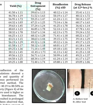
Percentage bioadhesion: The bioadhesion of the microspheres in the optimized formulations showed a significant change with the presence and quantity of bioenhancer. The bioadhesion study was performed (in triplicate) using a previously reported method. The percentage bioadhesion was found to be 40.19 to 80.50% as shown in Table 4. The bioadhesive property (Figure 4) of the microspheres in which bioenhancers were used is higher as compared to microspheres without bioenhancer. The bioadhesive property of microspheres resulted in prolonged retention in the small intestine. It has been observed that, the microspheres containing comparably higher amount of bioenhancer showed significant increase in bioadhesion about 86%. It has also been observed that, the percentage bioadhesion increases as the amount of bioenhancer increases as shown in Table 4. The bioadhesive property of these particles resulted in prolonged retention in the small intestine (17).
Figure 4: Bioadhesion study of Microspheres prepared by MEM and CCM
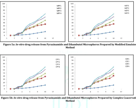
shown in figure 5 a & b respectively. Approximately, 8-10% of the drug was released in the SGF, pH 1.2 over a period of 2 h and about 35- 80% in SIF, pH 6.8 up to 12 h. It has been found that the microspheres containing bioenhancer show greater increase in drug release as compared to microspheres without bioenhancer. In Figure 5,
CRF1-formulation without bioenhancer and CRF2, CRF3 and CRF4 are the formulations with bioenhancers prepared by complex coacervation method whereas the fraction of bioenhancer were used in 5, 10 and 15 mg respectively in
2nd, 3rd and 4th formulations in both the methods as shown in
Figure 5 a and b.
Figure 5a: In-vitro drug release from Pyrazinamide and Ethambutol Microspheres Prepared by Modified Emulsion Method
Figure 5b: In-vitro drug release from Pyrazinamide and Ethambutol Microspheres Prepared by Complex Coacervation Method
CONCLUSION:
In this study, microspheres of pyrazinamide and ethambutol were prepared by modified emulsion and complex coacervation method. Effect of variables like drug-polymer ratio, cross linker concentration and the cross-linking time on in-vitro release of pyrazinamide and ethambutol was examined. The mean particle size of the microspheres increased with an increase in the concentration of polymer. The cross-linking time shorter than 30 minutes resulted in higher entrapment efficiencies. The microspheres were spherical and well formed. The mean diameter, entrapment efficiency and bioadhesion of the optimized microspheres were found to be 120-130 μm, 67.21 ± 1.25% and 68.12 ± 1.28% for pyrazinamide and 71.14 ± 1.49 and 72.40 ± 1.74 for ethambutol prepared by modified emulsion method and complex coacervation method respectively. . The release profiles of pyrazinamide and ethambutol from microspheres were examined in simulated gastric fluid (SGF pH 1.2) and simulated intestinal fluid (SIF pH 7.4). About 10% of the pyrazinamide and ethambutol was released in the SGF in first 2 hours and released quickly about 85% in 10 hrs. in SIF. The concentration of polymers and the presence of cross-linking agent had a great effect on the release of pyrazinamide and ethambutol. The most important finding
of this study relates to the very significant enhancement in drug release (35 to 70%), due to co-administration of 15 mg bioenhancer along with each dose of pyrazinamide and ethambutol microspheres.
REFERENCES:
1. Petri, W.A., 2001. Antimicrobial Agents. In: Hardmann, J.G., Limbird, L.E., Gilman, A.G. (Eds.), The Goodman and Gilmans: The Pharmacological Basis of Therapeutics. McGraw-Hill Publishing Division, New York, USA. 10th ed., pp. 1273-1294. 2. Rang H.P., Dale M.M., Ritter J.M., Flower R.J. 2007. Antibacterial
Drugs. In: Rang and Dale’s Pharmacology. Churchill Livingstone. Elsevier Health Science Rights Department, Philadelphia, USA. 6th ed., pp. 661-678.
3. WHO. 2003. Treatment of tuberculosis: Guidelines for national programmes, 3rd Edition published by World Health Organization, Geneva. Accessed on Dec. 15, 2012 at http://whqlibdoc.who.int/hq/2003/who_cds_tb_2003.313_eng. pdf
4. Atal N., Bedi K.L. 2010. Bioenhancers: Revolutionary concept to market. Journal of Ayurveda and Integrative Medicine. 1(2): 96-99.
5. Duncan K., Barry C.E. 2004. Prospects for new antitubercular drugs. Current Opinion in Microbiology. 7:460-465.
sustained drug delivery. International Journal of Pharmaceutics 334: 71-77.
7. Puvar A.N., Patil J. S., 2009. Comparative evaluation of Ethambutol HCl microspheres prepared by different methods. Journal of Pharmacy Research. 2(4):615-618.
8. Raman G., Gaikar V.G. 2002. Extraction of piperine from Piper nigrum (black pepper) by hydrotropic solubilization. Industrial and Engineering Chemical Research. 41:2966-2976.
9. Shaikh J., Ankola D.D., Beniwal V., Singh D., Kumar M.N. 2009. Nanoparticle encapsulation improves oral bioavailability of curcumin by at least 9-fold when compared to curcumin administered with piperine as absorption enhancer. European Journal of Pharmaceutical Sciences. 37:223-230.
10. Kadam P.V., Yadav K.N., Patel F.A., Karjikar F.A., Patil M.J., 2013. Pharmacognostic, phytochemical and physicochemical studies of Piper nigrum Linn. fruit (Piperaceae). International Research Journal of Pharmacy. 4(5):189-193.
11. Joshi K., Nishteswar K. Goyale M., Ladani S. 2014. Pharmacognostic evaluation of Pippali mula (root of Piper longum Linn.) W.S.R. to micrometric and isolation techniques. Ayurpharm - International Journal of Ayurveda and Allied Sciences. 3(6):162-170.
12. Kokate C.K., Purohit A.P., Gokhale S.B., 2012. Pharmacognosy. 46th edition. Nirali Prakashan, Pune. 1.55-1.57. 15. The Ayurvedic Pharmacopoeia of India.
13. Department of Ayush, Ministry of Health and Family Welfare, Government of India. Part I Volume II. 141.
14. Pingale P.L., Ravindra R.P. 2013. Effect of piper nigrum on in-vitro release of Isoniazid from oral microspheres. International Journal of Pharma and Bio Sciences. 4(1):1027-1036.
15. Lucinda-Silva R. M., Evangelista R. C., 2003. Microspheres of Alginate – Chitosan Containing Isoniazid. Journal of Microencapsulation. 20(2), 145-52.
16. Pingale P.L., Ravindra R.P. 2013. Effect of Piper nigrum on in-vitro release of Rifampicin microspheres. Asian Journal of Pharmaceutical and Clinical Research. 6(5):79-83.
17. Ranga Rao K.V., Buri P., 1989. A novel in situ method to test polymers and coated microparticles for bioadhesion. International Journal of Pharmaceutics. 52, 265–270.