http://www.sciencepublishinggroup.com/j/ajist doi: 10.11648/j.ajist.20180201.13
Plasmid Profile Analysis and Curing of Multidrug-Resistant
Bacteria Isolated from Hospital Environmental Surfaces in
Akure Metropolis, Ondo State, Nigeria
Anthony Kayode Onifade, Ogedegbe Gloria Palmer
*Department of Microbiology, The Federal University of Technology, Akure, Nigeria
Email address:
*Corresponding author
To cite this article:
Anthony Kayode Onifade, Ogedegbe Gloria Palmer. Plasmid Profile Analysis and Curing of Multidrug-Resistant Bacteria Isolated from Hospital Environmental Surfaces in Akure Metropolis, Ondo State, Nigeria. American Journal of Information Science and Technology. Vol. 2, No. 1, 2018, pp. 18-23. doi: 10.11648/j.ajist.20180201.13
Received: April 19, 2018; Accepted: May 2, 2018; Published: May 24, 2018
Abstract:
Plasmid profile analysis and curing of multidrug-resistant bacteria isolated from hospital environment in Akure metropolis, Ondo State Nigeria were investigated. Staphylococcus aureus, Streptococcus pyogenes, Escherichia coli, Pseudomonas aeruginosa, Klebsiella pneumoniae and Bacillus cereus were the bacterial isolated in the course of the research. The result revealed that Staphylococcus aureus and Pseudomonas aeruginosa showed multiple antibiotics resistance.Staphylococcus aureus and Pseudomonas aeruginosa are rapidly increasing as multidrug resistant strains worldwide. The results of antibiotics sensitivity test on the microorganisms subjected to plasmid analysis indicated that they were more susceptible to antibiotics after the plasmid curing of the microorganisms. The results from this investigative study have shown that resistance in S. aureus and P. aeruginosa are plasmid based as a result of its loss after curing.
Keyword:
Plasmid, Hospital, Environment, Multiple, Antibiotics1. Introduction
Hospital acquired infections (HAIs) caused by antibiotic resistant bacteria are a significant cause of morbidity and mortality worldwide. In recent decades the incidence of HAI with antibiotic resistant bacteria has increased remarkably. Emerging trends in nosocomial infections signal high alerts towards multidrug resistant pathogens. Studies show that 70% of nosocomial infections are due to antibiotic resistant strains [1, 2]. Major causative agents include antibiotic resistant strains of Staphylococcus aureus and Pseudomonas aeruginosa. The percentages of antimicrobial resistance continued to increase in Europe leading to mounting healthcare costs, failed treatment and deaths [3]. Some bacteria are naturally resistant to certain types of antibiotics. Recently, the probable involvement of surfaces and equipment from the hospital environment as a disseminating source of pathogens, including resistant bacteria, has been highlighted [4].
Antimicrobial resistance turns into a complex both ecological and clinical problems when considering the
genetic variability in microorganisms. Its contention is one of the greatest challenges of the 21st century, and originates appeals from several international health organizations asking for regional data in bacterial susceptibility patterns, especially for strains of nosocomial circulation [5].
Most outbreaks of infection associated with inanimate objects are caused by items that should be sterile but have been contaminated [6]. The surfaces near colonized patients, or those touched frequently by health professionals and by those who move within the area, can become contaminated by antibiotic resistant bacteria, especially methicillin resistant
Staphylococcus aureus MRSA [7, 8] and vancomycin resistant enterococcus-VRE [9].
genes can also be transferred between cells on plasmids or transposons by transductive or conjugative processes [13]. DNA elements which mediate integration of resistance genes (eg, integrons) may also be involved [14] resulting in the further spread of multidrug resistant bacteria.
The aim of this study was to examine plasmid profile analysis and curing of multidrug-resistant bacteria isolated from hospital environmental surfaces in Akure metropolis, Ondo State, Nigeria.
2. Materials and Methods
2.1. Description of Study Location
This research work was carried out from September 2016 to April, 2017 in Akure metropolis, Ondo state, Nigeria. Akure covers an area of 14,798.8,993.7 square kilometers and lies at latitude 7°15′0″N, 70 11′ N 5°11′42″E and longitude 5°11′42″E, 5O35’E. Akure is one of the 18 local government areas of Ondo State with a population of 484,798 based on the 2006 population census. It is situated in the peripheral zone of the rainforest of Ondo state. Akure is the administrative capital of Ondo state. Akure lies about 70°15’ north of the equator and 50°15’ east of the Meridian. It is about 700 km Southwest of Abuja and 311 km north of Lagos State. The town is situated in the tropic rainforest zone in Nigeria.
2.2. Collection of Samples
Swab Samples were collected by swab sticks from Male Accident and Emergency Bed, Female Accident and Emergency Bed, Male Toilet, Female Toilet, Male Surgical Ward Chair, Female Surgical Ward Chair, Male Medical Ward Floor, Female Medical Ward Floor, Male Ward Air flora, Female Ward Air flora, Theatre Couch, Injection Room Tables, Neonatal Ward Couch and Maternity Ward Couch from Health Centre FUTA, Don Bosco Catholic Hospital and State Specialist Hospital Akure. The date, time, conditions and sites of sampling were noted. Basically, swabs were used, at least, for each sampling site. For sampling, swabs were moistened in 2 mL sterile saline solution and rolled several times over a surface area of around 25 cm2, and the swab sticks were transported to the laboratory. Sampling was always done between 8-10am
2.3. Isolation of Microorganisms from Hospital Environmental Surfaces
Isolation of microorganisms from hospital environmental surfaces was carried out as described by Bakkali et al. [15] with slight modification. Basically, swabs were used, at least, for each sampling site. For sampling, swabs were moistened in 2 mL sterile saline solution and rolled several times over a surface area of around 25 cm2, and the swab sticks were transported to the laboratory. Sampling was always done between 8-10 am. A five-fold serial dilution was made and 0.1 mL of the 10-3 and 10-5 dilutions were uniformly pour-plated onto 14 cm diameter wide agar plates and of nutrient
agar, Potato dextrose agar, MacConkey agar and EMB agar.
2.4. Characterization of Bacterial Isolates
The pure culture of each isolate was examined. Microscopic examination, staining techniques and biochemical tests were carried out on the isolates according to the methods described by Olutiola et al. [16] and Cheesbrough [17].
2.5. Identification of Fungal Isolates
Fungal isolates were characterized and identified based on macroscopic and microscopic details with reference to Barnett and Hunter [18].
2.6. Antibiotic Sensitivity Assay
2.6.1. Standardization of Inoculum
A pure bacterial colony was touched with a loop, and then transferred aseptically into a tube containing 4 to 5 mL of Mueller Hinton broth medium. The broth culture was incubated at 37°C until it achieved the turbidity of the 0.5 McFarland standards which took up to six hours.
2.6.2. Antibiotic Sensitivity Profile Test
The antibiotic sensitivity profile was investigated in order to compare the sensitivity of the microorganisms to the different conventional antibiotics. The disc diffusion method described by Bauer et al. [19] was used to determine the susceptibility and resistance of the organisms to the antimicrobial drugs.
2.7. Plasmid Analysis
2.7.1. Determination of Plasmid Profile of Multiple Antibiotics Bacterial Isolates
The agarose was then poured into electrophoresis tank with the comb in place to obtain a gel thickness of about 4-5 mm and was allowed to solidify for about 20 minutes and the comb was removed, the tray was then placed in the electrophoresis tank. This was followed by the addition of 1X TBE buffer; this was then poured into the tank ensuring that the buffer covered the surface of the gel. The sample 15 ul was mixed with 2 ul of the loading dye and was carefully loaded into the wells created by the combs (marker was loaded in line 1). Electrodes were connected to the power pack in such a way that the negative terminal is at the end where the sample was loaded; electrophoresis was run at 60-100 V until loading dye has migrated about three-quarter of gel. Electrodes were disconnected and gel was removed from the tank and visualized in UV- trans-illuminator.
2.7.2. Procedure for Plasmid Curing
This was carried out based on the method described by Akinjogunla and Enabulele [21] with slight medication. Fifty microliter (50 µl) of Acridine orange (0.10 mg/mL) was added to 5 mL of Lysogeny broth (LB) followed by subsequent culture inoculation of resistant Staphylococcus aureus and Pseudomonas aeruginosa with plasmid into separate LB broth having acridine orange. These were then incubated at 37°C for 24 hrs in a shaker. After incubation, the cultures were swabbed in to the Muelller Hinton Agar (MHA) plates for confirmatory antibacterial assay.
2.8. Post Curing Susceptibility Testing
After incubation, the standardized inocula of these bacteria were swabbed in to the Muelller Hinton Agar (MHA) plates and incubated at 37°C for 18hrs as a confirmatory antibacterial assay. The plates were examined and the diameters of the zones of inhibition measured to the nearest whole millimeter with a ruler. The sizes of the zone of inhibition were then juxtaposed with those obtained before curing.
2.9. Statistical Analysis of Data
All experiments were carried out in triplicate, and data
obtained were subjected to one way analysis of variance, while the means were compared by Duncan’s New Multiple Range Test at 95% confidence interval using Statistical Package for Social Sciences version 16.0. Differences were considered significant at p≤0.05.
3. Results
Table 1. Rate of occurrence of different bacteria isolated from FUTA Health Centre, State Specialist hospital Akure and Don Bosco Hospital Akure.
Bacteria Number of surfaces
Tested Positive
Percentage positivity (%)
Staphylococcus aureus 39 22.81
Streptococcus pyogenes 24 14.04
Escherichia coli 21 12.28
Pseudomonas
aeruginosa 27 15.79
Klebsiella pneumonia 33 19.30
Bacillus cereus 27 15.79
Total 171 100.01
Table 1: The rate of occurrence of different bacteria isolated from different hospital environmental surfaces is presented in Table 1. It was observed that Staphylococcus aureus had the highest rate of occurrence, while Esherichia coli had the lowest rate of occurrence out of the bacteria isolated for different hospital environment surfaces.
Table 2. Rate of occurrence of different fungi isolated from FUTA Health Centre, State Specialist hospital Akure and Don Bosco Hospital Akure.
Fungi Number of surfaces
Tested Positive
Percentage positivity (%)
Aspergillus fumigatus 21 36.84
Aspergillus flavus 18 31.58
Candida albicans 18 31.58
Total 57 100
Table 2: The rate of occurrence of different fungi isolated from different hospital environmental surfaces is presented in Table 4. It was observed that Aspergillus fumigatus had the highest rate of occurrence followed by Candida albicans and
Aspergillus niger which share the same number percentage positivity.
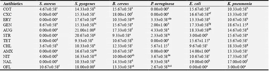
Table 3. Susceptibility pattern of bacteria isolated from hospital environmental surfaces.
Antibiotics S. aureus S. pyogenes B. cereus P. aeruginosa E. coli K. pneumonia
COT 4.67±0.58c 14.33±0.58b 15.67±0.58e 0.00±0.00a 15.67±0.58e 10.33±0.58b
CXC 0.00±0.00a 15.33±0.58c 18.00±1.00f 0.00±0.00a 16.67±0.58ef 13.33±0.58c
ERY 0.00±0.00a 17.67±0.58ef 10.33±0.58ab 3.33±0.58cde 13.33±0.58d 10.67±0.58b
GEN 8.67±0.58e 15.33±0.58bc 15.67±0.58e 2.00±1.00b 17.33±0.58fg 18.67±1.15g
AUG 0.00±0.00a 21.00±1.00g 17.33±0.58f 4.33±0.58e 18.33±0.58gh 14.67±0.58d
STR 0.00±0.00a 20.67±0.58g 9.33±0.58a 2.33±0.58bc 3.00±0.00b 15.67±0.58e
TET 8.00±0.00d 9.33±0.58a 10.67±0.58b 0.00±0.00a 15.67±1.15e 13.67±0.58c
CHL 3.67±0.58b 10.33±0.58a 12.33±0.58c 5.67±1.15f 9.67±0.58c 18.33±0.58g
AMX 0.00±0.00a 16.67±0.58de 10.67±0.58b 0.00±0.00a 14.00±1.00d 13.33±0.58c
NIT 4.00±0.00b 16.33±0.58cd 10.00±0.00ab 3.67±0.58de 10.67±0.58c 17.33±0.58f
NAL 0.00±0.00a 10.33±0.58a 14.33±0.58d 9.33±0.58g 19.00±0.00h 17.00±0.00f
OFL 10.67±0.58f 18.00±0.00f 13.33±0.58cd 2.67±0.58bcd 0.00±0.00a 3.00±0.00a
Data are presented as Mean±S.D (n=3). Values with the same superscript letter(s) along the same column are not significantly different (P<0.05).
Table 3: The susceptibility pattern of gram positive bacteria isolated from hospital environmental surfaces is presented in Table 3. It was observed that S. aureus and P. aeruginosa had multiple resistant against antibiotics.
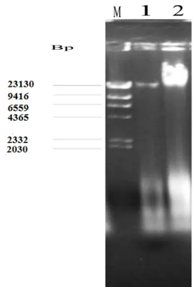
Figure 1. Electrophoretic patterns for plasmid profile of bacterial isolates from hospital environmental surfaces.
Keys: M=Molecular weight marker, 1= Staphylococcus aureus, 2=
Pseudomonas aeruginosa, bp= basepair, 1bp= 3.4Angstrom (Å) while 1000bp=1kilo base pairs.
Figure 1: The bacterial were investigated for the presence of DNA plasmids using Agarose-gel electrophoresis. The results obtained revealed the presence of plasmid bands of different molecular weights. The molecular weights of the plasmids were determined using DNA- Hind III molecular weight marker (Figure 1).
Table 4. Post-curing antibiotics sensitivity patterns of selected Gram positive bacteria.
Antibiotics Staphylococcus aureus
COT bf 4.67±0.58c
COT af 12.33±0.58i
CXC bf 0.00±0.00a
CXC af 11.00±0.00gh
ERY bf 0.00±0.00a
ERY af 11.67±0.58hi
GEN bf 8.67±0.58ef
GEN af 17.67±0.58jk
AUG bf 0.00±0.00a
AUG af 6.67±0.58d
STR bf 0.00±0.00a
STR af 8.67±0.58ef
TET bf 8.00±0.00e
TET af 17.33±0.58j
Antibiotics Staphylococcus aureus
CHL bf 3.67±0.58b
CHL af 9.67±1.15g
AMXbf 0.00±0.00a
AMXaf 9.00±0.00g
NITbf 4.00±0.00bc
NITaf 10.67±0.58h
NALbf 0.00±0.00a
NALaf 6.33±0.48d
OFLbf 10.67±0.58h
OFLaf 18.33±0.58k
Data are presented as Mean±S.D (n=3). Values with the same superscript letter(s) along the same column are not significantly different (P<0.05). LEGEND: bf = before curing, af = After curing, COT = Cotrimazole (25µg), CXC = Cloxacillin (5µg), ERY = Erythromycin (5µg), GEN = Gentamycin (10µg), AUG = Augmentin (30µg), STR = Streptomycin (10µg), TET = Tetracycline (10µg), CHL = Chloramphenicol (10µg), AMX = Amoxacillin (10µg), NIT = Nitrafuratoin (10µg), NAL = Nalixidic acid (30µg), OFL= Ofloxacin (10µg).
Table 4: Shows the comparison of antibiotics susceptibility pattern of selected Gram positive bacteria isolate before and after plasmid curing. It was observed that the bacteria were more susceptible to antibiotics after plasmid curing.
Table 5. Post-curing antibiotics sensitivity patterns of selected Gram negative bacteria.
Antibiotics Pseudomonas aeruginosa
AMX bf 0.00±0.00a
AMX af 3.33±0.58cde
COT bf 0.00±0.00a
COT af 6.00±1.00g
NIT bf 3.67±0.58def
NIT af 6.67±0.58h
GEN bf 2.00±1.00b
GEN af 4.67±1.15fg
NAL bf 9.33±0.58ij
NAL af 17.33±0.58l
OFL bf 2.67±0.58bcd
OFL af 8.33±0.58i
AUG bf 4.33±0.58ef
AUG af 9.33±0.58ij
TET bf 0.00±0.00a
TET af 8.33±0.58i
CXCbf 0.00±0.00a
CXCaf 5.67±0.58gh
ERYbf 3.33±0.58cde
ERYaf 10.33±0.58j
STRbf 2.33±0.58bc
STRaf 6.33±0.58h
CHLbf 5.67±1.15gh
CHLaf 15.67±0.58k
Data are presented as Mean±S.D (n=3). Values with the same superscript letter(s) along the same column are not significantly different (P<0.05). LEGEND: bf= before curing, af = After curing, AMX = Amoxacillin (10µg), COT = Cotrimazole (25µg), NIT= Nitrafuratoin (10µg), GEN = Gentamycin (10µg), NAL= Nalixidic acid (30µg), OFL = Ofloxacin (10µg), AUG = Augmentin (30µg), TET = Tetracycline (10µg), CXC = Cloxacillin (5µg), ERY = Erythromycin (5µg), STR= Streptomycin (10µg), CHL = Chloramphenicol (10µg).
more susceptible to antibiotics after plasmid curing.
4. Discussion
Plasmid profile multidrug-resistant bacteria isolated from hospital environmental surfaces in Akure metropolis, Ondo State, Nigeria was investigated. Bacteria were found to be more predominant than fungi, the bacteria isolated from the hospital environmental surfaces were Bacillus cereus,
Escherichia coli, Klebsiella pneumoniae, Pseudomonas aeruginosa, Staphylococcus aureus, and Streptococcus pyogenes while fungi include Aspergillus fumigatus,
Aspergillus niger and Candida albicans. Staphylococcus aureus was found to be predominant bacteria with the occurrence of 22.81%, this correlate with the report of Awosika et al. [22] who reported Staphylococcus aureus as the most frequently isolated bacterium from hospital surface.
Aspergillus funmigatus was found to be predominant fungi with frequency occurrence of 36.84%, this correlate with the report of Cagginao et al. [23] who reported that Aspergillus fumigatus was the most commonly isolated (68.5%).
Staphylococcus aureus and Pseudomonas aeruginosa had high multiple resistances to antibiotics than other bacteria isolated in the course of the research, this correlate with report of Hauser and Sriram [24], Maltezou and Giamarellou [25] reported that Infections caused by Staphylococcus aureus and Pseudomonas aeruginosa are increasing both in hospitals and in general community. The efficacy of many antibiotics for treatment of severe infections has become quite limited due to the development of resistance. Seza and Fatma [26] reported that among the Gram positive bacteria,
Staphylococci are the most frequently resistant pathogen to antibiotics. Infection caused by P. aeruginosa are often severe, life threatening and difficult to treat because of limited susceptibility to antimicrobial agents and high frequency of emergence of antibiotics resistance during therapy [27]. Multi Drug Resistance has usually been described as developing in a susceptible strain of P. aeruginosa exposed sequentially to various antibiotic agents [27]. According to the literature, the high level of antimicrobial resistance to drugs used in hospitals and in the community constitutes an important alert to this severe phenomenon, which is considered one of the great challenges to science and medicine in the 21st century [5]. The high levels of resistance shown by CNSs against penicillin, oxacillin, erythromycin, azithromycin and clindamycin are relevant since these antimicrobials are used in the hospitals and in the community. The Staphylococcus aureus and
Pseudomonas aeruginosa were subject to plasmid curing, and the microorganisms were susceptible to antibiotics after plasmid curing. Plasmids frequently carry genes for antibiotic resistance, toxigenicity and can as well confer extremophily status on microorganisms. The functions of these plasmids have classically been correlated with phenotypical properties, including drug resistance, carbohydrate metabolism, amino-acid metabolism, carotenoids, cholic amino-acid derivatives, organic acids and bacteriocins production. Plasmids are useful
markers in Recombinant DNA technology and as such this makes plasmids indispensible tool in Molecular Biology [28, 29]. The results from this investigative study have shown that resistance in S. aureus and P. aeruginosa are plasmid based as a result of its loss after curing. The result showed by the isolates was plasmid mediated.
5. Conclusion
This research has been able to investigate, identify and prove the sensitivity patterns of microorganism isolated from hospital environmental surfaces, plasmid profile analysis and curing of multidrug-resistant bacteria isolated from hospital environmental in Akure metropolis, Ondo State Nigeria. Staphylococcus aureus and Pseudomonas aeruginosa have high multiple resistances to antibiotics than other bacteria isolated in the course of the research and after the Staphylococcus aureus and
Pseudomonas aeruginosa were subject to plasmid curing the microorganisms were susceptible to antibiotics.
Regular surveillance of hospital and community associated
S. aureus and Pseudomonas aeruginosa and their susceptibility to antibiotics is necessary to prevent an outbreak and spread of resistant strains in the locality.
Acknowledgements
The Author wish to acknowledge Mr. ADEGOKE, Tosin Victor for his contribution towards the research.
Competing Interests
The authors declare that they have no competing interests.
References
[1] Safdar, N., Crnich, C. J. and Maki, D. G. (2001). Nosocomial infections in the intensive care unit associated with invasive medical devices. Current Infectious Disease Reports, 1: 487–495.
[2] Burke, J. P. (2003). Infection Control—A Problem for Patient Safety. New England Journal of Medicine, 348: 651–656
[3] Krzowska-Firych, J., Kozlowska, A., Sukhadia, T. and Al-Mosawi, L. K. (2014). Hospital-Acquired infection cause by antibiotic resistance. Postepy Nauk Medycynych, t. XXVII, nr 11: 783-786
[4] Sehulster, L. M., Chinn, R. Y. W., Arduino, M. J., Carpenter, J., Donlan, R. and Ashford, D. (2003). Guidelines for Environmental Infection Control in Health-Care Facilities. Recommendations from Centers for Disease Control and Prevention (CDC) and the Healthcare Infection Control Practices Advisory Committee (HICPAC). Chicago: American Society for Healthcare Engineering/American Hospital Association. http://www.cdc.gov/hicpac/pdf/guidelines/eic_in_HCF_03.pdf. Accessed 13 January 2012.
[6] Barrie, D., Hoffman, P. N., Wilson, J. A. and Kramer, J. M. (1994). Contamination of hospital linen by Bacillus cereus. Epidemiology and Infection, 113(2): 297-306.
[7] Hardy, K. J., Oppenheim, B. A., Gossain, S., Gao, F. and Hawkey, P. M. (2006). A Study of the Relationship between Environmental Contamination with Methicillin-Resistant Staphylococcus aureus (MRSA) and Patients’ Acquisition of MRSA. Infection Control and Hospital Epidemiology, 27: 127-132.
[8] Sexton, T., Clark, P., O’Neill, E., Dillane, T. and Humphreys, H. (2006). Environmental Reservoirs of Methicillin-Resistant Staphylococcus aureus in Isolation Rooms: Correlation with Patient Isolates and Implications for Hospital Hygiene. Journal of Hospital Infection, 62: 187-194.
[9] Hayden, M. K., Blom, D. W., Lyle, E. A., Moore, C. G. and Weistein, R. A. (2008). Risk of Hand or Glove Contamination after Contact with Vancomycin-Resistant Enterococcus or the Colonized Patients’ Environment. Infection Control and Hospital Epidemiology, 29: 149-154.
[10] Wise, R., Hart, T., Cars, O., Streulens, M., Helmuth, R., Huovinen, P., Sprenger, M. (1998). Antimicrobial resistance is a major threat to public health. Bristish Medical Journal, 317: 609-610.
[11] Mazel, D. and Davies, J. (1999). Antibiotic resistancein microbes. Cellular and Molecular Life Sciences, 56: 746-752.
[12] Islam, Q. (2011). Antimicrobial Resistance: A Man Made Crisis. J. Bangladesh Coll. Physicians. Surgeons, 29: 120-125.
[13] Berger-Bachi, B. (2002). Resistance mechanisms of gram-positive bacteria. International Journal of Medical Microbiology, 292: 27-35.
[14] Moura, A., Pereira, C., Henriques, I. and Correia, A. (2011). Novel gene cassettes and integrons in antibiotic-resistant bacteria from urban wastewaters. Research in Microbiology (early on-line).
[15] Bakkali, M. E., Hmid, K., Kari, K. E., Zouhdi, M., Mzibri, M. E. and Lalaoui, A. (2015). Characterization of bacterial strains and their resistance status in hospital environment. Journal of Tropical Disease, 4: 180.
[16] Olutiola, P. O., Famurewa, O. and Senntag, H. G. (2000). An introduction to General microbiology, Hygiene institute Der UniversitalHeideberg. Federal Republic of Germany. Pp 267.
[17] Cheesbrough, M. (2010). District Laboratory Practice in Tropical Countries, 2ndedition, Cambridge University press, Cambridge University Press, New York, pp. 70 -71.
[18] Barnett, H. L. and Hunter, B. B. (1998). Illustrated genera of imperfect fungi. 4th edition. St. Paul, Minn: APS Press. pp. 2, 70, 92-94.
[19] Bauer, A. W., Kirby, W. M., Sherris, J. C. and Turck, M. (1996). Antibiotic susceptibility testing by a standardized single disk method. American journal of Clinical Pathology, 459(4): 493-6.
[20] Molina-Aja, A., Garcia-Gasca, A., Abreu-Grobios, A., Bolan-Mejia, C., Roque, A. and Gomez-Grill, B. (2002). Plasmid profiling and antibiotic resistance of Vibrio strains isolated from cultural panied shrimp. FEMS Microbiology Letter, 213: 7- 12.
[21] Akinjogunla, O. J. and Enabulele, I. O. (2010). Virulence Factors, Plasmid Profiling and Curing analysis of Multi- drug Resistant Staphylococcus aureus and Coagulase negative Staphylococcus spp. isolated from Patients with Acute Otitis Media. Journal of American Science, 6(11):1022-1033
[22] Awosika, S. A., Olajubu, F. A., and Amusa, N. A. (2012). Microbiological assessment of indoor air of a teaching hospital in Nigeria. Asian Pacific Journal of Tropical Biomedicine, 2(6): 465-458
[23] Caggiano, G., Napoli, C., Coretti, C., Lovero, G., Scarafile, G., Gigilio, O. D. and Montagna, T. (2014). Mold contamination in the controlled hospital environment: A 3 years surveillance in southern Italy. BMC Infectious Diseases, 14: 595
[24] Hauser, A. R. and Sriram, P. (2005). Severe Pseudomonas aeruginosa infections. Tackling the conundrum of drug resistance. Postgraduate Medicine, 117: 41–48.
[25] Maltezou, H. C. and Giamarellou, H. (2006). Community-acquired methicillin-resistant Staphylococcus aureus infections.International Journal of Antimicrobial Agents, 27: 87–96.
[26] Seza, A, and Fatma, O. (2012). Antimicrobial resistant of Staphylococcus aureus isolated from human and food against linezolid, quinuprsiti-dalforpristin, quinolones and imipenem. African journal of microbiology research, 6(11): 2616–2621.
[27] Carmeli, Y., Troillet, N., Eliopoulos, G. and Samore. M. H. (1999). Emergence of antibiotic-resistant Pseudomonas aeruginosa: comparison of risks associated with different antipseudomonal agents. Antimicrobial Agents and Chemotherapy, 43: 1379-1382.
[28] Agbagwa, O. E., Otokunefor, T. V. and Frank-Peterside, N. (2012). Plasmid Profile Analysis of Bacteria Isolated From Selected Honey Samples in Nigeria. Journal of Animal Sciences Advances, 2(3.2): 338-343