Bosn J Basic Med Sci 2013; 13 (3): 197-202
Abstract
Fluoride release is important characteristic of glass-ionomer cements. Quantity of fl uoride ions released from the glass-ionomer cements has major importance in defi nition of their biological activity. Th e objectives of this study were to defi ne the quantity of fl uoride ions released from the experimental glass-ionomer cements and to defi ne the eff ect of fl uoride ions released from the experimental glass-ionomer cements on their cytotoxicity. Concentrations of the fl uoride ions released in the evaluated glass-ionomer cements were measured indirectly, by the fl uoride-selective WTW, F electrode potential, combined with reference R/D electrode. Statistical analyses of F-ion concentrations released by all glass-ionomers evaluated at two time points, after and after hours, show statistically higher fl uoride releases from RMGICs: Vitrebond, Fuji II LC and Fuji Plus, when compared to conventional glass-ionomer cements: Fuji Triage, Fuji IX GP Fast and Ketac Silver, both after and after hours. Correlation coeffi cient between concentrations of fl uoride ion released by evaluated glass-ionomer cements and cytotoxic response of UMR- osteoblast cell-line are relatively high, but do not reach levels of biological signifi cance. Correlation between concentrations of fl uoride ion released and cytotoxic response of NIHT mouse fi broblast cell line after hours is high, positive and statisti-cally signifi cant for conventional GICs, Fuji Triage and Fuji IX GP Fast, and RMGIC, Fuji II LC. Statististatisti-cally signifi cant Correlation coeffi cient between concentrations of fl uoride ion released and cytotoxic response of NIHT cell line after hours is defi ned for RMGIC Fuji II LC
only. © Association of Basic Medical Sciences of FB&H. All rights reserved
KEY WORDS: fl uoride release, glass-ionomers, resin modifi ed glass-ionomers, cytotoxicity.
conventional and resin modifi ed glass-ionomer cements
Mediha Selimović-Dragaš1*, Lajla Hasić-Branković2, Fehim Korać3, Nermin Đapo4,Amina Huseinbegović1, Sedin Kobašlija1, Meliha Lekić5, Šahza Hatibović-Kofman6
1Department of Preventive and Pediatric Dentistry and 2Department of Restorative Dentistry, Faculty of Dentistry, University of Sarajevo,
Bolnička 4a, 71 000 Sarajevo, Bosnia and Herzegovina. 3Department of Physical Chemistry, Faculty of Sciences, University of Sarajevo,
Zmaja od Bosne 33-35, 71 000 Sarajevo, Bosnia and Herzegovina. 4Department of Psychology, Faculty of Philosophy, University of
Sarajevo, Franje Račkog 1, 71 000 Sarajevo, Bosnia and Herzegovina. 5Department of Medical chemistry, Faculty of Medicine, Čekaluša
90, 71 000 Sarajevo, University of Sarajevo, Bosnia and Herzegovina. 6Divisions of Orthodontics & Paediatric Dentistry, Schulich School of
Medicine & Dentistry, University of Western Ontario, London, Canada.
INTRODUCTION
Th e ability of glass-ionomer cements to release fl uoride has been known for a long time [] and has been a signifi cant fac-tor in their increasing use in dentistry. Both in vivo [,] and in vitro [-] studies have shown that the release of fl uoride ions can continue over a long period of time. Fluoride ions released by glass-ionomer cements helped in reduction of demineral-ization of adjacent enamel, enhancement of his remineraliza-tion and prevenremineraliza-tion of secondary caries by inhibiremineraliza-tion of mi-crobial growth and metabolism [,]. Fluorides represent the basic component of glass powder and if it is to be effi ciently
extracted by the polyacid it has to be in crystalline form as fl uorite []. Two mechanisms have been proposed by which fl uoride may be released from glass-ionomer cements. One mechanism is short term reaction presented by rapid disso-lution from outer surface into sodisso-lution while second is more gradual, presented with the sustained diff usion of ions trough the bulk cement []. Quantity of fl uoride ions released from the glass-ionomer cements has major importance in defi ni-tion of their biological activity. It is claimed that the fl uoride release of resin modifi ed glass-ionomer cements (RMGICs) is comparable to that of conventional glass-ionomer cements (GICs) []. In view of the complex chemistry and physico-chemistry of GICs and RMGICs diff erences in the process-es rprocess-esponsible for the fl uoride release can be expected []. Th e objectives of this in vitro study were to defi ne the quantity of fl uoride ions released from the experimental glass-ionomer cements and to defi ne the eff ect of fl uoride ions released from the experimental glass-ionomer cements on their cytotoxicity.
* Corresponding author: Mediha Selimović-Dragaš, Department of Preventive and Pediatric Dentistry, Faculty of Dentistry, University of Sarajevo, Bolnička 4a, 71 000 Sarajevo, Bosnia and Herzegovina
Phone: +387 33 214-249 (138); e-mail: [email protected]
Bosn J Basic Med Sci 2013; 13 (3): 198-202MATERIALS AND METHODS
Materials
Th ree conventional glass-ionomer cements: GC Fuji IX GP fast, GC FUJI Triage (GC Corporation) and Ketac Silver (M/ ESPE) and three resin modifi ed glass-ionomer cements: GC Fuji II LC, GC Fuji plus (GC Corporation) and Vitrebond (M/ ESPE) were used as an experimental materials in this study.
Procedures
The concentration of fluoride ions of eluates of each ex-perimental GICs and RMGICs was assayed by means of an electrode potential of ion specific electrode (WTW, F ) in combination with referent electrode R / D, for all ion selective electrodes series . The electrode was calibrated with three standard solu-tions of . g/L; . g/L and . g/L of fluoride. Materials were prepared at room temperature according to manufacturer’s instructions, packed into open silicon-ized rings (internal diameter mm. and height mm) be-tween two celluloid sheets. Resin modified glass-ionomer cements: GC Fuji II LC, GC Fuji Plus (GC Corporation) and Vitrebond (MESPE) were polymerized for sec. on each surface with light activation lamp Elipar TM FreeLight
L (MESPE) []. Chemically cured, conventional glass-ionomer cements: GC Fuji IX GP Fast, GC FUJI Triage (GC Corporation) and Ketac Silver (M/ESPE), were al-lowed to set under transparent matrix strips for minutes. The whole sample consisted of discs, discs for each experimental material. Discs were divided in three groups and each group was divided in two subgroups, con-sisted of three specimens of each experimental material. After hours the test specimens were
immersed in a polypropylene cont a i n e r P P , x m m / m l ( S e m i -kem: Cat. No..) completely cov-ered with ml distilled, deionized water. Th e containers were hermetically closed. First measurement of fl uoride concentration in eluates of three samples of each tested ma-terial was conducted after hours. Th e other three specimens of each experimental glass-ionomer cement were eluted for hours at room temperature until the moment of sec-ond measurement of fl uoride concentration. After hours for the first measurements,
and after hours for the second mea-surements, ml of TISAB solution (total ionic strength adjustment buffer solu-tion- Modell: TISAB; Best.Nr.: ; WTW D- Weilheim) was added
to each container. The dishes were hermetically closed and agitated at the speed of Hz for two minutes. Elutes, prepared by this way, were set for minutes in or-der to achieve stabile solution before measurements []. A fluoride ion selective electrode WTW, F, combined with reference R/D electrode were used to quantify the amount of fluoride ion released from each specimen into the buff er solution. Th e fl uoride ion concentrations of elu-ates of each experimental GICs were measured in triplicate. A total amount of fluoride released (expressed in micrograms of fluoride released per gram of solu-tion) into the buffer solution, after hours and hours was calculated from the calibration curve []. Each data point was the average of three samples. Concentration of free F— ions was determined by
po-tentiometric methods based on mathematic formula:
Based on this formula and data obtained during the experi-ment, calibration curve for Fluoride selective electrode was constructed. For quantitative determination of F— ions in the
eluates (μg/g) standard calibration curve were obtained by plotting the peak heights of known concentration of the Fluor solutions and calibration curve for Fluoride selective electrode constructed previously. Calibration diagram constructed that way (Figure .) gave us a mathematic formula for calculation of concentration of Fluoride ions express in μg/g which is:
Bosn J Basic Med Sci 2013; 13 (3): 199-202
Statistical analysis
The collected data were analyzed by SPSS . statistics software program for Windows. Wilcoxon test was used to determine significant differences between the con-centrations of fluoride ions of each experimental mate-rial released after hours and hours of elution time. Statistical differences in concentration of fluoride ions re-leased by experimental conventional GICs and RMGICs for both elution times ( hours and hours) were evalu-ated by Kruskal Wallis test and Mann-Whitney test. Cytotoxicity and F— release were evaluated for significant
differences by Spearman’s rank correlation coefficient.
RESULTS
Fluoride release
Table . shows descriptive statistical values of released fl uo-ride ions from the respective GICs and RMGICs . Distribu-tions of results do not deviate signifi cantly from symmetry. To assess the equality of variances in six groups of experimental materials Levene's test was used and statistical diff erence be-tween the variances in the experimental materials was found (F (, ) =., p=.; F (. ) =. , p=.). Considering a statistical difference between the ho-mogeneity of variances, for the evaluation of statisti-cal difference in the amount of released F— ions
be-tween two time points ( and hours) and for the six different GICs non-parametric statistical tests were used. The Wilcoxon signed-rank test was used to test statis-tical difference in the amount of released F— ions
be-tween two time points ( and hours) and statistical difference is significant Z= -., p=. so we can conclude that the amount of released F— ions for all
ex-perimental GICs was greater after hours (Figure .).
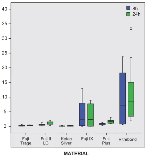
RMGIC Vitrebond released signifi cantly more F— ions at each
time interval than all other experimental GICs and RMGICs (Figure ).
Statistical test of differences of the amount of released F— ions between two time points ( and hours) showed
that RMGICs Vitrebond, Fuji II LC and Fuji plus released signifi cantly more fl uoride ions at both time points ( and hours) comparing with conventional GICs Fuji Triage, Fuji IX GP fast and Ketac Silver. Kruskal Wallis test showed statistically significant differences for hours and for hours, χ
h ()=., p=,; χ
h ()=., p=..
RMGIC Vitrebond (M/ESPE) released the greatest amount of F— ions which is significantly different
com-paring to all other GICs and RMGICs. Ketac Silver (M/ ESPE) released the smallest amount of F— ions which is
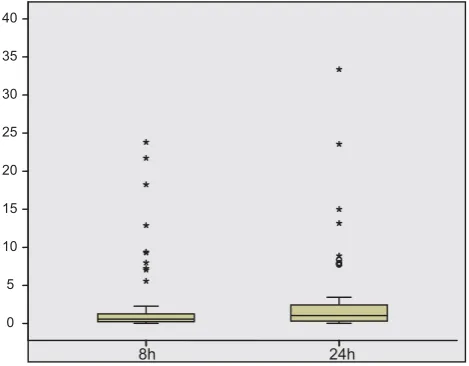
FIGURE 2. Box-plot distribution of the amount of released F- ions
between two time points (8 and 24 hours). Median of the amount of released F- ions after 8 hours is C=0.568, which is lower
com-paring with the median of the amount of released F- ions after 24
hours (C= 1.0485).
Material N Min Max Mean SD Skewness Kurtosis
Statisc Std. Error Statistic Std. Error
Fuji Triage_8h 9 0.098 0.682 0.333 0.200 0.205 0.717 -0.618 1.400
Fuji Triage_24h 9 0.144 0.812 0.392 0.201 0.988 0.717 1.478 1.400
Fuju LCII_8h 9 0.133 1.261 0.624 0.332 0.582 0.717 0.647 1.400
Fuju LCII_24h 9 0.312 2.092 1.265 0.568 -0.484 0.717 -0.627 1.400
Ketac Silver_8h 9 0.022 0.303 0.150 0.106 0.114 0.717 -1.348 1.400
Ketac Silver _24h 9 0.050 0.369 0.229 0.133 -0.676 0.717 -1.680 1.400
Fuji IX fast_8h 9 0.023 12.875 4.518 4.654 0.734 0.717 -0.751 1.400
Fuji IX fast_24h 9 0.027 8.894 3.425 3.726 0.620 0.717 -1.671 1.400
Fuji Plus_8h 9 0.243 1.318 0.855 0.385 -0.321 0.717 -1.528 1.400
Fuji Plus_24h 9 0.920 3.112 1.731 0.738 0.706 0.717 -0.254 1.400
Vitrebond_8h 9 0.625 23.808 9.962 9.146 0.514 0.717 -1.423 1.400
Vitrebond_24h 9 1.897 33.359 12.114 10.590 1.131 0.717 0.684 1.400
TABLE 1. Descriptive statistic values of concentration of F- ions (μg/g) in the 8 and 24 hours eluates
Values of concentration of F- ions (μg/g) released from experimental conventional and resin modifi ed glass-ionomer cements were much higher in the
Bosn J Basic Med Sci 2013; 13 (3): 200-202significantly different comparing to RMGICs Fuji plus and Vitrebond for both time points ( and hours) and GIC Fuji IX GP Fast after hours, only (Table .).
Correlation between cytotoxicity and fl uoride release
With the aim to investigate relationship between F— released
after hours and cytotoxic reaction of UMR – osteoblast-like cells for all experimental GICs, Spearman's rank cor-relation coeffi cient were obtained. Spearman's correlation coefficient indicated a moderate, negative and significant correlation between F— released after hours and cytotoxic
reaction of UMR- – osteoblast-like cells for all
experi-mental GICs (ρ=-.). Similar correlation was fi nd between F— released after hours and cytotoxic reaction of UMR-
– osteoblast-like cells for all experimental GICs (ρ= -.). Spearman's rank coeffi cient between F— released after hours
and cytotoxic reaction of NIHT mouse fibroblast cells indicated a low, negative and statistically insignifi cant cor-relation (ρ = -,) while corcor-relation between F— released
after hours and cytotoxic reaction of NIHT mouse
FIGURE 3. Box-plot distribution of the amount of released F- ions
after 8 hours and after 24 hours. Median of the amount of released F- ions from RMGIC Vitrebond is signifi cantly higher comparing
with all other GICs for the both time points (8 and 24 hours).
Time Material Statistical diff erence
8h
Fuji Triage (A) B, E, F
Fuji II LC (B) C, F
Ketac Silver (C) D, E, F
Fuji IX GP Fast (D) C
Fuji Plus (E) F
Vitrebond (F) A, B, C, E
24h
Fuji Triage (A) B, E, F
Fuji II LC (B) C, F
Ketac Silver (C) E, F
Fuji IX GP Fast (D) F
Fuji Plus (E) F
Vitrebond (F) A, B, C, D, E
TABLE 2. Pairs of statistical diff erences of the amount of released F- ions for all experimental materials after 8 hours and after 24
hours
RMGIC Vitrebond (3M/ESPE) released the greatest amount of F- ions
which is signifi cantly diff erent comparing to all other GICs and RMGICs. Conventional GIC Ketac Silver (3M/ESPE) released the smallest amount of F- ions.
Material F
— release
24 hrs
Cytotoxicity UMR-106 8hrs
Cytotoxicity UMR-106 24hrs
Cytotoxicity NIH3T3 8hrs
Cytotoxicity NIH3T3 24hrs
Fuji Triage F—8hrs 0.750* -0.050 0.717*
Fuji Triage F—24hrs -0.167 0.400
Fuji II LC F— 8hrs 0.800(**) -0.267 0.817(**)
Fuji II LC F—24hrs -0.383 0.750(*)
Ketac Silver F—8hrs 0.653 0.433 0.200
Ketac Silver F—24hrs -0.209 0.536
Fuji IX GP Fast F—8hrs 0.767(*) -0.067 0.800(**)
Fuji IX GP Fast F—24hrs -0.100 0.367
Fuji Plus F—8hrs -0.385 -0.583 -0.217
Fuji Plus F—24hrs 0.285 -0.561
Vitrebond F—8hrs 0.533 -0.083 -0.233
Vitrebond F—24hrs 0.167 0.250
TABLE 3. Matrices of correlation of F- release from experimental GICs and cytotoxic response of UMR-106 – osteoblast-like cells and
NIH3T3 mouse fi broblast cells.
** ρ statistically signifi cant at 0.01; * ρ statistically signifi cant at 0.05
No material, neither conventional nor resin modifi ed glass-ionomer cement showed statistically signifi cant correlation coeffi cient between F- released
in the both time points (8 and 24 hours) and cytotoxic reaction of UMR-106 – osteoblast-like cells. Correlation coeffi cient demonstrated a high, positive and signifi cant correlation between F- released after 8 hours and cytotoxic reaction of NIH3T3 mouse fi broblast cells for conventional GIC Fuji Triage (ρ
= 0.717), and Fuji IX GP Fast (ρ = 0.800) as well as RMGIC Fuji II LC (ρ = 0.817). Spearman's rank correlation coeffi cient between F- released after 24 hours
Bosn J Basic Med Sci 2013; 13 (3): 201-202
fibroblast cells was negative and significant (ρ = -.). Table . shows that, although some values of correla-tion coefficient are relatively high, no one reach the level of statistical significance of . probably because of in-sufficient numbers of specimens in the sample (n=). No material, neither conventional nor resin modifi ed glass-ionomer cement showed statistically signifi cant correlation coeffi cient between F— released after hours and cytotoxic
reaction of UMR- – osteoblast-like cells although cor-relation coefficient of conventional GIC Ketac Silver (ρ = .), and RMGIC Fuji Plus (ρ = -.) are relatively high. Correlation between F— released after hours and
cyto-toxic reaction of UMR- – osteoblast-like cells for each experimental GICs was low and not statistically signifi cant. Th e values of correlation coeffi cient between F— released
af-ter hours and cytotoxic reaction of NIHT mouse fi bro-blast cells were much higher. Spearman's rank correlation coefficient demonstrated a high, positive and significant correlation between F— released after hours and cytotoxic
reaction of NIHT mouse fi broblast cells for conventional GIC Fuji Triage (ρ = .), and Fuji IX GP Fast (ρ = .) as well as RMGIC Fuji II LC (ρ = .). Spearman's rank cor-relation coeffi cient between F— released after hours and
cytotoxic reaction of NIHT mouse fibroblast cells was statistically signifi cant for RMGIC Fuji II LC only (ρ = .).
DISCUSSION
Fluoride release by GICs and RMGICs is an important prop-erty of those materials and plays a major role in its selection for specifi c clinical application []. It seems that fl uoride ions released from GICs act in a dose-dependant manner. In vitro, at relatively high concentration, fl uoride acts as an enzyme inhibitor. In vivo, eliminates microorganisms, those which re-mains in the cavity after preparation, and reinforces deminer-alized enamel and dentin [].Th e pattern of released fl uoride is that the greatest amount of fl uoride being released during the fi rst days, than decreasing to the nearly constant level []. Th e present in vitro study evaluated the quantity of fl uoride ions released from the three conventional glass-ionomer cements: GC Fuji IX GP fast, GC FUJI Triage (GC Cor-poration) and Ketac Silver (M/ESPE) and three resin modified glass-ionomer cements: GC Fuji II LC, GC Fuji plus (GC Corporation) and Vitrebond (M/ESPE) and defined the effect of fluoride ions released from the ex-perimental glass-ionomer cements on their cytotoxicity. Results obtained in this study showed that the rate of F—
re-leased from RMGIC comparing with conventional GIC at both time points ( and hours) were signifi cantly higher. Kan et al. [] concluded that the greatest amount of F— release
occurs in the fi rst hours, which was confi rmed in present
study, were was the diff erence in fl uoride release for all experi-mental materials signifi cantly greater after hours (p= .). In the present study, the greatest amount of F— released
was showed by RMGIC Vitrebond (M/ESPE) and those results were significantly higher comparing with all experimental materials for both time points ( and hours). Results of this in vitro study coincide with the results of Mitra S.B., who has demonstrated that Vitre-bond was capable of long-term fluoride release without any degradation of physical properties over the time []. In the present in vitro study, the amount of F— in RMGIC
Fuji II LC (GC Corporation) hours. eluate was , ppm and demonstrated statistically signifi cant correlation with the cytotoxic response of NIHT mouse fibroblast cells after hours. (ρ= .) and hours immersion (ρ = .). Results obtained in this study demonstrated that there was no statistically significant relation with the cy-totoxic response of UMR- - osteoblast like cells and the amount of F— in RMGIC Fuji II LC (GC Corporation)
eluate after th and th hours immersion. Fuji II LC in the
present study demonstrated moderate cytotoxicity which coincide with the results of Kan et al. [], who pointed out that Fuji II LC behaved more like a resin composite. The fact that brand formulation and material type may influence the amount of F— ion released [], was
con-firmed in present study where RMGIC Vitrebond (M/ESPE) released significantly greater amount of F— ions comparing with other experimental RMGICs.
Capability of glass-ionomer cements to act as a fl uoride ion reservoir presents an important advantage in the process of prevention of secondary caries around restorative margins as well as surrounding surfaces []. Salar et al. [] showed that GC Fuji Triage, as a glass-ionomer sealant, have a relatively high fl uoride content and released enough fl uoride to local environment for a longer period of time which increased resistance to caries in the adjacent enamel. Results obtained in the study Marković et al. [] showed that the amount of fl uoride released by GC Fuji Triage in saline medium is much higher compared to the Fuji IX GP and Fuji II LC. Results ob-tained in present study showed that the GC Fuji Triage after hrours liberated . ppm of F— which was less
Bosn J Basic Med Sci 2013; 13 (3): 202-202by continuing release of F— ions over some time period.
Th e results obtained in present study indicated that Ketac-Silver® (M/ESPE) released . ppm F— after hours
im-mersion, which was the least amount of F— released in this
study. Th ose results corresponded with the results of Forsten [] who indicated that the amount fluoride released from the Ketac-Silver® is smaller than that of other conventional GICs. Th ose fi ndings have been confi rmed in clinical expe-rience because secondary caries is not as rare in connection with the Ketac-Silver® as with other conventional GICs []. Conventional GIC Fuji IX GP Fast (GC Corporation) with the average of . ppm of F— ions released after
hours showed minor cytotoxic effect which suggested that cytotoxicity cannot be explained by F— release alone.
With the exception of Fuji II LC (GC Corporation), the present in vitro study demonstrated that the concentration of F— released by experimental RMGICs and GICs after the
both and hours immersion had no signifi cant eff ect on cytotoxic response of UMR- – osteoblast-like cells and NIHT mouse fibroblast cells. It is more likely that cyto-toxic response of cell lines used in present study occurred due to unidentifi ed toxic components which were leached out during the immersion time. However, additional, more complex experimental studies comprising large number of factors could provide more valid conclusion. Having in mind that liberation of soluble components of experimental GICs can occur during the polymerization process or later on, fur-ther investigation should be based upon identifi cation of se-verely cytotoxic leachable substances and their quantifi cation.
CONCLUSION
Based on the results obtained in the present in vitro study it was concluded that:
Statistical analyses of F— ion concentrations released by all
glass-ionomers evaluated at two time points, after and af-ter hours, show statistically higher fl uoride releases from RMGICs: Vitrebond, Fuji II LC and Fuji Plus, when compared to conventional glass-ionomer cements: Fuji Triage, Fuji IX GP Fast and Ketac Silver, both after and after hours. Correlation coefficient between concentrations of fluo-ride ion released by evaluated glass-ionomer cements and cytotoxic response of UMR- osteoblast cell-line and NIHT mouse fibroblast cell line are relatively high, but do not reach levels of biological significance. According to the methodology employed in the present
study, it can be concluded that experimental GICs
liber-ated F— as well as other soluble components, which
dif-fused into the culture medium. These components can-not be dismissed as possible cytotoxic factors which contribute to the cytotoxicity observed in this study.
ACKNOWLEDGEMENTS
The authors gratefully acknowledge colleagues at the De-partment of Medical chemistry at the Faculty of Medicine, University of Sarajevo, for their skilful technical assistance.
DECLARATION OF INTEREST
Th e authors declare no confl ict of interest.
REFERENCES
[] Koch G, Hatibović-Kofman S. Glass ionomer cements as a fl uoride release system in vivo. Swed Dent J ;: -
[] Hatibović-Kofman S, Koch G. Fluoride release from glass ionomer cement in vivo and in vitro. Swed Dent J ;: - [] Hatibović-Kofman S, Koch G, Ekstrand J. Glass ionomer materials
as a rechargeable fl uoride- release system. Int J Paediatr Dent. ; (): -
[] Forsten L. Fluoride release and uptake by glass-ionomers and relat-ed materials and its clinical eff ect. Biomaterials. ;():-. [] Selimović-Dragaš M, Jurić H. Glass-ionomer cements [Glas jono-mer cementi] In: Vuličević Z.R. (ed.) Materials for clinical
appli-cation in children’s dentistry [Klinička primena materijala u dečjoj
stomatologiji] Beograd: Beobook ;.
[] Wiegand A, Buchalla W, Attin T. Review on fl uoride-releasing re-storative materials--fl uoride release and uptake characteristics, an-tibacterial activity and infl uence on caries formation. Dent Mater. ; ():-.
[] Verbeeck RMH, De Maeyer EAP, Marks LAM, De Moor RJG, De Witte AMJC, Trimpeneers LM. Fluoride relelase process of (resin-modifi ed) glass-ionomer cements versus (polyacid-(resin-modifi ed) com-posite resins. Biomaterials ;:-
[] Costa CAS, Hebling J, Garcia-Godoy F, Hanks CT. In vitro cyto-toxicity of fi ve glass-ionomer cements. Biomaterials ; : -
[] Kan KC, Messer LB, Messer HH. Variability in cytotoxicity and fl uoride release of resin–modifi ed glass-ionomer cements. J Dent Res ; ():-
[] Mitra SB. In vitro fl uoride release from a light-cured glass-ionomer liner / base. J Dent Res. ; ():-.
[] Basso Romanini G, Della Bona A, Gobbi DL, Cecchetti D. Fluoride release from restorative materials. Braz Dent J ; (): - [] Mousavinasab SM, Meyers I. Fluoride release by glass ionomer
ce-ments, compomer and giomer. Dent Res J (Isfahan). ; ():-.
[] Salar DV, Garcia-Godoy F, Flaitz CM, Hicks MJ. Potential inhi-bition of demineralization in vitro by fl uoride-releasing sealants. JADA ; : -