R E S E A R C H
Open Access
The significance of ectopic crypt formation in the
differential diagnosis of colorectal polyps
Mi-Jung Kim
1*, Eun-Jung Lee
2, Sung-Min Chun
3, Se-Jin Jang
3, Do Sun Kim
2, Doo Han Lee
2and Eui Gon Youk
2Abstract
Background:Ectopic crypts, defined as abnormally positioned crypts that have lost their orientation toward the muscularis mucosae, have been suggested to be the best defining histologic feature of traditional serrated adenoma (TSA). However, the significance of ectopic crypt formation (ECF) in the distinction between TSA and conventional adenoma (CA) has rarely been studied.
Methods:We designed this study to determine if ECF can be found in CA and its presence is exclusive to TSA. We studied 107 TSAs and 191 CAs including 106 tubular adenomas (TAs), 66 tubulovillous adenomas (TVAs), and 19 villous adenomas (VAs).
Results:ECF was identified in most (79.4%) but not all TSAs. Additionally, ECF was not infrequent in CA (62
of 191, 32.5%), and its presence correlated with the presence of a villous component and larger tumor size (each p <0.001).
Conclusions: Based on its strong association with the presence of a villous component and larger tumor size,
ECF appears to be involved in the protuberant growth of colorectal CA. Because ECF can be found in CA, particularly in cases with a villous component, the possibility of CA should be considered before making a diagnosis of TSA when encountering colorectal polyps with ECF.
Virtual Slides: The virtual slide(s) for this article can be found here: http://www.diagnosticpathology. diagnomx.eu/vs/13000_2014_212
Keywords: Traditional serrated adenoma, Conventional adenoma, Colon, Ectopic crypt formation, BRAF, KRAS
Background
The term traditional serrated adenoma (TSA) refers to a distinct subtype of serrated polyps and was initially pro-posed for polyps showing a mixture of hyperplastic and adenomatous features [1]. The original definition of TSA referred to a lesion characterized by its overall protuber-ant exophytic configuration, complex villous growth pat-tern, and tall columnar cells with abundant eosinophilic cytoplasm and elongated pencillate nuclei [1-3].
Recently, the presence of ectopic crypt formation (ECF), defined as development of abnormally positioned crypts that have lost their orientation toward the muscularis mu-cosae, was suggested to be the best defining histologic fea-ture to diagnose TSAs based on the report of Torlakovic
et al. that the presence of ECF was almost exclusive to TSAs among serrated polyps and was observed in all TSA cases [4,5]. However, the diagnostic reliability of ECF in the diagnosis of colorectal polyps, particularly in the dis-tinction of TSAs from other much more common polyps such as conventional adenoma (CA), has rarely been stud-ied [3]. We performed this study to determine whether ECF can be present in CA and whether its presence is a pathognomonic feature in the diagnosis of TSA.
Methods
Study sample and histologic evaluation
The present study included 107 TSA samples from 104 patients, all of which were retrieved from the Department of Pathology of Daehang Hospital in Seoul, Korea, be-tween November 2011 and September 2012, as previously described [6]. Of 104 patients, three patients had two TSAs in different location. The lesions were obtained by * Correspondence:mjkimdoc@gmail.com
1
Department of Pathology, Daehang hospital, 481-10 BangBae3-dong, Seoul, Seocho-gu 137-820, Republic of Korea
Full list of author information is available at the end of the article
polypectomy, endoscopic mucosal resection (EMR), endo-scopic submucosal dissection (ESD), or transanal excision. Consultation or referral cases from other hospitals were not included in the study. We also studied 191 consecu-tive CA samples obtained from 188 patients, including 106 tubular adenomas (TAs), 66 tubulovillous adenomas (TVAs), and 19 villous adenomas (VAs), which were completely removed by endoscopic ESD (n = 182) or EMR (n = 9) between January 2011 and September 2012. Among 188 patients with CA, three of them had syn-chronous CA in different location and underwent ESD for both lesions. None of the CA cases were obtained from patients with TSA. All TSA and CA samples were entirely submitted irrespective of size in order to allow evaluation of histopathologic features without any missing areas.
ECF was defined as abnormal development of crypts with loss of orientation toward the muscularis mucosae [4]. In brief, based on previously published histologic cri-teria, TSA was characterized by a serrated architecture and at least a focal area containing tall columnar cells with elongated nuclei and abundant eosinophilic cyto-plasm [2]. We also investigated features of conventional epithelial dysplasia such as extensive nuclear crowding, nuclear enlargement, loss of nuclear polarity, pseudos-tratification extending to the upper half of the neo-plastic cell, and increased mitoses, according to criteria of previously published studies [7,8]. The size and loca-tion of polyps were recorded. The sizes of the polyps were obtained from the pathologist’s measurements. The anatomical sites of polyps were identified and classified as proximal colon (up to the splenic flexure) versus distal colon/rectum.
CA was defined as an adenomatous polyp arranged in a tubular and/or villous architecture. Cytologic dysplasia characterized by the presence of cells with nuclear enlarge-ment, elongation, hyperchromasia, pseudostratification, in-creased mitoses, and crowding was diffusely identified in the polyp in a top-down manner. The degree of dysplasia was determined according to the WHO classification cri-teria [3]. CAs were subdivided into TA, TVA, and VA ac-cording to the percentage of villous component (<25%, 25-75%, >75%, respectively) [3].
Patients with familial adenomatous polyposis, heredi-tary nonpolyposis colorectal cancer syndrome, or inflam-matory bowel disease were excluded from this study. Patients were assessed for clinicopathologic features, including age and sex, anatomic site and size of the polyp, and the presence and type of synchronous or metachronous sessile serrated adenoma/polyp (SSA/P) upon follow-up by reviewing the patients’medical records and endoscopy and pathology reports. The slides were stained with hematoxylin and eosin. Tissue collection was approved by the Institutional Review Board at Daehang Hospital.
DNA extraction
A total of 107 TSA cases containing an adequate amount of tissue in the paraffin blocks were used for molecular studies. In brief, five sections of 10μm thickness samples were used for genomic DNA extraction. Genomic DNA was extracted using the QIAamp DNA formalin-fixed paraffin-embedded (FFPE) tissue kit (#56404; Qiagen, Hilden, Germany) according to the manufacturer’s instruc-tions. In three CAs with significant numbers of ectopic crypts, we also performed microdissection. Microdissec-tion was performed with great cauMicrodissec-tion using the marked slide as a guide. A razor blade was used to scrape off the cells of interest into a 1.5 ml microcentrifuge tube.
Detection of mutations forKRASandBRAFgenes using MassArray technology
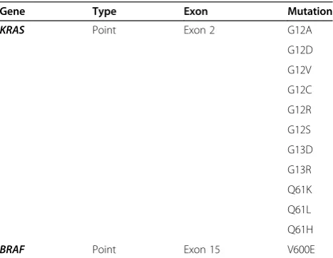
As previously described, mutation detection was car-ried out using an ASAN Panel under the Sequenom MassArray technology platform with the iPLEX-Pro chemistry (Sequenom, San Diego, USA) following the manufacturer’s instructions with minor modifications [6]. In brief, specific assay pools (ASAN Panel) were designed using Assay Designer software in the MassArray Typer package (v4.0). The list of genes and mutations assessed by Sequenom are presented in Table 1. After multiplex PCR with the following program; 95°C for 15 min; 45 cy-cles of incubations at 95°C for 20 sec, 56°C for 30 sec, 72°C for 30 sec; a final 72°C for 3 min, PCR-amplified DNA was cleansed using a shrimp alkaline phosphatase (SAP) mixture from the iPLEX-Prokit (Cat# 10142-2, Sequenom), and primer was extended by the iPLEX chem-istry, desalted using a cation exchange resin (Sequenom) and spotted onto SpectroCHIP II matrix chips using a MassArray nanodispenser. Mass determination was done with the MassArray Analyzer Compact MALDI-TOF mass spectrometer. The MassArray Typer 4.0 software
Table 1 Detectable mutations using the MassArray method (ASAN Panel)
Gene Type Exon Mutation
KRAS Point Exon 2 G12A
G12D
G12V
G12C
G12R
G12S
G13D
G13R
Q61K
Q61L
Q61H
was used for data acquisition and analysis. Genotypes were called after cluster analysis using the default setting of a Gaussian mixture model. Genotype calls were then further reviewed manually to undo any uncertain calls due to clustering artifact. Mutations for a subset of samples and targets were confirmed by Sanger sequencing [6].
Direct DNA sequencing forKRASandBRAF
Direct DNA sequencing was performed to verify mutations in KRAS and BRAF. Polymerase chain reaction (PCR) amplification was performed according to the previ-ously published condition [6,10]. The amplified DNA products were then purified using Montage centrifugal fil-ters (Millipore, Bedford, MA). After purification, direct sequencing was carried out using an ABI PRISM 310 se-quence analyzer (Applied Biosystem, Foster city, CA).
Statistical analysis
Analysis of data was done using SPSS version 21.0 (SPSS Inc., Chicago, IL, USA). For comparing parametric dis-tributions, a Student t-test was used, and for frequency distributions, aχ2test or Fisher exact test was used. A p value <0.05 was considered statistically significant.
Results
Patient demographics and tumor characteristics
A total of 107 TSA cases were obtained from 104 patients comprising 55 males and 49 females with a mean age of 61.2 years. The majority (74.8%) of polyps occurred in the distal colon and rectum, and approximately one-quarter
(25.2%) of cases were obtained from the proximal colon. The sizes of the polyps ranged from 0.3 cm to 5.2 cm, with a mean size of 1.3 cm. The size of the lesion differed sig-nificantly according to the status of conventional epithelial dysplasia, which was noted in 23 TSA lesions (21.5%); tumors were larger in cases with conventional epithelial dysplasia than in those without (mean; 2.1 cmvs. 1.1 cm, p <0.001). Mean age, sex, and tumor location did not dif-fer according to the status of conventional epithelial dys-plasia (p = 0.235, p = 1.000, and p = 0.178, respectively).
In total, 191 CAs were obtained from 188 patients: 105 males and 83 females with a mean age of 62.7 years. There was no significant difference between patients with TSA and those with CA with respect to mean age or sex (p = 0.339 and p = 0.229, respectively). Moreover, there were no differences in age or sex among patients with TA, TVA, and VA (p = 0.382 and p = 0.506, respect-ively). The mean tumor size of CAs was larger than that of TSAs (2.9 cmvs. 1.3 cm, p <0.001), most likely because we included CAs obtained by ESD or EMR. Three patients with CA had a prior history of right-sided SSA/Ps, and 10 other patients with CAs had synchronous or metachro-nous SSA/P. All 13 cases of SSA/P were histologically proven. The clinicopathologic features of the patients with TSA and CA are summarized in Table 2.
Histologic findings
All of the cases of TSA included in this study exhibited a serrated architecture in at least 20% of the tumor area and at least one focal area of tall columnar cells with
Table 2 Patient demographics and tumor characteristics
CA group
Parameters TSA group
(n = 107)a
TA (n = 106)b
TVA (n = 66)b
VA (n = 19)
p
Mean age (y) 61.2 63.3 62.6 59.7 0.345
(range) (30-81) (31-85) (37-80) (34-79)
Gender 0.634
Woman 49 42 31 10
Man 55 62 34 9
Tumor location <0.001
Proximal 27 (25.2%) 62 (58.5%) 30 (45.5%) 2 (10.5%)
Distal 80 (74.8%) 44 (41.5%) 36 (54.5%) 17 (89.5%)
Mean tumor size (cm) 1.3 2.7 3.2 3.6 <0.001
Procedure NA
Polypectomy 37 (34.6%) 0 0 0
EMR 57 (53.3%) 5 (4.7%) 4 (6.1%) 0
ESD 12 (11.2%) 101 (95.3%) 62 (93.9%) 19 (100%)
Transanal excision 1 (0.9%) 0 0 0
TSA, traditional serrated adenoma; CA, conventional adenoma; TA, tubular adenoma; TVA, tubulovillous adenoma; VA, villous adenoma; NA, not applicable; EMR, endoscopic mucosal resection; ESD, endoscopic submucosal dissection.
a
107 TSAs from 104 patients. b
abundant eosinophilic cytoplasm (Figure 1A). The low-power images of TSAs were somewhat variable depend-ing on the growth pattern, the extent of peculiar tall columnar cells with eosinophilic cytoplasm, the status of precursor lesions, and the amount of stromal com-ponent. ECF was identified in most (79.4%) but not all cases of TSA (Figure 1B). The frequency of ECF varied de-pending on the case or even the area within the same case.
ECF was numerous in 15.0% of cases, whereas some cases (5.7%) displayed minimal ECF.
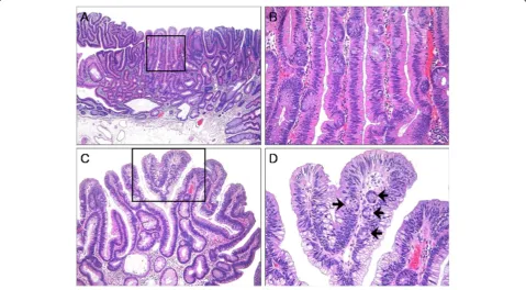
ECF was present in 62 (32.5%) cases of CA (Figures 2 and 3). The frequency of ECF in TAs, TVAs, and VAs was 22.6% (24 cases), 37.9% (25 cases), and 68.4% (13 cases), respectively (p <0.001) (Table 3). ECF was sparse in all but three cases of CA. Most CAs had ampho-philic or basoampho-philic cytoplasm, although a minority (5.2%) Figure 1Histologic findings of traditional serrated adenoma (TSA). (A), TSA showing a protuberant growth pattern and serrated architecture (hematoxylin and eosin; original magnification × 40).(B), TSA composed of tall columnar cells with diffuse serration and abundant eosinophilic cytoplasm. Note the abundant ectopic crypts (hematoxylin and eosin; original magnification × 200).
showed abundant eosinophilic cytoplasm in focal areas that was significantly associated with the presence of ECF (p <0.001) (Figure 2A and B). The presence of ECF sig-nificantly correlated with the presence of villosity and large tumor size (each p <0.001; Figure 2C and D). ECF was also noted in CA with high-grade dysplasia (HGD) as well as that with low-grade dysplasia (LGD) (Figure 3A and B). Serration of crypts was noted in 20 (10.5%) cases of CA; however, epithelial serration accounted for less than 5% of the polyp area in all but one CA, in which serration of crypts occupied approxi-mately 20% of the area. In that case, the epithelial serration was concentrated in the middle and lower portions of crypts associated with dilatation. Three CA cases showed Paneth cell metaplasia in areas with ECF (Figure 4A and B). In 16 CA cases, ECF, focal serration, and cytoplasmic eosinophilia occurred in the same polyp. The presence of ECF was significantly associated with focal serration of crypts and cytoplasmic eosinophilia in CA (each p <0.001).
Mutation results forBRAFandKRASgenes
BRAFmutation was observed in 59 (55.1%) TSA lesions, andKRASmutations were identified in 36 (33.6%) cases. Mutations of BRAFand KRAS were mutually exclusive. Twelve (11.2%) TSA lesions showed neitherBRAF600E nor KRAScodon 12, 13, or 61 mutations. Among three cases of CA showing significant numbers of ectopic crypts, two showedKRASmutation, while the other dis-played neitherBRAFnorKRASmutation (Figure 5).
Discussion
TSA is a distinct subtype of serrated neoplasm that is usually characterized by a protuberant growth pattern with an overall complex and often villous growth pat-tern, epithelial serration, and tall columnar cells with abundant eosinophilic cytoplasm and elongated pencillate nuclei [1-3]. However, the diagnosis of TSA is sometimes
challenging because of overlap with other polyps in terms of both gross and histologic features. More specifically, the gross morphology does not distinguish TSAs from other polyps because TSAs vary from flat elevated and sessile to pedunculated forms [9]. A pedunculated config-uration, which is known to be frequent in TSAs, is also found in other polyps, in particular the so-called‘ pedun-culated serrated polyp with SSA/P features’and CA [10]. The former lesion is a newly described serrated polyp that has clinicopathologic features of SSA/P except for the pedunculated gross configuration. The complex villous growth pattern can also be identified in CA. Serration of crypts can be observed in various conditions such as in-flammatory bowel disease, juvenile polyps, inin-flammatory polyps, and CAs, as well as other kinds of serrated polyps [11-13]. In addition, the characteristic cytological features such as abundant eosinophilic cytoplasm and pencillate nuclei can be found in other serrated polyps such as SSA/ P or sessile serrated adenoma with dysplasia (SSA/D), as well as in CA [14]. In summary, none of these histopatho-logic features alone is considered specific for TSA. There-fore, in this study, we defined TSA as a lesion combining a serrated architecture and at least one focal area contain-ing tall columnar cells with elongated nuclei and abundant eosinophilic cytoplasm [2].
Recently, the presence of ECF has been suggested to be the best defining histologic feature to diagnose TSA [4,5]. Figure 3Tubulovillous adenoma (TVA) with high grade dysplasia. (A), TVA with a villous architecture and columnar cells showing diffuse high grade dysplasia (hematoxylin and eosin; original magnification × 40).(B), Box in panel A, displaying high grade dysplasia and ectopic crypts (hematoxylin and eosin; original magnification × 100).
Table 3 Comparison of histologic features according to the subtype of CAs
Parameters TA (106), n(%)
TVA (66), n(%)
VA (19), n(%)
p
ECF (%) 24 (22.6%) 25 (37.9%) 13 (68.4%) < 0.001
Focal serration (%) 7 (6.6%) 7 (10.6%) 6 (31.6%) 0.005
Focal cytoplasmic eosinophilia (%)
1 (0.9%) 4 (6.1%) 5 (26.3%) < 0.001
However, the presence of ectopic crypts, which was ini-tially suggested to be exclusive to TSA, was reported to be present in other serrated polyps such as HP or SSA/P [14]. Similar to the result by Ensari et al. [14], ECF was noted in approximately 80% of TSA cases in the present study. In addition, a considerable fraction of CA showed ECF, albeit most cases were sparse. Al-though TSA showed a higher frequency of ECF than CA (p <0.001), the frequency of ECF in CA (32.5%) was higher than we expected, particularly in cases with a villous component (p <0.001). The actual rate of ECF, par-ticularly in polypectomy specimens, might be lower than that of the present study because we included large CAs obtained by ESD or EMR. At any rate, ECF itself does not seem to be a sine qua nonfor the diagnosis of TSA be-cause ECF can be found in CA, although its identification might be very helpful in the diagnosis of TSA. Therefore, if one encounters colorectal polyps with ECF, the possi-bility of CA should always be considered. In such cases,
histologic evaluation for the presence and/or extent of crypt serration, extent of ECF, and cytoplasmic character-istics are necessary to distinguish TSA from CA with ECF (Figure 6). Absence of extensive ECF, prominent serra-tion, serrated dysplasia, and/or diffuse cytologic eosino-philia are histologic features favoring a diagnosis of CA over TSA. According to the results of this study, the ex-tent of serration did not exceed 20% of crypts in CA.
In addition to the histopathologic evaluation, the re-sults of molecular studies for BRAF mutation are also important in the differentiation of CA from TSA be-cause BRAFmutation is rare in CA (<1%) but common in TSA [15-17]. Mutation study forKRASseems to be less useful than BRAF testing in the distinction between CA and TSA, although the frequency of KRAS mutations is lower in TSA than in CA [17]. In the present study, we performed mutation study forKRAS and BRAF in order to investigate whetherBRAFmutation was present in CA with frequent ECF. As expected, none of these cases Figure 4Tubulovillous adenoma (TVA) with ectopic crypts and Paneth cell metaplasia. (A), TVA with frequent ectopic crypts (hematoxylin and eosin; original magnification × 100).(B), Higher magnification of the box in panel A, displaying ectopic crypts showing Paneth cell metaplasia (hematoxylin and eosin; original magnification × 200).
exhibited a BRAF V600E mutation. Instead, two of three CAs with frequent ECF showed KRASmutations. Muta-tion study of the APC gene might be also helpful in the distinction between CA and TSA because, in contrast to CA,APCgene mutation is infrequent in TSA [18]. How-ever, we could not perform molecular study of the APC gene in this study.
The presence of ECF, serration of crypts, and cytoplas-mic eosinophilia in some CAs suggest the possibility that such polyps share a series of molecular alterations with TSAs that are responsible for the histological differenti-ation toward serrated neoplasia. Accordingly, Pai et al. [19] reported that a subset of CA shows atypical features such as cytoplasmic eosinophilia with or without focal serrations, usually in patients predisposed to developing SSA/P. On molecular analysis, these atypical CAs showed a higher frequency of CpG island methylation than did typical CA (35.7% vs. 4.2%). We did not find any signifi-cant difference in the rates of ECF, cytoplasmic eosino-philia, or serration of crypts between CA that developed SSA/P and CA that did not (p = 0.228, p = 0.515, and p = 1.000, respectively), although this might be because of the small number of patients that developed SSA/P among the patients with CA.
We believe that CA with ECF is not uncommon, al-though there is some controversy over whether such cases represent TSA with diffuse conventional dysplasia [5]. Indeed, most CAs in this study were otherwise typical, except for three cases that showed easily recognizable ECF, none of which showed prominent serration of crypts (more than 20%), serrated dysplasia, diffuse cytoplasmic eosinophilia, orBRAF mutation. According to a previous study, ECF is considered one of the mechanisms that in-crease crypt numbers as a result of complex biologic pro-cesses involving a sequential proliferation-differentiation process that depends on the time point and
epithelial-mesenchymal interaction [20]. It is presumed that inhib-ition of bone morphogenic protein (BMP) signaling results in the formation of ectopic crypt units perpendicular to the crypt-villus axis [21]. The concentration of Ki-67+cells in ectopic crypts supports our contention that ectopic crypts play an important role in the protuberant growth of polyps [4]. Accordingly, the presence of ECF significantly correlated with large tumor size in this study.
Conclusions
In conclusion, ECF appears to be associated with villous or protuberant growth of colorectal adenoma. Although identification of ECF seems to be very helpful in the diagnosis of TSA, ECF is also found in CA, particularly in cases with a villous component. Therefore, the possi-bility of CA should be considered before making a diag-nosis of TSA in cases of colorectal polyp with ECF.
Abbreviations
BMP:Bone morphogenic protein; CA: Conventional adenoma; ECF: Ectopic crypt formation; EMR: Endoscopic mucosal resection; ESD: Endoscopic submucosal dissection; FFPE: Formalin-fixed paraffin-embedded; HGD: High grade dysplasia; LGD: Low grade dysplasia; PCR: Polymerase chain reaction; SSA/P: Sessile serrated adenoma/polyp; TA: Tubular adenoma;
TSA: Traditional serrated adenoma; TVA: Tubulovillous adenoma; VA: Villous adenoma.
Competing interests
The authors declare that they have no competing interests.
Authors’contributions
MJK is the principal researcher and drafted the manuscript. EJL participated in the design of the study. DSK, DHL, and EGU were responsible for the clinical data. SMC and SJJ carried out the mutation analysis. All authors read and approved the final version submitted.
Author details
1Department of Pathology, Daehang hospital, 481-10 BangBae3-dong, Seoul, Seocho-gu 137-820, Republic of Korea.2Department of Surgery, Daehang hospital, Seoul, Republic of Korea.3Department of Pathology, University of Ulsan College of Medicine, Asan Medical Center, Poongnap 2-Dong, Seoul, Songpa-Gu 138-736, Republic of Korea.
Received: 30 June 2014 Accepted: 26 October 2014
References
1. Longacre TA, Fenoglio-Preiser CM:Mixed hyperplastic adenomatous polyps/serrated adenomas. A distinct form of colorectal neoplasia.Am J Surg Pathol1990,14:524–537. Review.
2. Torlakovic E, Skovlund E, Snover DC, Torlakovic G, Nesland JM:Morphologic reappraisal of serrated colorectal polyps.Am J Surg Pathol2003,27:65–81. 3. Bosman FT, Carneiro F, Hruban RH, Theise ND:World Health Organization
Classification of Tumours of the Digestive System.4th edition. Lyon, France: IARC Press; 2010:160–165.
4. Torlakovic EE, Gomez JD, Driman DK, Parfitt JR, Wang C, Benerjee T, Snover DC: Sessile serrated adenoma (SSA/P) vs. traditional serrated adenoma (TSA). Am J Surg Pathol2008,32:21–29.
5. Snover DC:Update on the serrated pathway to colorectal carcinoma. Hum Pathol2011,42:1–10.
6. Kim MJ, Lee EJ, Suh JP, Chun SM, Jang SJ, Kim do S, Lee DH, Lee SH, Youk EG: Traditional Serrated Adenoma of the Colorectum: Clinicopathologic Implications and Endoscopic Findings of the Precursor Lesions.Am J Clin Pathol2013,140:898–911.
7. Fu B, Yachida S, Morgan R, Zhong Y, Montgomery EA, Iacobuzio-Donahue CA: Clinicopathologic and genetic characterization of traditional serrated adenomas of the colon.Am J Clin Pathol2012,138:356–366.
8. Snover DC, Jass JR, Fenoglio-Preiser C, Batts KP:Serrated polyps of the large intestine: a morphologic and molecular review of an evolving concept.Am J Clin Pathol2005,124:380–391. Review.
9. Jaramillo E, Tamura S, Mitomi H:Endoscopic appearance of serrated adenomas in the colon.Endoscopy2005,37:254–260.
10. Kim MJ, Lee EJ, Chun SM, Jang SJ, Kim CH, Seo JP, Kim Do, Lee DH, Lee SH, Youk EG:Pedunculated Serrated Polyp With Histologic Features of Sessile Serrated Adenoma: A Clinicopathologic and Molecular Study.Am J Surg Pathol2013,37:1039–1043.
11. Srivastava A, Redston M, Farraye FA, Yantiss RK, Odze RD:Hyperplastic/ serrated polyposis in inflammatory bowel disease: a case series of a previously undescribed entity.Am J Surg Pathol2008,32:296–303. 12. Huang CC, Frankel WL, Doukides T, Zhou XP, Zhao W, Yearsley MM:
Prolapse-related changes are a confounding factor in misdiagnosis of sessile serrated adenomas in the rectum.Hum Pathol2013,44:480–486. 13. Bariol C, Hawkins NJ, Turner JJ, Meagher AP, Williams DB, Ward RL:
Histopathological and clinical evaluation of serrated adenomas of the colon andrectum.Mod Pathol2003,16:417–423.
14. Ensari A, Bilezikçi B, Carneiro F, Doğusoy GB, Driessen A, Dursun A, Flejou JF, Geboes K, de Hertogh G, Jouret-Mourin A, Langner C, Nagtegaal ID, Offerhaus J, Orlowska J, Ristimäki A, Sanz-Ortega J, SavaşB, Sotiropoulou M, Villanacci V, Kurşun N, Bosman F:Serrated polyps of the colon: how reproducible is their classification?Virchows Arch2012,461:495–504. 15. Spring KJ, Zhao ZZ, Karamatic R, Walsh MD, Whitehall VL, Pike T, Simms LA,
Young J, James M, Montgomery GW, Appleyard M, Hewett D, Togashi K, Jass JR, Leggett BA:High prevalence of sessile serrated adenomas with BRAF mutations: a prospective study of patients undergoing colonoscopy.Gastroenterology2006,131:1400–1407.
16. Burnett-Hartman AN, Newcomb PA, Potter JD, Passarelli MN, Phipps AI, Wurscher MA, Grady WM, Zhu LC, Upton MP, Makar KW:Genomic aberrations occurring in subsets of serrated colorectal lesions but not conventional adenomas.Cancer Res2013,73:2863–2872.
17. Rex DK, Ahnen DJ, Baron JA, Batts KP, Burke CA, Burt RW, Goldblum JR, Guillem JG, Kahi CJ, Kalady MF, O’Brien MJ, Odze RD, Ogino S, Parry S, Snover DC, Torlakovic EE, Wise PE, Young J, Church J:Serrated lesions of the colorectum: review and recommendations from an expert panel. Am J Gastroenterol2012,107:1315–1329.
18. Fu X, Li J, Li K, Tian X, Zhang Y:Hypermethylation of APC promoter 1A is associated with moderate activation of Wnt signalling pathway in a subset of colorectal serrated adenomas.Histopathology2009,55:554–563. 19. Pai RK, Mackinnon AC, Joseph L, Noffsinger A, Hart J:Identification of
histologically distinct conventional adenomas that arise predominately in patients with sessile serrated adenomas.Am J Surg Pathol2010, 34:355–363.
20. Del Buono R, Lee CY, Hawkey CJ, Wright NA:Isolated crypts form spheres prior to full intestinal differentiation when grown as xenografts: an in vivo model for the study of intestinal differentiation and crypt
neogenesis, and for the abnormal crypt architecture of juvenile polyposis coli.J Pathol2005,206:395–401.
21. Haramis AP, Begthel H, van den Born M, van Es J, Jonkheer S, Offerhaus GJ, Clevers H:De novo crypt formation and juvenile polyposis on BMP inhibition in mouse intestine.Science2004,303:1684–1686.
doi:10.1186/s13000-014-0212-x
Cite this article as:Kimet al.:The significance of ectopic crypt formation in the differential diagnosis of colorectal polyps.Diagnostic Pathology
20149:212.
Submit your next manuscript to BioMed Central and take full advantage of:
• Convenient online submission
• Thorough peer review
• No space constraints or color figure charges
• Immediate publication on acceptance
• Inclusion in PubMed, CAS, Scopus and Google Scholar
• Research which is freely available for redistribution