RVC OPEN ACCESS REPOSITORY – COPYRIGHT NOTICE
This is the author’s accepted manuscript of an article published in Seminars in Cell & Developmental Biology.
© 2018. This manuscript version is made available under the CC-BY-NC-ND 4.0 license http://creativecommons.org/licenses/by-nc-nd/4.0/.
The full details of the published version of the article are as follows:
TITLE: Like a hole in the head: Development, evolutionary implications and diseases of the cranial foramina
AUTHORS: Imelda M. McGonnell and Sophia E. Akbareian JOURNAL: Seminars in Cell & Developmental Biology PUBLISHER: Elsevier
1
Like a hole in the head: Development, evolutionary implications and
diseases of the cranial foramina
Imelda M. McGonnell * and Sophia E. Akbareian
Dept. Comparative Biomedical Sciences, Royal Veterinary College, Royal College
St, London NW1 0TU.
*Author for correspondence: [email protected]
Abstract
Cranial foramina are holes in the skull through which nerves and blood vessels pass to reach both deep and superficial tissues. They are often overlooked in the literature; however they are complex structures that form within the developing cranial bones during
embryogenesis and then remain open throughout life, despite the bone surrounding them undergoing constant remodelling. They are invaluable in assigning phylogeny in the fossil record and their size has been used, by some, to imply function of the nerve and/or blood vessel that they contained. Despite this, there are very few studies investigating the development or normal function of the cranial foramina. In this review, we will discuss the development of the cranial foramina and their subsequent maintenance, highlighting key gaps in the knowledge. We consider whether functional interpretations can be made from fossil material given a lack of knowledge regarding their contents and maintenance. Finally, we examine the significant role of malformation of foramina in congenital diseases such as craniosynostosis.
1. Cranial Foramina
2
The term foramen means an “opening” and is used to describe a number of other anatomical structures both within skeletal tissue, such as the apical foramen of the tooth, and non-skeletal tissues such as the foramen ovale of the foetal heart and the foramina of Monro which are channels connecting the ventricles of the brain. With regards to the cranial foramina, the largest is the foramen magnum which allows the passage of the spinal cord, spinal and vertebral arteries and veins through the skull base. This cavity forms in the embryo at the boundary between 4 individual elements that comprise the occipital bone (basi-occipital, supra-occipital and paired exo-occipital bones) rather than within a skeletal element itself. These separate elements later fuse to form the occipital bone. In contrast, the majority of other cranial foramina are found within individual skeletal elements.
The cranial foramina develop in precise topographical locations that are typical to specific species. The literature contains a considerable array of nomenclature for the adult cranial foramina and there is variation in the described nerve and blood vessel contents between different species, particularly in birds. However, most cranial foramina appear to contain both a nerve and a blood vessel [1-5].
2. Development of the Cranial Foramina
2.1 Cranial foramina develop in skeletal elements of differing embryological origin The vertebrate skull is a complex structure; whilst seemingly one unit, it comprises the viscerocranium (facial skeleton), the chondrocranium (the skull base) and the
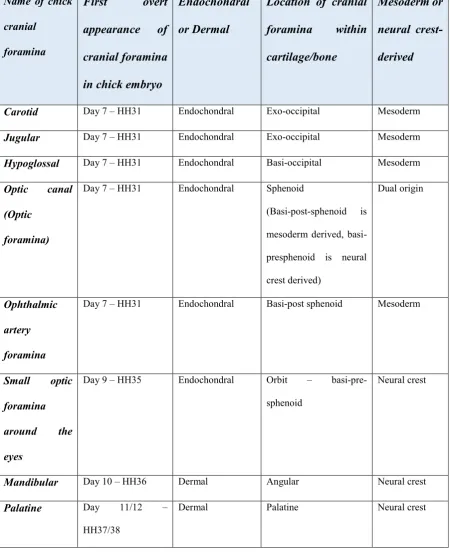
dermatocranium (skull vault), which have different developmental origins. In contrast to the mesodermally derived axial and appendicular skeletons, the skull has contributions from the neural crest as well as somitic and cranial paraxial mesoderm [6-7]. A number of fate
mapping studies using quail/chick chimeras [8-9] as well as retroviral labelling [10] have shown that the majority of the chick facial skeleton (viscerocranium) and part of the rostral frontal bone (a component of the dermatocranium) are of neural crest origin with the
remaining dermatocranium being of cranial mesodermal origin (Fig. 1). Similar studies using genetic labelling of neural crest with wnt1-cre and somitic mesoderm with Mesp1-cre in mice [11-13] and zebrafish neural crest with Sox10-cre [14] have shown the conserved evolution of this pattern in other vertebrates. The chondrocranium (skull base) has also been
3
In addition to the complexity of tissue of origin, there are also two different mechanisms of ossification at play in the vertebrate skull. The first; endochondral ossification occurs in a number of cranial skeletal elements as well as those in the axial and appendicular skeleton. It involves the formation of bone via a cartilage template that is laid down early in
development [18]. This process is initiated by the condensation of uncommitted mesenchyme, driven by the expression of the SRY transcription factor Sox9 [19-22].
Commitment to the cartilage phenotype involves the expression of Sox 5 and 6 [23] followed by expression of genes such as aggrecan which drive chondrocyte differentiation and production of extracellular matrix [24]. The final step is hypertrophy of this template to allow the invasion of blood vessels and the deposition of osteoblasts that will differentiate into bone [25]. It has recently been discovered that some of the hypetrophic cartilage cells
transdifferentiate into osteoblasts rather than being removed through apoptosis or autophagy [26, 27].
In contrast, intramembranous ossification involves direct differentiation of mesenchyme into osteoblasts without a cartilage template. The mesenchyme is initially dormant for a long period of time in development before the differentiation process commences. BMP, Wnt, Ihh and PTHrP (parathyroid hormone related protein) signalling play roles in intramembranous ossification [28-29].
However, both models of ossification are very similar when it comes to the formation of bone. The osteoblasts mature into osteocytes and secrete a bony matrix. The molecular pathways involved in osteocyte differentiation involve the expression of runx2, osterix, osteopontin and osteocalcin [30-32].
Therefore, cranial foramina in the head can be sub-divided depending on the origin of the tissue they develop in and the mode of ossification. Table 1 details the location, subtype and timing of development of key cranial foramina in the chick embryo as an illustration of the variety of cranial foramina. Thus examples of foramina within neural crest derived
4
controlling the formation of each sub-type of cranial foramina and may also have impact upon the mechanisms that maintain them throughout life. This differing origin could also explain why sub-sets of foramina may develop abnormally or close in specific congenital syndromes and diseases while others are unaffected. However, currently little is known about the overall mechanisms of development of foramina and sub-type differences have not been investigated.
2.2 Mesenchymal clearance in the developing cranial foramina.
At present, there is little published literature available concerning the development of foramina in any species. We have used the chick embryo model to examine the development of the foramina of the occipital bones; the jugular, the carotid and the hypoglossal that develop in mesodermally derived skeletal elements formed through
endochondral ossification (Fig. 2, Table 1). They are first visible in section at HH31 (Day 7 of development) as a region of mesenchyme surrounding a blood vessel and/or nerve that is less dense than the surrounding mesenchyme that will form the basi- and exo-occipital cartilages [43]. By HH 34 (Day 8 of development), the mesenchyme immediately adjacent to the nerve and blood vessel has become more sparse and is bordered by cells with a
flattened morphology that resemble a perichondrium. The jugular foramen appear more distinct than the hypoglossal foramen at this stage of development (Fig 3 A, B). By HH 35 (Day 9), there is a distinct foramen with a boundary between the condensing mesenchyme (that will become cartilage) and the sparse undifferentiated cartilage of the foramen itself (Fig 3 C, D). The shaping of the jugular foramen conforms to that of the blood vessel
however the relationship between hypoglossal foramen shape and contents does not appear to be as close at this stage of development. The foramen at HH 43 (Day 17) is well
developed with little mesenchyme now present in the cavity (Fig3 E, F).
A similar clearing of mesenchyme is also seen in the development of the limb synovial joint, a skeletal cavity that has been studied in some detail. This cavity is thought to develop by either cavitation of proliferating cells, apoptosis of mesenchymal cells within the joint space or a combination of these mechanisms [33-36]. In contrast, in the cranial foramina, analysis of cell proliferation patterns indicate that increased cell proliferation does not coincide with formation of the cavity but that it may play a role in refinement of the size of the cavity once formed. Similarly, apoptosis does not seem to play a role in clearing mesenchyme from the foramina [37]. Thus there appears to be other, as yet unidentified, cellular mechanisms involved in the clearance of mesenchyme of the foramina space.
5
The question of what is directing this cavitation process is still unanswered, however the nerves and blood vessel are likely candidates. The innervation of the head is complex and comprises motor, sensory, parasympathetic and sympathetic innervation [38-39]. The timing of axonal outgrowth from each nerve population is varied but the majority of major nerves have grown into the developing chick head regions by HH19-23 [40], prior to mesenchymal condensation or differentiation and foramina development. In contrast, myelination of nerves occurs later in development, after condensation of the mesenchyme (e.g. around HH29 for the hypoglossal nerve) [37, 41]. Thus it is clear that the nerves are present prior to
skeletogenesis and do not tunnel through condensing, differentiating or differentiated mesenchyme, suggesting a role for them in creating a “zone of inhibition” of developing cartilage and bone to allow a cavity to develop. This zone of inhibition can be seen in the developing occipital foramina using the expression of the prechondrogenic gene Sox9. The mesenchyme that is adjacent to the cranial nerves blood vessels initially expresses Sox9 prior to any changes in the mesenchyme (between HH 21-27, Day 3.5-5.5 of development in the chick embryo). However, just prior to the change in mesenchyme density, Sox9
expression is lost in this population while it is maintained in the denser mesenchyme, more distant to the blood vessel and nerve [37]. These Sox9 negative cells are subsequently cleared during foramina development. This may suggest that the blood vessel and nerve have some role in controlling the fate of mesenchymal cells closest to them, preventing them from progressing along the cartilage differentiation pathway.
We have used the developing chick model to directly test whether nerves play a role in foramina induction. Unilateral ablation of the hypoglossal nerve, through removal of one half of the neural tube at the level of somite3/4 to the base of the heart loop, results in loss of the ventral nerve rootlets that normally traverse the basi-occipital through the hypoglossal foramina, a group of 3 foramina each side of the midline. We found that this ablation also resulted in maintained Sox9 expression in the mesenchyme, a lack of mesenchymal clearing and cavitation and the absence of foramina (unpublished observation). This suggests that nerves are likely to play a significant role in foramina development.
The direct role of the vasculature in the development of the cranial foramina is less clear as similar ablation experiments have not been performed in the chick model. However,
6
meningeal artery normally traverses the foramen spinosum however in PAS syndrome, this foramina is absent which is attributed to the loss of the artery [42]. Congenital agenesis or hypoplasia of the internal carotid artery also occurs in humans, which can involve narrowing or even absence of this artery and this is accompanied by a narrowed or absent carotid canal (foramen) [43], again suggesting that the vasculature plays a direct role in foramina development and maintenance. However, this evidence should be viewed with some caution as the cause of these vascular abnormalities are typically unknown and an underlying
abnormality in the developing bone that could account for the abnormal foramina cannot be completely ruled out.
Interestingly, a recent paper investigating the postnatal development of meningeal lymphatic vessels in mice, has shown that these vessels form around the cranial foramina before extending [44]. This could mean that there is an intriguing interplay between the vascular system and foramina such that the development of the foramina may be regulated by arteries and veins and in turn, the foramina may act to direct formation of lymphatic vessels, although more functional work is required to substantiate this theory.
There may also be multiple mechanisms controlling the formation and maintenance of the foramina cavity at different stages of development. Evidence points to this in the
endochondral occipital foramina (jugular, carotid and hypoglossal) where there is a steady increase in diameter of each from the time they first appear in the mesenchyme on day 6/7 of incubation (HH 29/30) up to day 9/10 (HH35/36) in the chick embryo. This is followed by a considerable reduction in diameter for several days before the foramen widens again [37]. This period of narrowing coincides with the transition from cartilage to bone in the occipital skeleton (Fig. 2D), suggesting that the mechanisms preventing cartilage formation may not be sufficient to initially prevent the encroachment of bone and other mechanisms may be required to keep the endochondral foramina patent.
3. Do cranial foramina play a role in interpreting function in fossil evidence?
7
the animal. For example, the size of the carotid foramen has been employed as a proxy for blood flow to the brain to determine if disproportionate increases in the blood flow rate, and thus metabolism, contributed to increased brain volume during hominin evolution [49]. The carotid foramen houses the internal carotid artery that carries blood to the brain; it has been proposed that internal carotid arterial size (and thus blood flow and metabolic rate) is closely linked to cognitive ability [49]. While the internal carotid artery diameter is itself dynamically regulated by the rate of blood flow, this analysis relies on the assumption that the size of the bony foramen is determined solely by the diameter of the artery and that other contents (nerve and/or extracellular matrix) play no role in the regulation of foramen size in the adult. However, we know little of how foramina size is regulated in normal developing or adult bone and how dynamic this process may be.
Similarly, Bird et al have investigated foramina as a proxy for olfactory ability in the fossil record [50]. The cribriform plate of the ethmoid bone contains numerous small foramina that allow passage of olfactory sensory neuron axons from the olfactory epithelium of the nose to the olfactory bulb of the brain. These neurons undergo constant turnover in adult life and are replaced by neurons generated by stem cells in the olfactory epithelium [51]. The axons of these regenerating neurons are thought to track through existing foramina, established during development of the cribriform plate, to reach the olfactory bulb. Bird et al concluded that cumulative cribriform plate foramina area measured in fossil skulls could be a useful correlate to olfactory ability in carnivora. However, these foramina may close due to loss of neurons, such as with head trauma, neurodegenerative disease [52] or with age [53] and an age related decrease in cribriform plate foramina area has been identified in humans [54]. Thus without detailed information about the age of the animal and any potential injuries that could have led to significant neuronal loss, it is difficult to conclude that there is a direct relationship.
8
between the two, indicating that infraorbital foramen size is a good proxy for maxillary nerve size and by implication, function. However this foramina only contains a small artery and vein so it is not clear if this relationship would hold in species where there are variations in the contents of this foramen. Equally, it is not clear how strong this relationship would be in other foramina, where there are larger blood vessels in addition to the nerve. In many species, neither the actual contents nor the sizes of the blood vessels and nerves within the foramina are well documented which can lead to misinterpretation. Indeed Muchlinki points out that the size of the hypoglossal canal that houses axons of the motor hypoglossal nerve innervating the tongue had previously been implied as an anatomical proxy for the
appearance of humanlike speech in the fossil record [55]. It was suggested that the larger size of this foramen in humans was due to increased hypoglossal nerve area allowing for fine motor control of the tongue [55]. However, further investigation in humans demonstrated that the nerve was a minor component of the content, with the venous plexus being a much bigger contributor [56]. Thus, the vascular contents and not the nerve likely dictate the overall size of this foramen.
4. Diseases and Syndromes in involving the cranial foramina
A number of congenital syndromes and other diseases involve the cranial foramina. Failure to form in the embryo, abnormal morphological development or narrowing/closure during adulthood can lead to blindness, deafness, facial paralysis and raised intracranial pressure which can result in death, depending on which foramina are affected [57-59].
Understanding the contribution of the cranial foramina to disease can be complicated by the large amount of normal anatomical variation in location, shape, size and content of the cranial foramina in humans. These variations are best described for cranial foramina that have surgical significance such as the lingual [60-61]and the mental foramen [62-63], where significant variations are reported. It is likely that there are similar normal variations in other cranial foramina.
4.1 Craniosynostosis and achondroplasia
Craniosynostosis syndromes involve premature fusion of cranial sutures. The majority of these are due to FGFR mutations, although mutations in other genes such as TWIST1 are also reported [64-68]. What is less well known is that both humans with these syndromes and animal models of craniosynostosis typically present with either smaller and/or
9
Crouzons) and this typically results in raised intracranial pressure that is not alleviated by surgery upon the fused sutures [69]. Fgf signalling is involved in multiple steps of both cartilage and bone development and the dominant active receptor mutations that cause these syndromes result in premature differentiation of skeletal tissue [70]. Thus these foramina phenotypes suggests that the dynamic interplay between the bony foramina and the vascular contents that normally ensure that the vessels are not compressed is lost in these syndromes and must involve aspects of FGF signalling.
Individuals with TWIST1 mutations present with Saethre-Chotzen syndrome which features synostosis of the coronal suture; raised intracranial pressure is also commonly associated with this condition although this had not been specifically attributed to jugular vein or foramen stenosis [71]. A recent paper by Tischfield et al 2017 [72] highlighted the complex relationship between the developing cranial skeleton and vasculature in craniosynostosis. In this study they demonstrate that both humans with Saethre-Chotzen syndrome and mouse models of the disease have abnormal cranial venous development secondary to abnormal cranial bone development with associated jugular foramen stenosis and features of raised intracranial pressure. Using mouse models they demonstrated that TWIST1 normally regulates BMP signalling in skeletal progenitor cells, which in turn, directs the formation of the cranial venous vessels through paracrine signalling. This interplay is lost in Saethre-Chotzen syndrome where there is TWIST1 haploinsufficiency causing abnormal cranial skeletal development, resulting in cerebral vein malformations. However, from these models, it is not clear if the jugular foramen malformations identified are due to skeletal malformation, vascular abnormalities or both and further investigations are required to identify the detailed mechanisms that underlie raised intracranial pressure in this syndrome.
Achondroplasia is a form of short limb dwarfism in humans and over 95% of cases are due to a gain of function mutation in the FGFR3 gene which, similar to craniosynostosis, leads to premature differentiation of cartilage. As a consequence, individuals with achondroplasia have reduced stature and other pathologies. Both humans with achondroplasia and mouse models of this disease have a high incidence of cranial foramina malformations [73, 59] such as stenosed jugular and hypoglossal foramina which can lead to jugular venous
hypertension and associated difficulties with breathing, amongst other symptoms.
10 4.2 Hyperostosis and Sclerosis
Mutations in the Sclerostin (SOST) gene lead to different forms of hyperostosis, a group of pathologies associated with excessive bone deposition. The SOST gene codes for a secreted protein that is produced by osteocytes and binds to the LRP5/6 receptor on the surface of osteoblast cells. The resulting signalling inhibits bone formation through
suppression of Wnt signalling. Therefore loss of function of SOST results in increased Wnt signalling, which in turn promotes differentiation of osteoblasts and the formation of bone [31].Thus SOST is a potent regulator of bone formation.
The most severe hyperostosis syndrome associated with SOST, craniodiaphyseal dysplasia, is a rare loss of function autosomal dominant disease that manifests as hyperostosis of the skull and facial bones with excessive deposition of calcium in the cranial bones [74]. In this syndrome, cranial foramina are narrowed from early childhood and eventually close, resulting in compression of cranial nerves, typically leading to blindness and deafness, as well as raised intracranial pressure [75]. Sclerosteosis and Van Buchem Disease are loss of function autosomal recessive diseases caused by different mutations in the SOST gene but present with milder symptoms than craniodiaphyseal dysplasia, although individuals with these syndromes can also manifest with narrowed cranial foramina and compressed nerves.
4.3 Chiari malformation
Chiari malformation type I is characterised as a protrusion of cerebellar tonsils towards the spinal canal via the foramen magnum, due to a reduced volume of the posterior fossa of the skull [76]. It is often associated with syringomyelia, a cavity in the spinal cord filled with fluid [77]. It is a relatively common disease in humans with suggested incidence between 1:1000 to 1:5000 [78] but can be asymptomatic. The pathophysiology of Chiari malformation type 1 is complex. It is believed to be a polygenic trait and it often presents with other conditions, such as craniosynostosis [79], thus it has proved difficult to dissect the pathological
mechanisms underlying the condition. Dogs also suffer from Chiari-like malformation [80-81]. In particular, specific breeds such as the Cavalier King Charles Spaniels have a very high incidence; over 95% of this breed are thought to have this condition thus facilitating
11
Hydrocephalus and raised intracranial pressure also occurs in a subset of human Chiari malformation type 1 patients and it has been hypothesised that stenosis of the jugular foramen may contribute to these symptoms [83]. However a comprehensive study of foramina morphology in humans with Chiari malformation type 1 has yet to be performed.
4.3.1 Osteopetrosis
Osteopetrosis is a disease in which bone resorption is defective due to abnormal osteoclast function or a total absence of osteoclasts. There are at least 8 genes that have been linked to an osteopetrotic pathology in humans (reviewed in [84]). There are both autosomal recessive and dominant forms of the disease with the most severe phenotypes seen in the recessive forms; some of these manifest in early childhood. The cranial bones seem to be particularly affected and there are few treatment options other than haemopoetic stem cell transplants [84].
One of the prominent features of osteopetrosis is the closure of the cranial foramina which leads to compression of cranial nerves and blood vessels resulting in loss of vision and hearing, other neuronal deficits and increased intracranial pressure [85]. This demonstrates a significant role for the osteoclast in maintaining the patency of the foramina once formed. However, what controls the function of these osteoclasts is not clear but, similar to the developing foramina, the blood vessels and nerves are likely to exert some influence, either through secreted molecules, variations in blood pressure or other mechanisms.
We have investigated whether osteoclasts also play a role in development of the occipital foramina in the chick embryo. Tartrate Resistant Alkaline Phosphatase (TRAP) staining was used to identify osteoclasts between HH31 to HH43 (Day 7-17). There was little TRAP positive staining in the foramina at any stage except for HH34 (Day 8), where multi-nucleated TRAP positive cells could be seen within the mesenchyme of the jugular, hypoglossal and carotid foramina. At later stages, HH38-43 (D12-17) TRAP positive cells were clearly visible in other areas of cranial bone, resorbing the bone matrix (unpublished observations). Thus there does not appear to be a significant role for the osteoclast in the development of the occipital foramina in the chick. In keeping with this, c-fos knockout mice, which lack functional osteoclasts, still develop cranial foramina [86].
.
Concluding remarks
12
nerves and blood vessels leading to blindness, deafness and raised intracranial pressure which can be fatal. This warrants further investigations into the mechanisms of development and maintenance both in model species such as the chick embryo and in man.
Acknowledgements
13 References
[1] Nickel RR, Schummer AA (1977) Anatomy of the Domestic Birds. Stuttgart, Germany: Parey Im MVS.
[2] BAUMEL J, J. (1979) Nomina anatomica avium; an annotated anatomical dictionary of birds. London, Academic Press.
[3] DE BEER G, R. (1985) The Development of the vertebrate skull. London, University of Chicago Press.
[4] BERGE, J. K. & BERGMAN, R. A. (2001) Variations in size and in symmetry of foramina of the human skull. Clin Anat, 14, 406-13.
[5] PASQUINI, SPURGEON & PASQUINI (2003) Anatomy of Domestic Animals. Systemic and Regional Approach., Pilot Point, Texas, Sudz publishing, USA.
[6] COULY, G. F., COLTEY, P. M. & LE DOUARIN, N. M. (1993) The triple origin of skull in higher vertebrates: a study in quail-chick chimeras. Development, 117, 409-29.
[7] NODEN, D. M. & TRAINOR, P. A. (2005) Relations and interactions between cranial mesoderm and neural crest populations. J Anat, 207, 575-601.
[8] CREUZET, S., COULY, G. & LE DOUARIN, N. M. (2005) Patterning the neural crest derivatives during development of the vertebrate head: insights from avian studies. J Anat, 207, 447-59.
[9] LE DOUARIN, N. M., ZILLER, C. & COULY, G. F. (1993) Patterning of neural crest derivatives in the avian embryo: in vivo and in vitro studies. Dev Biol, 159, 24-49.
14
[11] MCBRATNEY-OWEN, B., ISEKI, S., BAMFORTH, S. D., OLSEN, B. R. & MORRISS-KAY, G. M. (2008) Development and tissue origins of the mammalian cranial base. Dev Biol, 322, 121-32.
[12] YOSHIDA, T., VIVATBUTSIRI, P., MORRISS-KAY, G., SAGA, Y. & ISEKI, S. (2008) Cell lineage in mammalian craniofacial mesenchyme. Mech Dev, 125, 797-808.
[13] JIANG, X., ISEKI, S., MAXSON, R. E., SUCOV, H. M. & MORRISS-KAY, G. M. (2002) Tissue origins and interactions in the mammalian skull vault. Dev Biol, 241, 106-16.
[14] KAGUE E, GALLAGHER M, BURKE S, PARSONS M, FRANZ-ODENDAAL T, & FISHER, S. (2012) Skeletogenic Fate of Zebrafish Cranial and Trunk Neural Crest. PLOS ONE, 7(11): e47394.https://doi.org/10.1371/journal.pone.0047394).
[15] NODEN, D. M. (1988) Interactions and fates of avian craniofacial mesenchyme. Development, 103 Suppl, 121-40.
[16] HUANG, R., ZHI, Q., PATEL, K., WILTING, J. & CHRIST, B. (2000) Contribution of single somites to the skeleton and muscles of the occipital and cervical regions in avian embryos. Anat Embryol (Berl), 202, 375-83.
[17] THOMPSON H, TUCKER A.S. (2013). Dual origin of the epithelium of the mammalian middle ear. Science. 339:1453-6.
[18] MACKIE, E. J., AHMED, Y. A., TATARCZUCH, L., CHEN, K. S. & MIRAMS, M. (2008) Endochondral ossification: How cartilage is converted into bone in the developing skeleton. Int J Biochem Cell Biol, 40, 46-62.
[19] HELMS, J. A. & SCHNEIDER, R. A. (2003) Cranial skeletal biology. Nature, 423, 326 31.
15
steps of the chondrocyte differentiation pathway and is required for expression of Sox5 and Sox6. Genes Dev, 16, 2813-28.
[21] HEALY, C., UWANOGHO, D. & SHARPE, P. T. (1999) Regulation and role of Sox9 in cartilage formation. Dev Dyn, 215, 69-78.
[22] MORI-AKIYAMA, Y., AKIYAMA, H., ROWITCH, D. H. & DE CROMBRUGGHE, B. (2003) Sox9 is required for determination of the chondrogenic cell lineage in the cranial neural crest. Proc Natl Acad Sci U S A, 100, 9360-5.
[23] DE CROMBRUGGHE, B., LEFEBVRE, V., BEHRINGER, R. R., BI, W., MURAKAMI, S. & HUANG, W. (2000) Transcriptional mechanisms of chondrocyte differentiation. Matrix Biol, 19, 389-94.
[24] SEKIYA, I., TSUJI, K., KOOPMAN, P., WATANABE, H., YAMADA, Y., SHINOMIYA, K., NIFUJI, A. & NODA, M. (2000) SOX9 enhances aggrecan gene promoter/enhancer activity and is up-regulated by retinoic acid in a cartilage-derived cell line, TC6. J Biol Chem, 275, 10738-44.
[25] SUN, M.M & BEIER, F. (2014) Chondrocyte hypertrophy in skeletal development, growth, and disease. Birth Defects Res C Embryo Today. 102, 74-82. doi:
10.1002/bdrc.21062.
[26] ZHOU X, VON DER MARK K, HENRY S, NORTON W, ADAMS H, DE CROMBRUGGHE B.(2014) Chondrocytes transdifferentiate into osteoblasts in
endochondral bone during development, postnatal growth and fracture healing in mice. PLoS Genet. 10 (12):e1004820. doi: 10.1371/journal.pgen.1004820.
[27]YANG L, TSANG KY, TANG HC, CHAN D, CHEAH KS. (2014)
16
[28] ABZHANOV, A., RODDA, S. J., MCMAHON, A. P. & TABIN, C. J. (2007) Regulation of skeletogenic differentiation in cranial dermal bone. Development, 134, 3133-44.
[29] EAMES, B. F. & HELMS, J. A. (2004) Conserved molecular program regulating cranial and appendicular skeletogenesis. Dev Dyn, 231, 4-13.
[30] VAES, B. L., DUCY, P., SIJBERS, A. M., HENDRIKS, J. M., VAN SOMEREN, E. P., DE JONG, N. G., VAN DEN HEUVEL, E. R., OLIJVE, W., VAN ZOELEN, E. J. & DECHERING, K. J. (2006) Microarray analysis on Runx2-deficient mouse embryos reveals novel Runx2 functions and target genes during intramembranous and endochondral bone formation. Bone, 39, 724-38.
[31] DAY, T. F. & YANG, Y. (2008) Wnt and hedgehog signaling pathways in bone development. J Bone Joint Surg Am, 90 Suppl 1, 19-24.
[32] SUDA, N., BABA, O., UDAGAWA, N., TERASHIMA, T., KITAHARA, Y., TAKANO, Y., KURODA, T., SENIOR, P. V., BECK, F. & HAMMOND, V. E. (2001) Parathyroid hormone-related protein is required for normal intramembranous bone development. J Bone Miner Res, 16, 2182-91.
[33] ARCHER, C. W., DOWTHWAITE, G. P. & FRANCIS-WEST, P. (2003) Development of synovial joints. Birth Defects Res C Embryo Today, 69, 144-55.
[34] PITSILLIDES, A. A. & ASHHURST, D. E. (2008) A critical evaluation of specific aspects of joint development. Dev Dyn, 237, 2284-94.
[35] ITO, M. M. & KIDA, M. Y. (2000) Morphological and biochemical re-evaluation of the process of cavitation in the rat knee joint: cellular and cell strata alterations in the interzone. J Anat, 197 Pt 4, 659-79.
17
[37] AKBAREIAN S.E., PITSILLIDES, A.A., MACHARIA, R.G. & McGONNELL, I.M. (2015) Occipital foramina development involves localised regulation of mesenchyme proliferation and is independent of apoptosis J. Anat. 226, 560—574.
[38] DUFOUR, H. D., CHETTOUH, Z., DEYTS, C., DE ROSA, R., GORIDIS, C., JOLY, J. S. & BRUNET, J. F. (2006) Precraniate origin of cranial motoneurons. Proc Natl Acad Sci U S A, 103, 8727-32.
[39] SCHWARZ, Q., VIEIRA, J. M., HOWARD, B., EICKHOLT, B. J. & RUHRBERG, C. (2008) Neuropilin 1 and 2 control cranial gangliogenesis and axon guidance through neural crest cells. Development, 135, 1605-13.
[40] KURATANI, S., TANAKA, S., ISHIKAWA, Y. & ZUKERAN, C. (1988) Early development of the hypoglossal nerve in the chick embryo as observed by the whole-mount nerve staining method. Am J Anat, 182, 155-68.
[41] KURATANI, S. C., MIYAGAWA-TOMITA, S. & KIRBY, M. L. (1991) Development of cranial nerves in the chick embryo with special reference to the alterations of cardiac branches after ablation of the cardiac neural crest. Anat Embryol (Berl), 183, 501-14.
[42] SILBERGLEIT R., QUINT, D. J., Mehta, B .A., PATEL, S. C., METES, J.J., & NOUJAIM, S.E (2000). The persistent stapedial artery. Am J Neuroradiol, 21, 572–77.
[43] MAKOWICZ, G., PONIATOWSKA, R. & LUSAWA, M. (2013). Variants of cerebral arteries – anterior circulation. Pol J Radiol., 78, 42–47.
[44] ANTILA, S., KARAMAN, S., NURMI, H., AIRAVAARA, M., VOUTILAINEN, M.
H., MATHIVET, T., CHILOV, D., LI, Z., KOPPINEN, T., PARK, J.H., FANG, S., ASPELUND, A., SAARMA, M., EICHMANN, A., THOMAS, J. L. & ALITALO K. (2017) Development and plasticity of meningeal lymphatic vessels. J Exp Med., 214, 3645-3667.
18
[46] FERRETTI, M.P. & DEBRUYNE, R (2011). Anatomy and phylogenetic value of the mandibular and coronoid canals and their associated foramina in proboscideans
(Mammalia). Zoological Journal of the Linnean Society, 161. DOI 10.1111/j.1096-3642.2010.00637.x
[47] MADDIN H.C., JENKINS, F.A. Jr & ANDERSON, J.S. (2012). The braincase of Eocaecilia micropodia (Lissamphibia, Gymnophiona) and the origin of Caecilians. PLoS One., 7(12):e50743. doi: 10.1371/journal.pone.0050743.
[48] PISOVA, H., RANGEL DE LAZARO, G., VELEMINSKY, P., & BRUNER, E. (2017) Craniovascular traits in anthropology and evolution: from bones to vessels. J. Anthrop. Sci., 95; 1-32.
[49] SEYMOUR, R.S., BOSIOCIC, V. & SNELLING, E.P (2016)Fossil skulls reveal that blood flow rate to the brain increased faster than brain volume during human evolution. Soc Open Sci. 3: 160305. doi: 10.1098/rsos.160305.
[50] BIRD, D.J., AMIRKHANIAN, A., PANG, B., & VAN VALKENBURGH, B. (2014). Quantifying the cribriform plate: influences of allometry, function, and phylogeny in Carnivora. Anat Rec (Hoboken). 297: 2080-92. doi: 10.1002/ar.23032.
[51] SULTAN-STYNE, K., TOLEDO,R., WALKER, C., KALLKOPF, A., RIBAK, C.E. & GUTHRIE, K.M. (2009). Long-term survival of olfactory sensory neurons after target depletion. J Comp Neurol. 515; 696–710. doi: 10.1002/cne.22084.
[52] DOTY, R.L. & KAMATH V(2014). The influences of age on olfaction: a review. Front Psychol. 5:20. doi: 10.3389/fpsyg.2014.00020.
[53] ZOU, Y-M., LU, D., LIU, L-P., ZHANG, H-H. & ZHOU, Y-Y. (2016). Olfactory dysfunction in Alzheimer’s disease. Neuropsychiatr DisTreat. 12: 869–875. doi: 10.2147/NDT.S104886
19
[55] KAY, R.F., CARTMILL, M. & BALOW, M. (1998). The hypoglossal canal and the origin of human vocal behaviour. Proc Natl Acad Sci USA 95:5417–5419.
[56] JUNGERS, W., POKEMPNER, A., KAY, R., & CARTMILL, M. (2003). Hypoglossal canal size in living hominoids and the evolution of human speech. Hum Biol 75:473–484.
[57] ROBSON, C. D., MULLIKEN, J. B., ROBERTSON, R. L., PROCTOR, M. R., STEINBERGER, D., BARNES, P. D., MCFARREN, A., MULLER, U. & ZURAKOWSKI, D. (2000) Prominent basal emissary foramina in syndromic craniosynostosis: correlation with phenotypic and molecular diagnoses. AJNR Am J Neuroradiol, 21, 1707-17.
[58] RICH, P. M., COX, T. C. & HAYWARD, R. D. (2003) The jugular foramen in complex and syndromic craniosynostosis and its relationship to raised intracranial pressure. AJNR Am J Neuroradiol, 24, 45-51.
[59] HORTON, W. A., HALL, J. G. & HECHT, J. T. (2007) Achondroplasia. Lancet, 370, 162-72.
[60] BERNARDI, S., BIANCHI, S., CONTINEZA, M.A., MACCHIARELLI, G. (2017). Frequency and anatomical features of the mandibular lingual foramina:
systematic review and meta-analysis. Surg Radiol Anat. doi: 10.1007/s00276-017-1888-x.
[61] HE, P., TRUONG, M.K., ADEEB, N., TUBBS, R.S. & IWANAGA J (2017). Clinical anatomy and surgical significance of the lingual foramina and their canals. Clin Anat. 30,194-204. doi: 10.1002/ca.22824.
[62] CHU, R.A., NAHAS, F.X., DI MARTINO, M., SOARES, F.A., NOVO, N.F., SMITH, R.L., & FERREIRA, L.M. (2014). The enigma of the mental foramen as it relates to plastic surgery. J Craniofac Surg. 25:238-42. doi: 10.1097/SCS.0000000000000445.
20
Accessory mental foramen: A rare anatomical variation detected by cone-beam computed tomography. Imaging Sci Dent. 45:61-5. doi: 10.5624/isd.2015.
[64] HAJIHOSSEINI, M. K., WILSON, S., DE MOERLOOZE, L. & DICKSON, C. (2001). A splicing switch and gain-of-function mutation in FgfR2-IIIc hemizygotes causes
Apert/Pfeiffer-syndrome-like phenotypes. Proc Natl Acad Sci U S A, 98, 3855-60.
[65] WANG, Y., XIAO, R., YANG, F., KARIM, B. O., IACOVELLI, A. J., CAI, J., LERNER, C. P., RICHTSMEIER, J. T., LESZL, J. M., HILL, C. A., YU, K., ORNITZ, D. M., ELISSEEFF, J., HUSO, D. L. & JABS, E. W. (2005) Abnormalities in cartilage and bone development in the Apert syndrome FGFR2(+/S252W) mouse. Development, 132, 3537-48.
[66] MORRISS-KAY, G. M. & WILKIE, A. O. (2005) Growth of the normal skull vault and its alteration in craniosynostosis: insights from human genetics and experimental studies. J Anat, 207, 637-53.
[67] HOWARD, T.D., PAZNEKAS, W.A., GREEN, E.D., CHIANG, L.C., MA, N., ORTIZ DE LUNA, R.I., GARCIA DELGADO, C., GONZALEZ-RAMOS, M., KLINE, A.D., & JABS, E.W. (1997) Mutations in TWIST, a basic helix-loop-helix transcription factor, in Saethre-Chotzen syndrome. Nat Genet.15, 36-41.
[68] EL GHOUZZI, V., LE MERRER, M., PERRIN-SCHMITT, F., LAJEUNIE, E., BENIT, P., RENIER, D., BOURGEOIS, P., BOLCATO-BELLEMIN, A.L., MUNNICH, A., &
BONAVENTURE J (1997) Mutations of the TWIST gene in the Saethre-Chotzen syndrome. Nat Genet., 15, 42-6.
[69] RICH, P.M., COX, T. C. S. & HAYWARD, R. D. (2003) The Jugular Foramen in Complex and Syndromic Craniosynostosis and Its Relationship to Raised Intracranial Pressure. American Journal of Neuroradiology, 24; 45-51.
[70] TEVEN CM, FARINA EM, RIVAS J, REID RR (2014). Fibroblast growth factor (FGF) signaling in development and skeletal diseases. Genes Dis. 1:199-213.
[71] WOODS, R. H., UL-HAQ, E., WILKIE, A. M. O., JAYAMOHAN, J., RICHARDS, P. G., JOHNSON, D., LESTER, T., & WALL, S. A (2009).
Reoperation for intracranial
21
[72] TISCHFIELD, M.A., ROBSON, C.D., GILETTE, N.M., CHIM, S.M., SOFELA,
F.A., DELISLE, M.M., GELBER, A., BARRY, B.J., MACKINNON, S., DAGI, L.R., NATHANS, J. & ENGLE, E.C., (2017). Cerebral Vein Malformations Result from Loss of Twist1
Expression and BMP Signalling from Skull Progenitor Cells and Dura. Dev Cell., 42, 445-461.e5. doi: 10.1016/j.devcel.2017.07.027.
[73] REYNOLDS, K. K., MODAFF, P. & PAULI, R. M. (2001) Absence of correlation between infantile hypotonia and foramen magnum size in achondroplasia. Am J Med Genet, 101, 40-5.
[74] KIM, S. J., BIEGANSKI, T., SOHN, Y. B., KOZLOWSKI, K., SEMENOV, M., OKAMOTO, N., KIM, C. H., KO, A.-R., AHN, G. H., CHOI, Y.-L., PARK, S. W., KI, C.-S., KIM, O.-H., NISHIMURA, G., UNGER, S., SUPERTI-FURGA, A., & JIN, D.-K.(2011). Identification of signal peptide domain SOST mutations in autosomal dominant craniodiaphyseal
dysplasia. Hum. Genet. 129: 497-502.
[75] SHIVANAND, G. & KANDPAL, H. (2007). Craniodiaphyseal dysplasia: an unusual cause of recurrent dacryocystitis. Indian J Ophthalmol.55:219–221..
[76] MEADOWS J, KRAUT M, GUARNIERI M, HAROUN RI, & CARSON BS. (2000) Asymptomatic Chiari Type I malformations identified on magnetic resonance imaging. J Neurosurg. 92:920-6.
.
[77] SHOJA MM, JOHAL J, OAKES WJ, TUBBS RS. (2017) Embryology and Pathophysiology of the Chiari I and II Malformations: A Comprehensive Review. Clin Anat. 2017 Jun 13. doi: 10.1002/ca.22939
[78] SPEER MC, ENTERLINE DS, MEHLTRETTER L, HAMMOCK P, JOSEPH
22
[79] KANAVAKI A1, JENNY B, & HANQUINET S. (2013) Chiari I malformation associated with premature unilateral closure of the posterior intraoccipital synchondrosis in a preterm infant. J Neurosurg Pediatr. 11:658-60. doi: 10.3171/2013.3.PEDS12549. Epub 2013 Apr 12.
[80] RUSBRIDGE C, KNOWLER SP. (2003) Hereditary aspects of occipital bone hypoplasia and syringomyelia (Chiari type I malformation) in cavalier King Charles spaniels.
Vet Rec.;153:107-12.
[81] SHAW T.A., MCGONNELL I.M., DRIVER C.J., RUSBRIDGE C., & VOLK HA. (2013) Caudal cranial fossa partitioning in Cavalier King Charles spaniels. Vet Rec., 30, 172 :341. doi: 10.1136/vr.101082.
[82] SCHMIDT M.J, ONDREKA N., SAUERBREY M., VOLK H.A., RUMMEL C., & KRAMER M. (2012) Volume reduction of the jugular foramina in Cavalier King Charles Spaniels with syringomyelia. BMC Vet Res., 8, 158. doi: 10.1186/1746-6148-8-158.
[83] DI ROCCO, C., FRASSANITO, P., MASSIMI, L., & PERAIO, S. (2011). Hydrocephalus and Chiari type I malformation. Childs Nerv. Syst. 27, 1653-64. doi: 10.1007/s00381-011-1545-3.
[84] COUDERT, A.E., DE VERNEJOUL, M-C., MURACA,M., & DEL FATTORE (2015) Osteopetrosis and Its Relevance for the Discovery of New Functions Associated with the Skeleton. Int J Endocrinol. 372156. doi: 10.1155/2015/372156
[85] STEWARD C.G. (2003). Neurological aspects of osteopetrosis. Neuropathol Appl Neurobiol. 2003;29:87–97.
23
Table 1 Location and stages of development of key cranial foramina in the chick embryo
Name of chick
cranial
foramina
First
overt
appearance
of
cranial foramina
in chick embryo
Endochondral
or Dermal
Location of cranial
foramina
within
cartilage/bone
Mesoderm or
neural
crest-derived
Carotid Day 7 – HH31 Endochondral Exo-occipital Mesoderm
Jugular Day 7 – HH31 Endochondral Exo-occipital Mesoderm
Hypoglossal Day 7 – HH31 Endochondral Basi-occipital Mesoderm
Optic canal
(Optic
foramina)
Day 7 – HH31 Endochondral Sphenoid
(Basi-post-sphenoid is
mesoderm derived,
basi-presphenoid is neural
crest derived)
Dual origin
Ophthalmic
artery
foramina
Day 7 – HH31 Endochondral Basi-post sphenoid Mesoderm
Small optic
foramina
around the
eyes
Day 9 – HH35 Endochondral Orbit –
basi-pre-sphenoid
Neural crest
Mandibular Day 10 – HH36 Dermal Angular Neural crest
Palatine Day 11/12 –
HH37/38
24
Figure 1
25
Figure 2
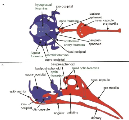
Figure 2 Subtypes of cranial foramina in the developing chick embryo.
Alcian blue and Alizarin red stained whole chick embryo skulls. A) The intramembranous neural crest derived palatine foramen (pf) can first be identified at Day 12 (HH38) as a prominent hole in the maxillary bone of the viscerocranium. B) Later in development, these holes appear more discrete, suggesting remodelling occurs. C) Endochondral mesoderm derived foramina form in the chick occipital skull of the chondrocranium from day 8 of development – here we can see 2 of the 3 hypoglossal foramina (hf) and the jugular foramina (jf). D) While the occipital foramina initially develop in the cartilaginous template (Figure 2C), they remain open as the template (blue) is replaced by bone (red). However there is temporary but significant narrowing of the foramina during the cartilage to bone transition suggesting that there may be different mechanisms controlling
foramina development at different developmental stages.
D
C
26 Figure 3
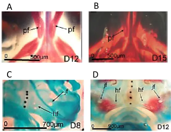
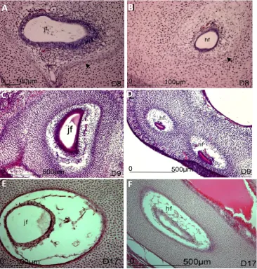
Figure 3 Histological sections of the developing occipital foramina in the chick embryo
Sections through the developing chick embryo head between HH34 and 43 (Day 8-17) stained with H&E. Jugular foramina (jf) and hypoglossal foramina (hf) are shown A, B) Jugular and hypoglossal foramina at HH34, just after onset of foramina development. The prominent blood vessel with adjacent nerves are surrounded by mesenchyme. The mesenchyme more distant to the blood vessel and nerve is denser. The intersection between the more sparse and dense mesenchyme is demarcated by flattened cells (black arrowheads), reminiscent of a developing perichondrium.
A
B
C
D
27
C, D) Jugular and hypoglossal foramina at HH35. The mesenchyme adjacent to the blood vessel and nerve is now much sparser than at a distance and the edge of the foramen is distinct in each case. The shape of each foramen is also distinct. In the case of the jugular foramen, the shape conforms to that of the blood vessel (see also images in [37]) although this relationship is less clear in hypoglossal foramina. Note that the blood vessel is not always central within the developing foramina.