Harpenden, Herts, AL5 2JQ
Telephone: +44 (0)1582 763133
Web: http://www.rothamsted.ac.uk/
Rothamsted Research is a Company Limited by Guarantee Registered Office: as above. Registered in England No. 2393175. Registered Charity No. 802038. VAT No. 197 4201 51. Founded in 1843 by John Bennet Lawes.
Rothamsted Repository Download
A - Papers appearing in refereed journals
Helliwell, J. R., Sturrock, C. J., Miller, A. J., Whalley, W. R. and Mooney,
S. J. 2019. The role of plant species and soil condition in the structural
development of the rhizosphere. Plant, Cell & Environment. 42 (6), pp.
1974-1986.
The publisher's version can be accessed at:
•
https://dx.doi.org/10.1111/pce.13529
The output can be accessed at: https://repository.rothamsted.ac.uk/item/8w964.
© 4 February 2019. Licensed under the Creative Commons CC BY.
O R I G I N A L A R T I C L E
The role of plant species and soil condition in the structural
development of the rhizosphere
Jon R. Helliwell
1,3|
Craig J. Sturrock
1|
Anthony J. Miller
2|
W. Richard Whalley
3|
Sacha J. Mooney
11
Division of Agricultural and Environmental Sciences, Gateway Building, Sutton Bonington Campus, University of Nottingham, Leicestershire LE12 5RD, UK 2
Metabolic Biology, John Innes Centre, Norwich Research Park, Norwich NR4 7UH, UK
3
Sustainable Soils and Grassland Systems Department, Rothamsted Research, West Common, Harpenden, Hertfordshire AL5 2JQ, UK
Correspondence
S. Mooney, Division of Agricultural and Environmental Sciences, Gateway Building, Sutton Bonington Campus, University of Nottingham, Leicestershire LE12 5RD, UK. Email: sacha.mooney@nottingham.ac.uk
Funding information
BBSRC, Grant/Award Numbers: Designing Future Wheat and BB/JJ004553/1; BBSRC Designing Future Wheat project, Grant/Award Number: BB/P016855/1; John Innes Founda-tion; University of Nottingham; Lawes Agri-cultural Trust studentship
Abstract
Roots naturally exert axial and radial pressures during growth, which alter the
struc-tural arrangement of soil at the root
–
soil interface. However, empirical models
sug-gest soil densification, which can have negative impacts on water and nutrient
uptake, occurs at the immediate root surface with decreasing distance from the root.
Here, we spatially map structural gradients in the soil surrounding roots using non
‐
invasive imaging, to ascertain the role of root growth in early stage formation of soil
structure. X
‐
ray computed tomography provided a means not only to visualize a root
system in situ and in 3
‐
D but also to assess the precise root
‐
induced alterations to soil
structure close to, and at selected distances away from the root
–
soil interface. We
spatially quantified the changes in soil structure generated by three common but
con-trasting plant species (pea, tomato, and wheat) under different soil texture and
com-paction treatments. Across the three plant types, significant increases in porosity at
the immediate root surface were found in both clay loam and loamy sand soils and
not soil densification, the currently assumed norm. Densification of the soil was
recorded, at some distance away from the root, dependent on soil texture and plant
type. There was a significant soil texture × bulk density × plant species interaction
for the root convex hull, a measure of the extent to which root systems explore the
soil, which suggested pea and wheat grew better in the clay soil when at a high bulk
density, compared with tomato, which preferred lower bulk density soils. These
results, only revealed by high resolution non
‐
destructive imagery, show that although
the root penetration mechanisms can lead to soil densification (which could have a
negative impact on growth), the immediate root
–
soil interface is actually a zone of
high porosity, which is very important for several key rhizosphere processes occurring
at this scale including water and nutrient uptake and gaseous diffusion.
K E Y W O R D S
rhizosphere, root diameter, soil structure, structural development, X‐ray computed tomography
(CT)
-This is an open access article under the terms of the Creative Commons Attribution License, which permits use, distribution and reproduction in any medium, provided the original work is properly cited.
© 2019 The Authors Plant, Cell & Environment Published by John Wiley & Sons Ltd DOI: 10.1111/pce.13529
1
|I N T R O D U C T I O N
The dynamic nature of the rhizosphere (the zone of soil surrounding a
growing root, which is influenced by it) provides a niche environment,
which exhibits biophysical and chemical gradients very different to
those found away from the soil immediately influenced by the root,
referred to as the bulk soil. These gradients control root activity
through a combination of root‐derived exudations and physical
struc-tural alterations, influencing water and nutrient uptake, gaseous
exchange, particle rearrangement, and wettability at the immediate
root surface. Carminati et al. (2010) revealed the influence of mucilage
on the water holding capacity of the soil immediately around the root,
and its implications for hydraulic continuity around the root system
was demonstrated by (Moradi et al., 2011). In compacted soils, the
influence of plant derived exudates have been highlighted to improve
mechanical conditions for root penetration (Oleghe, Naveed, Baggs, &
Hallett, 2017). Carminati and Vetterlein (2013) proposed the concept
of rhizosphere plasticity to help understand the bimodal hydraulic
responses found at the root–soil interface under different bulk soil
moisture conditions. However, soil structural dynamics, particularly
around an actively growing root, have been largely limited to
theoret-ical models (Dexter, 1988) or root analogue approaches (Aravena,
Berli, Ghezzehei, & Tyler, 2011) due to the inherent difficulties in
observing a fragile, opaque system in situ.
As impeded roots elongate, they undergo radial and axial
elonga-tions (Misra, Dexter, & Alston, 1986), exerting compressive and shear
forces on the surrounding soil in horizontal and vertical directions
(Bengough & MacKenzie, 1994; Kolb, Hartmann, & Genet, 2012). It
is known that root diameter varies in response to compaction and soil
strength, with many studies demonstrating an increased radial
expan-sion of the root axes in dense soil (Atwell, 1988; Materechera, Dexter,
& Alston, 1991; Tracy et al., 2012). These pressures, generated by the
root, are partly responsible for soil structural alterations in the
rhizo-sphere, and they in turn affect the hydraulic continuity of the pore
system (Aravena et al., 2011). However, the exact effect of root
growth on soil structure, especially at the scale of the pore, is
uncer-tain, in large part due to the limited number of studies, which have
compared root responses under contrasting physical soil conditions
for different plant species (Iijima & Kato, 2007; Materechera, Dexter,
& Alston, 1992). Aravena et al. (2011) reported decreased porosity
around growing roots using a root analogue technique, which showed
the radial forces in wet soil reduce interaggregate pore space,
impacting on the hydraulic contact between aggregates. Contrary to
this, Helliwell et al. (2017) recently reported an increase in porosity
at the immediate root surface at a resolution of 12μm, surrounding
the growing roots of tomato in both coarse and fine soil textures, with
a decrease in porosity observed away from the root in the bulk soil.
The functioning of the rhizosphere and, in particular, its role in
regulating the hydraulic behaviour of plants have been active areas
of research for many years. Carminati et al. (2013) showed the
impor-tance of gap formation around roots in decreasing transpirational
demand in lupin. Likewise, Berli, Carminati, Ghezzehei, and Or (2008)
highlighted the potentially beneficial role of rhizosphere densification
in increasing hydraulic contact and connectivity between neighbouring
aggregates. Hence, understanding how plants influence the precise
arrangement of soil around a root in terms of densification, gap
forma-tion, and the resulting impact on water and nutrient flow towards
roots is very important from a plant developmental perspective.
Ascer-taining the role of root growth on the structure of the rhizosphere is
challenging due to the fragile nature of soils. Previous attempts to
address this have employed thin‐section microscopy through resin
impregnation, to“fix”and preserve the root and soil systems prior to
analysis (Mooney, Morris, Craigon, & Berry, 2007; Veen,
Vannoordwijk, Dewilligen, Boone, & Kooistra, 1992). However, these
techniques are very laborious, still allow for substantial root and soil
disturbance, and do not readily enable the study of the system in 3‐
D. Non‐invasive imaging such as X‐ray computed tomography (CT),
X‐ray radiography, neutron radiography, and magnetic resonance
imaging are now accepted methods that are assisting us overcoming
these limitations having been successfully employed in studies of
plant–soil interactions over the last decade (see reviews by Helliwell
et al., 2013; Mooney, Pridmore, Helliwell, & Bennett, 2012; Pires,
Bor-ges, Bacchi, & Reichardt, 2010; Taina, Heck, & Elliot, 2008). Recent
advances in X‐ray detector efficiencies, X‐ray source power, and
image analysis methodologies have also highlighted X‐ray CT as an
exciting tool for mapping microscale alterations to root architectures
and soil structures (Helliwell et al., 2013; Mooney et al., 2012), with
previous limitations of coarse resolutions and poor image quality
greatly reduced.
The objective of this study was to take advantage of the recent
advances in imaging methodology to visualize the root‐mediated soil
structure in 3D (e.g., Helliwell et al. 2017) and gain a new insight into
root‐induced physical transformations in the rhizosphere. The first aim
was to assess how three different plant species with contrasting root
architecture modify the soil structure at the immediate soil surface in
comparison with the bulk soil. Second, we sought to investigate how
the root response to the soil was influenced by soil texture (or particle
size) as this has often been ignored in previous studies that have
tended to focus on one soil type. Finally, we examined the root
response to soil structuring when grown in soils at different bulk
den-sities to assess the impact of compaction. Based on previous work, we
hypothesized that while root growth mechanisms would generate
zones of higher soil density, the root–soil interface, a key zone for
water and nutrient exchange, would be a zone of higher porosity
con-sistent across all species.
2
|M A T E R I A L S A N D M E T H O D S
2.1
|Soil core preparation and sampling
Four replicate columns (80‐mm height × 25‐mm diameter) per soil
tex-ture and per bulk density were uniformly packed to 1.2 and 1.5 Mg m−3
with air‐dried sieved (<2 mm) Newport series loamy sand (sand 83.2%,
silt 4.7%, and clay 12.1%; pH 6.35; organic matter 2.93%; Food and
Worcester series clay loam (sand 35.6%, silt 31.5%, and clay 32.9%;
pH 6.50; organic matter 5.19%; FAO Argillic Pelosol) soil from the
Uni-versity of Nottingham farm at Bunny (Nottinghamshire, UK—52.52°N,
1.07°W). The water retention curves for these soils can be found in
Helliwell, Miller, Whalley, Mooney, and Sturrock (2014). To ensure
homogeneity in sample preparation and reduce any effects of soil
slumping following packing into the cores, the samples underwent
one wetting and drying cycle using tension table apparatus, before
being maintained at a tension of−5 kPa on the tension table throughout
seedling establishment and growth. Previous work in Helliwell et al.
(2017) showed that this was optimal for soil structure stabilization
without inducing noticeable cracking through shrinkage. Surgical
micropore tape (3M United Kingdom PLC, Bracknell) was placed over
the columns during soil preparation to reduce soil surface evaporation
and prevent sample contamination, while still enabling gaseous
exchange. Seeds of tomatoSolanum lycopersicum cv.“Ailsa Craig,”
win-ter wheat Triticum aestivum cv.“Cordiale,” and common pea Pisum
sativum cv.“Kelvedon Wonder”were germinated in the dark on wetted
filter paper for 48 hr before being planted 5 mm below the soil surface
in the replicate columns for each soil texture and bulk density
combina-tion (n = 48). Plants were grown under controlled conditions (22°C
day/16°C night); 40% relative humidity; a 12‐hr photoperiod with a
photosynthetic photon flux density at plant level of 330μmol m−2s−1
in a climate chamber for a period of 8 days. During this 8‐day period,
the plants are mainly using nutrient seed reserves to support growth
(Bouaziz & Hicks, 1990), and there was insufficient time for the
devel-opment of nitrogen‐fixing nodules on the pea roots.
2.2
|X
‐
ray CT scanning procedure
The samples were scanned using two X‐ray microtomography
sys-tems, in order to assess plant‐induced structural development across
two different spatial resolutions. All samples were initially scanned
using a Phoenix Nanotom 180NF X‐ray micro‐CT scanner (GE Sensing
and Inspection Technologies, Wunstorf, Germany). The source had a
3‐μm focal spot, with the centre of the sample 5.4 cm from the X‐
ray source and a resultant imaged voxel size of 12μm. The entire
sam-ple was imaged with a field of view of 2,308 × 2,308 pixels using an X‐
ray energy of 110 kV, a current of 110μA and an exposure time of
750 ms. A 0.2‐cm Cu filter was used, and 1,600 image projections
were taken, with each scan taking 70 min to complete. Each sample
was scanned once 8 days after planting, exposing each plant to a
cal-culated dose of 6.33 Gy (Zappala et al., 2013).
A subsection of two replicates per plant × soil texture × soil bulk
density treatment were further scanned using a Phoenix v|tome|x m
240 kV X‐ray micro‐CT scanner (GE Sensing and Inspection
Technolo-gies, Wunstorf, Germany). Due to an improved detector efficiency
(allowing enhanced X‐ray projection image collection) and higher X‐
ray flux in this system, scans at a voxel spatial resolution of 8.5μm were
possible, with each scan taking 43 min to complete. An X‐ray energy of
120 kV and current of 60μA was used, with 1,981 projections taken at
a timing of 333 ms per projection. The centre of the sample was
3.48 cm from the X‐ray source. Each sample was scanned once, also
after 8 days, exposing each plant to a calculated dose of 7.52 Gy.
2.3
|Image processing, segmentation, and analysis
Image processing was performed in VG StudioMax® 2.2 software,
using procedures largely detailed in Helliwell et al. (2017). Briefly,
seg-mentation of soil, root, and pore phases was undertaken after applying
a median filter of Radius 3 pixels to remove noise but preserve
struc-tural borders. To segment pore and soil phases, the greyscale
histo-gram was calibrated (individually for each sample) against pore space
and a common aluminium reference object, segmenting solid material
from pore and organic (including root) material. At this high resolution
and early growth stage, the roots were readily segmented using an
adaptive region growing algorithm, starting from the greyscale value
of the user‐selected voxel and selecting all connected voxels within
the user defined range. The entire segmented root architecture from
this point was analysed as a whole. To assess changes to soil structure
with distance from the root surface, the surface mesh of the root
region was three‐dimensionally (3‐D) dilated, creating discreet regions
moving away from the root in which pore and soil volumes could be
calculated. The first one‐voxel dilation was subtracted from all
subse-quent dilations to prevent any mischaracterization at the immediate
root surface due to partial volume effects or noise. The “Volume
Analyser”tool was used to assess the volume of pore and soil material
within each dilated region, giving porosity profiles (where one
voxel = 12μm) for each zone moving away from the root surface. This
could be compared with a bulk soil value, taken as the porosity of a
large volume of soil observed at the furthest distance away from the
root, but without being influenced by the container wall (i.e., in most
cases, c. 1 cm from the edge). The short growth period of the
experi-ment meant that roots did not interact with the boundary of the
con-tainer; however, to minimize any potential impact of this, we excluded
material from the edges (c. 2 mm) from the analysis. By this method,
we analysed the full root system of each plant. No roots overlapped
for the imaging undertaken at 8.5μm; however, for the wheat plants
scanned at 12μm, two samples had instances of roots in close
proxim-ity or overlapping, which were excluded from the study; however, as
extra samples had been prepared and scanned,n= 4 for each
treat-ment was maintained.
Root diameter was assessed by the novel application of an
existing image analysis protocol. A binary image stack of thresholded
root material was exported from the VG Studio Max v2.2 volume
and imported into Image J 1.47 (http://rsbweb.nih.gov/ij/). Here, 3‐
D thickness measurements were made on root systems using the
BoneJ plugin (Doube et al., 2010). This plugin places sequentially
smaller spheres inside the object of interest, and each sphere never
overlaps the object border or each other. The mean diameter of these
spheres is deemed the“thickness,”giving a single value for each root
system. A subsequent colour heat map can be used to illustrate
changes to relative sphere size to give an indication of soil pore
Root convex hull can be used to provide a measure of potential soil
exploration by different plant root systems (Iyer‐Pascuzzi et al., 2010),
by assigning straight vertices between the outer most points of the
root system. Convex hull was determined by importing the segmented
root systems intoRooTraksoftware (Mairhofer et al., 2013) using the
QuickHull algorithm (Barber, Dobkin, & Huhdanpaa, 1996) and
estimat-ing hull volume usestimat-ing Monte Carlo Integration (Rubinstein, 1981).
2.4
|Statistical analysis
All data were analysed in GenStat Release 15.1 (VSN International)
using a single‐variate linear mixed model restricted maximum
likeli-hood (REML), containing all possible interactions as explanatory
vari-ables and sample as a random effect. For soil porosity analysis, a
REML analysis containing plant species, soil texture, the distance from
the root surface, and soil bulk density as the fixed model and sample
as a random effect was used. Standard residual plots were examined
in GenStat to check data normality, with comparisons of means based
on least significant differences at theP= 0.05 andP= 0.01 levels.
3
|R E S U L T S
3.1
|The influence of root growth on rhizosphere
porosity
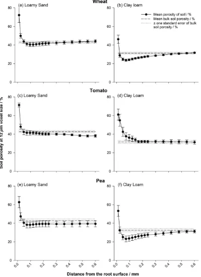
There was a clear gradient in porosity surrounding the root systems in
all treatments after 8 days of growth (Figures 1 and 2), with an
enhanced porous zone at the immediate root surface in all samples
and treatment specific localized compaction/densification at increased
distance from the root.“Densification”was considered as the point at
which the porosity of an individual dilated region became statistically
the same or lower than that of the bulk soil. The interaction of bulk
density × plant species × soil texture × distance from the root surface
was significant (P< 0.001).
When averaged over all treatments, there was a significant
increase in soil porosity at the immediate root surface compared with
48 μm away from the root (mean porosity of 47.3% and 26.8%
respectively; Figures 1 and 2; P < 0.001; SE's available in
Tables S1–S4), with a significant interaction for plant species ×
dis-tance from the root (P< 0.05), soil texture × distance from the root
(P< 0.001), and bulk density × distance from the root (P< 0.001).
Scanning at a higher resolution revealed a clear gap formation around
tap and lateral roots in both soil textures (Figure 3), the diameter of
which approximately equalled the zones of increased porosity
quanti-fied in Figures 1 and 2. Beyond this initial gap formation, changes to
porosity at increased distance from the root surface were explained
by soil texture and bulk density.
At a bulk density of 1.2 Mg m−3, the loamy sand soil exhibited no
further significant change to porosity compared with the bulk soil at
increasing distances away from the root surface for any plant species
(Figure 1a,c,e). At the same bulk density in the clay loam, there was no
significant change in porosity from the bulk soil value for the tomato
treatment (Figure 1d), but significant reductions in porosity of 7.5
and 9.5% compared with the bulk soil value to 23.6% and 23.1% in
the wheat and pea species, respectively (Figure 1b,f;P< 0.001). This
localized densification compared with the bulk soil extended to 0.36
and 0.42 mm from the root surface in the wheat and pea species,
respectively, with the soil particularly compressed at the 0.1 mm
loca-tion for both species compared with the root–soil interface.
At 1.5 Mg m−3, the tomato plants exhibited no further changes in
porosity following the initial increase at the immediate root surface in
either soil texture (Figure 2c,d), although the differences in the soil
porosity profile between the two textures were the most pronounced
observed. However, there were significant decreases in porosity in
wheat and pea plants in both soil textures (Figure 2a,b,e,f;
P< 0.001), the magnitude of which were texture specific. In the loamy
sand, the wheat and pea plants exhibited decreases in porosity
com-pared with the bulk soil of 5.6% and 4.0%, respectively, with localized
soil densification extending to 0.14 and 0.12 mm from the root
sur-face. In the clay loam, the wheat and pea plants exhibited greater
decreases in porosity of 8.1% and 7.6%, respectively, compared with
the bulk soil than in the loamy sand. Densification of the soil
surround-ing the root extended further than in the loamy sand, to 0.42 and
0.22 mm from the root surface in the clay loam for the wheat and
pea treatments, respectively.
The zone of influence of the root (i.e., the spatial degree of any
change in porosity away from the bulk soil) as an isolated dependent
variable was significantly influenced by plant species (P< 0.001) in
the following the order wheat > pea > tomato (means of 694.7,
483.9, and 21.2 mm3, respectively). Soil texture also significantly
influ-enced the zone of influence (P< 0.05), with clay loam having a much
higher volume of 511.7 mm3compared with 288.2 mm3in the loamy
sand. The bulk density × texture interaction was significant (P= 0.05),
with a larger zone of influence in the clay at 1.5 than 1.2 Mg m−3
(mean values of 630.3 and 402.9 mm3, respectively), but in sand, it
was the converse (mean values of 225.2 and 334.5 mm3, respectively).
In comparison with the lower density soil, the porosity at the root–soil
interface and the bulk soil was reduced by between 25% and 50% in
the 1.5 Mg m−3of treatment.
3.2
|Impact of soil physical properties on root
characteristics
Representative images of root system architecture segmented from
the X‐ray CT images for the three plant species are provided in
Figure 4. Mean root thickness increased with increasing bulk density
(0.58 mm vs. 0.74 mm at bulk densities of 1.2 and 1.5 Mg m−3,
respec-tively;P= 0.001), with a significant interaction of bulk density × plant
species (Figure 5; P < 0.005). Root thickness significantly differed
between plant species with the following the order:
pea > tomato > wheat (mean thickness values of 1.16, 0.49 and
0.34 mm, respectively; Figure 5;P< 0.001). Root thickness varied
sig-nificantly with soil type (P< 0.001), increasing in the finer textured
loamy sand textures, respectively). The interaction of species × texture
was significant (P= 0.01). Averaged across all treatments, there was no
significant effect of root thickness on porosity of the defined
rhizo-sphere region, but a significant interaction of plant species × root zone
of influence (P< 0.005) and bulk density × plant species × root zone of
influence (P= 0.001). Note, this is based on analysis of the soil around
the roots hence where root architecture varied so did the volume of
soil assessed.
Mean values for convex hull volume were higher in the clay loam
than loamy sand (5,607 vs. 4,060 mm3; Figure 6;P< 0.005) and were
significantly affected by plant species (convex hull volumes of 7,077,
3,940, and 3,483 mm3 in the wheat, pea, and tomato, respectively;
FIGURE 1 Porosity distributions at a bulk density of 1.2 Mg m−3for wheat (a,b), tomato (c,d), and pea (e,f) roots: (a,c,e) loamy sand and (b,d,f) clay
P< 0.001). There were significant interactions of bulk density × soil
texture (P< 0.05) and bulk density × species × texture (P< 0.05).
There was a significant relationship between convex hull volume and
the volume of the root zone of influence (P< 0.001), with mean total
volumes of both convex hull and the volume of root zone of influence
differing dramatically between plant species (P< 0.001) and soil
tex-ture (P< 0.005; Figure 6).
4
|D I S C U S S I O N
Root growth has a significant impact on soil structure in the
rhizo-sphere, which we observed here after very early growth. The extent
of soil reorganization is influenced not only by the plant but also by
the soil's physical properties. Previous work has indicated that soil
structure in the rhizosphere has key consequences for soil physical
FIGURE 2 Porosity distributions at a bulk density of 1.5 Mg m−3for wheat (a,b), tomato (c,d), and pea (e,f) roots: (a,c,e) loamy sand and (b,d,f) clay
(Gregory, 2006; Hinsinger, Bengough, Vetterlein, & Young, 2009) and
hydraulic processes that directly influence root system development
(Carminati et al., 2013; Hallett et al., 2009). Although previous work,
such as Aravena et al. (2011), used root analogues to try disentangle
the consequences of root growth on structural development in the
rhi-zosphere, an assessment of real growing roots in field soil on rhizosphere
structure evolution has previously been considered not possible. In this
study, we used X‐ray CT to observe the structural development of the
rhizosphere across multiple plant species and soil treatments at scales
down to 8.5μm on soil from the field that was structure less. This
approach offer new opportunities to study in situ how plants influence
the soil environment to their advantage/disadvantage and how this is
affected by different abiotic stresses.
4.1
|Impact of root growth on rhizosphere porous
architecture
There was a plant species independent increase in porosity
immedi-ately at the root surface, which subsequently declined with distance
from the root previously measured by Helliwell et al. (2017). This
con-trasts with the previous work using root analogues (Aravena et al.,
2011), which demonstrated a soil densification gradient at the
imme-diate root surface, increasing in porosity with distance from the root.
Aravena et al. (2011) acknowledge limitations to their balloon root
analogue, in that, it consists of an unreactive nondynamic interface,
isolating lateral compressive forces due to radial expansion. Therefore,
in a real root system, more dynamic differences in the structural
gradi-ents from the root to the bulk soil are expected. Beyond this zone of
increased porosity, an increase in densification of the soil was
observed, governed by soil texture (Figures 1 and 2). Figure 3b,c
high-lights the development of cracking behaviour in the clay loam soil,
with root‐derived cracks radiating from the root surface in all plant
species. This is almost certainly due to shrinkage induced by soil drying
(Hallett & Newson, 2005) and not a sample preparation artefact
because great care was taken to ensure the samples were packed as
homogenously as possible following the method of Helliwell et al.
(2017). The plastic nature of the clay loam can lead to the formation
of localized microcracks during root growth, corresponding to and
accounting for the increases in porosity quantified at the immediate
root surface (Figure 1). The loamy sand texture, which has a much
smaller capacity to shrink than clay loam, exhibited a smaller but
mea-surable shrinkage upon drying at the root surface, linked to a loss of
contact, which was particularly pronounced in the thicker pea roots
(Figure 3a). However, as this soil did not crack, the magnitude of
porosity increase, estimated from the CT images, was smaller
(Figure 2).
New lateral root growth was observed in crack shaped pores in
the soil, with an apparent preference for growth into pre‐existing pore
space as opposed to forging new pathways. New root proliferation is
known to exploit existing pore channels and fissures where possible
(Bengough et al., 2006), due to the relatively unimpeded pathways in
these regions compared with denser surrounding soil, although the
extent of this can be regulated by the overall soil bulk density
FIGURE 5 The influence of bulk density and plant species on root thickness after 8 days of growth. Error bars represent standard errors of four replicates. Significance:*P< 0.05 and **
P< 0.01
(Colombi, Braun, Keller, & Walter, 2017). Hence, root growth often
becomes clustered in these channels that bypass stronger regions of
the soil (White & Kirkegaard, 2010), creating hotspots of intense
water and nutrient uptake and zones of relatively unaffected soil in
poorly explored impenetrable areas (Passioura, 2002). It is likely that
the increased yield observed in some zero tillage systems is due to
enhanced root penetration at depth due to an increased frequency
of biopores and enhanced pore connectivity (Pittelkow et al., 2015).
Roots can also proliferate to locally exploit patches of nutrients (Drew,
1975). However, as the soil was homogenized before packing into
col-umns in this investigation, we can discount root exploitation of pre‐
existing nutrient patches. We observed that roots exhibited a clear
strategy where lateral roots explore newly formed fissures, potentially
as an energy conservation mechanism. This also accounts for a degree
of gap formation immediately around the tap and lateral roots
(Figure 3b,c), as the roots often failed to fully fill the pores. The
impor-tance of gap formation around growing roots was highlighted by
Carminati et al. (2013), with the shrinkage of roots responsible for
air‐filled gaps particularly pronounced around the tap root. However,
Carminati et al. (2013) and other previous investigations (Carminati,
Vetterlein, Weller, Vogel, & Oswald, 2009) demonstrated shrinkage
of the root as opposed to the soil was the driver for the gap
develop-ment dynamics. It is possible that shrinkage of the soil was overlooked
in previous work due to the coarser resolution (~100μm); thus,
micro-scale structural changes were not observed. Also, the high sand
con-tent (92%) used by Carminati et al. (2013) would limit shrinkage of
the soil itself, a likely factor influencing rhizosphere structure
develop-ment. The role of root hairs in structural formation is not considered
FIGURE 6 The influence of plant species and soil type on (a) mean root zone of influence at 1.2 Mg m−3; (b) convex hull volume for 1.2 Mg m−3;
(c) mean root zone of influence at 1.5 Mg m−3; and (d) convex hull volume for 1.5 Mg m−3. Error bars associated with the histograms represent
here due to an inability to observe them in these soils at the
pre-scribed moisture content (due to an overlap in X‐ray attenuation
rather than resolution), although Koebernick et al. (2017) has shown
this is possible in a coarse textured soil via synchrotron imaging when
considering air‐filled pores only.
Beyond the initial increase in porosity at the immediate root
sur-face, the contrasting porosity changes at distances further from the
root surface are also likely to be influenced by the different cohesive
properties of the soil. It follows that an apparent lack of densification
surrounding roots growing in coarser, less cohesive soil is due to its
relative ductility, with freely mobile particles able to be reorganized
as the root grows. Conversely, the plastic nature of the clay soil
cre-ates a readily compressible mass, clearly influenced by root size. The
root effect on increasing densification away from the root interface
was greater in the highest bulk density treatment and was consistent
between the two soil types, though Figure 5 shows that this cannot
be explained by root diameter alone.
We hypothesized a relationship between the thickness of a root,
soil bulk density, and the degree and size of its impact on the
sur-rounding physical soil environment, with thicker roots under increased
soil bulk density thought to contribute to an increased deformation of
rhizosphere soil. The nonsignificant effect of root thickness as a factor
determining the “zone of influence” shows that root diameter,
although reported to increase the ability of roots to penetrate
compacted soil, (Bengough, 1997), does not account for the changes
to structure we have observed once the rhizosphere has developed.
The combination of thicker (Figure 4) and blunter pea roots under
the appropriate soil texture exhibited increased soil deformation
com-pared with tomato and wheat (Figures 1f and 2f). Although the degree
of structural change was independent of root thickness, the
displace-ment of particles was less than one root diameter in all treatdisplace-ments.
This contrasts with Vollsnes, Futsaether, and Bengough (2010) who
showed compression of sand in front of the root tip extending up to
eight times the root diameter in maize using particle image
velocimetry. Aravena et al. (2011) reported lateral densification of
~8–12% extending to one root diameter in wet aggregates at a
reso-lution of 4.4μm. In this investigation, we observed a similar degree
of deformation of~4–9% depending on the soil texture and plant
spe-cies, extending~0.5× the root diameter (although root‐induced
crack-ing often extended beyond this; Figure 4b,c). It is therefore clear that
investigations using artificial sand or a saturated medium may cause
differences in the size and magnitude of structural change observed
not representative of field soil conditions.
A common feature we observed was that immediately adjacent
to the root; there was a region of increased porosity. This was most
likely due some combination of both soil and root shrinkage
along-side the thigmotropic response of root development. The way in
which particles, especially in structure‐less samples, are arranged at
the root–soil interface has been proposed to account for the zone
of higher porosity (Koebernick et al., 2018), and although we cannot
discount that this as a contributing factor, it is clear from Figure 3b–
d where a particulate structure is not observed, that this is unlikely
to explain our findings. At greater distances from the root, there
was a compacted region (except for Figure 1d), which was due to
either (a) a legacy of soil deformation at the root tip or (b) microscale
soil shrinkage due to water uptake by the root. Differences in root
exudate composition between the plant species are also thought to
be important in modifying the physical properties soil (Naveed
et al., 2017).
4.2
|Implications for modelling of rhizosphere
densification
Dexter (1987) developed a model for the compression of soil
sur-rounding a growing root by assuming soil porosity is reduced
adja-cent to the root where compression is greatest. This was based on
work considering a metal probe as a root analogue entering the soil
and expanding to cause a porosity gradient, which increased
expo-nentially from the object surface (Dexter & Tanner, 1972). This
was later supported by experimental work using particle image
velocimetry in pure sand at a spatial resolution of 0.5 mm (Vollsnes et al.,
2010), where the displacement of sand particles into pores in their
immediate vicinity was facilitated by root growth. Our work confirmed
the predictions by Dexter (1987) that following root compression of soil
to a minimum porosity and an example of this behaviour is seen in
Figure 3d. However, we more commonly observed a dual‐zone impact
of root growth on soil structure in the rhizosphere (Figures 1 and 2),
with the first corresponding to the increase in porosity at the immediate
root surface to an approximate distance of 50μm, only observable by
high resolution imaging and not previously considered in similar
model-ling approaches. This high porosity zone where root–soil contact is
somewhat reduced could have profound implications for soil root
inter-action: reduced hydraulic conductivity and water flow to the root due
to a loss of hydraulic connection, lower nutrient flux to the root
espe-cially nitrate, and increased aeration. The improved aeration could be
of considerable benefit to the root whereas the effects related to
reduced water flux might be compensated by root mucilage production
(Carminati et al., 2009).
Plant roots donate carbon to encourage the development of
beneficial populations of microbes in the rhizosphere. For example,
phosphate‐solubilizing microorganisms can mobilize previously
inaccessi-ble pools of this important nutrient for plants (Wang, Shi, Jiang, Zhang, &
Feng, 2016). Microorganisms growing on the root surface contribute to
the disruption of soil structure at the root surface that can aid aeration
and the pathway for nutrient and water delivery to the root surface
(Helliwell et al., 2014). Our finding that the extent of this root surface
phenomenon, the zone of influence, differs between species and depends
on soil type and density (Figures 6 and 7) is worthy of further
investiga-tion. For example, pea showed more sensitivity to the soil type when
compared with wheat and tomato at higher bulk density (Figures 6c,d).
In the thicker pea roots (Figure 5), the production of specialized exudates
particularly rich in hydroxyproline‐rich cell wall glycoprotein when
compared with cereals (Knee et al., 2001) may be depend on soil type.
There may be the potential to improve this trait in future crop breeding
programmes by manipulating root exudate composition. In addition, the
considerable differences in root‐induced structure around and away from
the root surface and the varied response to soil texture and bulk density
highlight the needs for plants breeders to undertake studies under more
natural conditions when screening for beneficial root traits.
5
|C O N C L U S I O N S
Plants modify the soil environment in the rhizosphere very early on
dur-ing plant root growth. Soils with contrastdur-ing textures are deformed by
roots in different ways, depending on initial soil bulk density and plant
species. X‐ray microtomography of loamy sand and clay loam soils
showed an increase in the porosity of soil immediately adjacent to the
root in all three plant species examined, which was independent of root
diameter. Multiscale scanning at higher resolutions revealed
consider-able microcrack formation around roots, attributconsider-able to soil
shrink-age. However, subsequent deformation and compaction created by
root growth was spatially highly heterogeneous, and dependent on
a combination of root thickness, higher soil bulk density, and finer
textured soils. Imaging approaches, such as those demonstrated
here, could provide a basis for the future development of conceptual
root–soil interaction models, especially important as the soil
struc-ture in the rhizosphere has implications for the acquisition of water
and nutrients by plant roots as they engineer new hydraulic
path-ways through soils. In addition, they could be used to support the
efforts of plant breeders when seeking to identify idealized root
traits as the root‐modulated soil porous architecture is likely to play
as an important role in root development as the root system itself.
A C K N O W L E D G E M E N T S
JRH was funded by a Lawes Agricultural Trust studentship and the
University of Nottingham. AJM is supported by grant funding [BB/
JJ004553/1] from the BBSRC and the John Innes Foundation.
WRW is supported at Rothamsted Research by the BBSRC
Designing Future Wheat project [grant number BB/P016855/1].
SJM and CJS are also supported by the BBSRC Designing Future
Wheat project [grant number BB/P016855/1].
O R C I D
Craig J. Sturrock https://orcid.org/0000-0002-5333-8502
Anthony J. Miller https://orcid.org/0000-0003-0572-7607
W. Richard Whalley https://orcid.org/0000-0003-0755-2943
Sacha J. Mooney https://orcid.org/0000-0002-9314-8113
R E F E R E N C E S
Aravena, J. E., Berli, M., Ghezzehei, T. A., & Tyler, S. W. (2011). Effects of root‐induced compaction on rhizosphere hydraulic properties—X‐ray microtomography imaging and numerical simulations. Environmental
Science & Technology, 45, 425–431. https://doi.org/10.1021/
es102566j
Atwell, B. (1988). Physiological responses of lupin roots to soil compaction.
Plant and Soil,111, 277–281. https://doi.org/10.1007/bf02139953
Barber, C. B., Dobkin, D. P., & Huhdanpaa, H. (1996). The QuickHull algo-rithm for convex hulls. ACM Trans. Math. Software, 22, 469–483. https://doi.org/10.1145/235815.235821
Bengough, A. G. (1997). Modelling rooting depth and soil strength in a dry-ing soil profile.Journal of Theoretical Biology,186, 327–338. https://doi. org/10.1006/jtbi.1996.0367
Bengough, A. G., Bransby, M. F., Hans, J., McKenna, S. J., Roberts, T. J., & Valentine, T. A. (2006). Root responses to soil physical conditions: Growth dynamics from field to cell.Journal of Experimental Botany,
57, 437–447. https://doi.org/10.1093/jxb/erj003
Bengough, A. G., & MacKenzie, C. J. (1994). Simultaneous measurement of root force and elongation for seedling pea roots.Journal of Experimental
Botany,45, 95–102. https://doi.org/10.1093/jxb/45.1.95
Berli, M., Carminati, A., Ghezzehei, T. A., & Or, D. (2008). Evolution of unsaturated hydraulic conductivity of aggregated soils due to compres-sive forces.Water Resources Research, 44, W00C09. https://doi.org/ 10.1029/2007WR006501
Carminati, A., Moradi, A. B., Vetterlein, D., Vontobel, P., Lehmann, E., Weller, U.,…Oswald, S. E. (2010). Dynamics of soil water content in the rhizosphere. Plant and Soil, 332, 163–176. https://doi.org/ 10.1007/s11104‐010‐0283‐8
Carminati, A., & Vetterlein, D. (2013). Plasticity of rhizosphere hydraulic properties as a key for efficient utilization of scarce resources.Annals
of Botany,112, 277–290. https://doi.org/10.1093/aob/mcs262
Carminati, A., Vetterlein, D., Koebernick, N., Blaser, S., Weller, U., & Vogel, H. J. (2013). Do roots mind the gap? Plant and Soil, 367, 651–661. https://doi.org/10.1007/s11104‐012‐1496‐9
Carminati, A., Vetterlein, D., Weller, U., Vogel, H.‐J., & Oswald, S. E. (2009). When roots lose contact.Vadose Zone Journal,8, 805–809. https://doi. org/10.2136/vzj2008.0147
Colombi, T., Braun, S., Keller, T., Walter, A. (2017). Artificial macropores attract crop roots and enhance plant productivity on compacted soils.
Sci. Total Environ. 574, 1283–1293. https://doi.org/10.1016/j.
scitotenv.2016.07.194
Dexter, A. R. (1987). Compression of soil around roots.Plant and Soil,97, 401–406. https://doi.org/10.1007/BF02383230
Dexter, A. R. (1988). Advances in characterization of soil structure.Soil and
Tillage Research, 11, 199–238. https://doi.org/10.1016/0167‐
1987(88)90002‐5
Dexter, A. R., & Tanner, D. W. (1972). Soil deformations induced by a mov-ing cuttmov-ing blade, an expandmov-ing tube and a penetratmov-ing sphere.Journal
of Agricultural Engineering Research, 17, 371–375. https://doi.org/
10.1016/S0021‐8634(72)80045‐3
Doube, M., Kłosowski, M. M., Arganda‐Carreras, I., Cordelières, F. P., Dougherty, R. P., Jackson, J. S.,…Shefelbine, S. J. (2010). BoneJ: Free and extensible bone image analysis in ImageJ.Bone,47, 1076–1079. https://doi.org/10.1016/j.bone.2010.08.023
Drew, M. (1975). Comparison of the effects of a localized supply of phos-phate, nitrate, ammonium and potassium on the growth of the seminal root system, and the shoot, in barley.New Phytol 75, 479–490.
Gregory, P. J. (2006). Roots, rhizosphere and soil: The route to a better understanding of soil science? European Journal of Soil Science, 57, 2–12. https://doi.org/10.1111/j.1365‐2389.2005.00778.x
Hallett, P. D., Feeney, D. S., Bengough, A. G., Rillig, M. C., Scrimgeour, C. M., & Young, I. M. (2009). Disentangling the impact of AM fungi versus roots on soil structure and water transport. Plant and Soil, 314, 183–196. https://doi.org/10.1007/s11104‐008‐9717‐y
Hallett, P. D, Newson, T. A., (2005). Describing soil crack formation using elastic plastic fracture mechanics.European Jounral of Soil Science,56, 31–38.
Helliwell, J., Sturrock, C. J., Craigon, J., Ashton, R., Mairhofer, S., Miller, A.,
…Mooney S. J. (2017). The emergent rhizosphere: Imaging the devel-opment of the porous architecture at the root‐soil interface.Scientific
Reports. https://doi.org/10.1038/s41598-017-14904-w
Helliwell, J. R., Miller, A. J., Whalley, W. R., Mooney, S. J., & Sturrock, C. J. (2014). Quantifying the impact of microbes on soil structural develop-ment and behaviour in wet soils. Soil Biology and Biochemistry, 74, 138–147. https://doi.org/10.1016/j.soilbio.2014.03.009
Helliwell, J. R., Sturrock, C. J., Grayling, K. M., Tracy, S. R., Flavel, R. J., Young, I. M.,…Mooney, S. J. (2013). Applications of X‐ray computed tomography for examining biophysical interactions and structural development in soil systems: A review.European Journal of Soil Science,
64, 279–297. https://doi.org/10.1111/ejss.12028
Hinsinger, P., Bengough, A. G., Vetterlein, D., & Young, I. M. (2009). Rhizo-sphere: Biophysics, biogeochemistry and ecological relevance. Plant
and Soil,321, 117–152. https://doi.org/10.1007/s11104‐008‐9885‐9
Iijima, M., & Kato, J. (2007). Combined soil physical stress of soil drying, anaerobiosis and mechanical impedance to seedling root growth of four crop species.Plant Production Science,10, 451–459. https://doi. org/10.1626/pps.10.451
Iyer‐Pascuzzi, A. S., Symonova, O., Mileyko, Y., Hao, Y., Belcher, H., Harer, J.,…Benfey, P. N. (2010). Imaging and analysis platform for automatic phenotyping and trait ranking of plant root systems.Plant Physiology,
152, 1148–1157. https://doi.org/10.1104/pp.109.150748
Knee, E. M, Gong, F‐C., Gao, M., Teplitski, M., Jones, A. R., Foxworthy, A.,
…Bauer, W. D. (2001). Root Mucilage from Pea and Its Utilization by Rhizosphere Bacteria as a Sole Carbon Source.Mol Plant‐Microbe Inter-act,14(6), 775–784. https://doi.org/10.1094/MPMI.2001.14.6.775
Koebernick, N., Daly, K. R., Keyes, S. D., Bengough, A. G., Brown, L. K., Cooper, L. J.,…Roose, T. (2018). Imaging microstructure of the barley rhizosphere: particle packing and root hair influences.New Phytologist. https://doi.org/10.1111/nph.15516
Koebernick, N., Daly, K. R., Keyes, S. D., George, T. S., Brown, L. K., Raffan, A.,…Roose, T. (2017). High‐resolution synchrotron imaging shows that root hairs influence rhizosphere soil structure formation. New
Phytologist 216(1), 124–135. https://doi.org/10.1111/nph.14705
Kolb, E., Hartmann, C., & Genet, P. (2012). Radial force development during root growth measured by photoelasticity.Plant and Soil,360, 19–35. https://doi.org/10.1007/s11104‐012‐1316‐2
Mairhofer, S., Zappala, S., Tracy, S., Sturrock, C., Bennett, M. J., Mooney, S. J., & Pridmore, T. P. (2013). Recovering complete plant root system architectures from soil via X‐ray mu‐computed tomography. Plant
Methods,9, 8. https://doi.org/10.1186/1746‐4811‐9‐8
Materechera, S. A., Dexter, A. R., & Alston, A. M. (1991). Penetration of very strong soils by seedling roots of different plant species.Plant
and Soil,135, 31–41. https://doi.org/10.1007/BF00014776
Materechera, S. A., Dexter, A. R., & Alston, A. M. (1992). Formation of aggregates by plant‐roots in homogenized soils.Plant and Soil, 142, 69–79. https://doi.org/10.1007/BF00010176
Misra, R. K., Dexter, A. R., & Alston, A. M. (1986). Maximum axial and radial growth pressures of plant roots.Plant and Soil,95, 315–326. https:// doi.org/10.1007/BF02374612
Mooney, S., Pridmore, T., Helliwell, J., & Bennett, M. (2012). Developing X‐ ray computed tomography to non‐invasively image 3‐D root systems architecture in soil. Plant and Soil, 352, 1–22. https://doi.org/ 10.1007/s11104‐011‐1039‐9
Mooney, S. J., Morris, C., Craigon, J., & Berry, P. (2007). Quantification of soil structural changes induced by cereal anchorage failure: Image anal-ysis of thin sections.Journal of Plant Nutrition and Soil Science, 170, 363–372. https://doi.org/10.1002/jpln.200622042
Moradi, A. B., Carminati, A., Vetterlein, D., Vontobel, P., Lehmann, E., Weller, U., … Oswald, S. E. (2011). Three‐dimensional visualization and quantification of water content in the rhizosphere.New Phytologist,
192, 653–663. https://doi.org/10.1111/j.1469‐8137.2011.03826.x
Naveed, M., Brown, L., K., Raffan, A. C., George, T. S., Bengough, A. G., Roose, T., … Hallett, P. D. (2017). Plant exudates may stabilize or weaken soil depending on species, origin and time.Eur J Soil Sci,68, 806–816. https://doi.org/10.1111/ejss.12487
Oleghe, E., Naveed, M., Baggs, E. M., & Hallett, P. D. (2017). Plant exudates improve the mechanical conditions for root penetration through compacted soils.Plant and Soil,421, 19–30. https://doi.org/10.1007/ s11104‐017‐3424‐5
Passioura, J. B. (2002). Soil conditions and plant growth.Plant, Cell & Environ-ment,25, 311–318. https://doi.org/10.1046/j.0016‐8025.2001.00802.x
of the Brazilian contribution. Soil & Tillage Research, 110, 197–210. https://doi.org/10.1016/j.still.2010.07.013
Pittelkow, C. M., Linquist, B. A., Lundya, M. E., Liang, X. K., Groenigenc, J. van., Lee, J.,…Kessel, C. (2015). When does no‐till yield more? A global meta‐analysis. Field Crops Research. https://doi.org/10.1016/j. fcr.2015.07.020
Rubinstein, R. Y. (1981). Simulation and the Monte Carlo methods. New York: John Wiley and Sons.
Taina, I. A., Heck, R. J., & Elliot, T. R. (2008). Application of X‐ray computed tomography to soil science: A literature review.Canadian Journal of Soil
Science,88, 1–20. https://doi.org/10.4141/CJSS06027
Tracy, S. R., Black, C. R., Roberts, J. A., Sturrock, C., Mairhofer, S., Craigon, J., & Mooney, S. J. (2012). Quantifying the impact of soil compaction on root system architecture in tomato (Solanum lycopersicum) by X‐ ray micro‐computed tomography. Annals of Botany, 110, 511–519. https://doi.org/10.1093/aob/mcs031
Veen, B. W., Vannoordwijk, M., Dewilligen, P., Boone, F. R., & Kooistra, M. J. (1992). Root–soil contact of maize, as measured by a thin‐section technique. 3. Effects on shoot growth, nitrate and water‐uptake effi-ciency. Plant and Soil, 139, 131–138. https://doi.org/10.1007/ BF00012850
Vollsnes, A. V., Futsaether, C. M., & Bengough, A. G. (2010). Quantifying rhizosphere particle movement around mutant maize roots using time‐lapse imaging and particle image velocimetry. European Journal
of Soil Science, 61, 926–939. https://doi.org/10.1111/j.1365‐
2389.2010.01297.x
Wang, F., Shi, N., Jiang, R., Zhang, F., & Feng, G. (2016). In situ stable iso-tope probing of phosphate‐solubilizing bacteria in the hyphosphere.
Journal of Experimental Botany, 67(6), 1689–1701. https://doi.org/
10.1093/jxb/erv561
White, R. G., & Kirkegaard, J. A. (2010). The distribution and abundance of wheat roots in a dense, structured subsoil—Implications for water uptake. Plant, Cell & Environment, 33, 133–148. https://doi.org/ 10.1111/j.1365‐3040.2009.02059.x
Zappala, S., Helliwell, J. R., Tracy, S. R., Mairhofer, S., Sturrock, C. J., Pridmore, T.,…Mooney, S. J. (2013). Effects of X‐ray dose on rhizo-sphere studies using X‐ray computed tomography. PLoS ONE, 8, e67250. https://doi.org/10.1371/journal.pone.0067250
S U P P O R T I N G I N F O R M A T I O N
Additional supporting information may be found online in the
Supporting Information section at the end of the article.
Table S1. Raw porosity data for clay loam samples at a soil bulk
den-sity of 1.2 Mg m‐3
Table S2. Raw porosity data for loamy sand samples at a soil bulk
den-sity of 1.2 Mg m‐3
Table S3. Raw porosity data for clay loam samples at a soil bulk
den-sity of 1.5 Mg m‐3
Table S4. Raw porosity data for loamy sand samples at a soil bulk
den-sity of 1.5 Mg m‐3
How to cite this article: Helliwell JR, Sturrock CJ, Miller AJ, Whalley WR, Mooney SJ. The role of plant species and soil
condition in the structural development of the rhizosphere.
Plant Cell Environ. 2019;42:1974–1986. https://doi.org/


![FIGURE 4Example root system architectures at a bulk density of 1.5 Mg m−3 for (a) tomato, (b) wheat, and (c) pea [Colour figure can be viewedat wileyonlinelibrary.com]](https://thumb-us.123doks.com/thumbv2/123dok_us/1074950.1608168/9.595.228.540.270.467/figure-example-architectures-density-tomato-colour-viewedat-wileyonlinelibrary.webp)


