Case Report 1
ERBB1 and ERBB2 Positive Medullary Thyroid Carcinoma:
2
A Case Report
3
4
Michele Minuto1, Emanuela Varaldo1, Gianluca Marcocci1,2, Amleto de Santanna2, Ermanno 5
Ciccone2, Katia Cortese2* 6
7
1 DISC, Department of Surgical Sciences, University of Genoa, Largo R. Benzi, 8, Genoa, Italy; 8
[email protected] (MM); [email protected] (EV);
9
[email protected] (GM)
10
2 DIMES, Department of Experimental Medicine, University of Genoa, Via Antonio de Toni 14,
11
16132, Genoa, Italy; [email protected] (ADS); [email protected] (EC); [email protected] 12
(KC)
13
* Correspondence: [email protected], +39 0103537864 14
15 16
Abstract: Medullary thyroid carcinomas (MTCs) are rare thyroid tumors occurring in both 17
sporadic and hereditary forms and whose pathogenesis is related to RET proto-oncogene 18
alterations. MTCs originate from parafollicular cells, which produce calcitonin that 19
represents the biochemical activity of MTC. Total thyroidectomy is the main treatment for 20
MTC and often cures patients with confined diseases. In cases of metastasis, the approach 21
depends on the rate of progression of disease. We report a case of a 54 years old female with 22
a single, incidentally discovered, thyroid nodule of 1 cm, classified as suspicious MTC after a 23
stimulation test with i.v. calcium. After surgery, we examined the nodule using 24
immunohistochemistry, immunofluorescence and electron microscopy. In addition to 25
calcitonin, we found that it expressed intracellular positivity for the RTK receptors ERBB1 26
and ERBB2. Consistently with MTC features, ultrastructural examination of the tumor 27
displayed heterogeneous spindle-shaped cells containing two groups of secretory granules. 28
Due to the significant correlation found between high ERBB1/ERBB2 levels in MTCs and 29
extrathyroidal growth, the detection of ERBB1 and ERBB2 expression suggests that the two 30
oncoproteins may possibly be involved in tumor proliferative responses and/or 31
differentiation of C-cells. The biological, prognostic and therapeutic significance of these 32
patterns would merits further investigations. 33
Keywords: MTC; calcitonin; parafollicular C cells; secretory granules; immunofluorescence; 34
ultrastructure; transmission electron microscopy; ERBB1; ERBB2 35
36
37
38
1. Introduction 39
Medullary thyroid cancers (MTCs) are rare neuroendocrine tumors arising from 40
differentiated parafollicular cells (C cells) of the thyroid gland. Current understanding on C 41
cells propose that their precursors arise in the foregut endoderm, and not in the neural crest, 42
arguing that MTC should be reclassified to the family of neuroendocrine tumors with 43
endodermal origin [1]. MTC accounts for less than 5% of all thyroid cancers worldwide. Its 44
prognosis, although worse than that of differentiated thyroid cancers arising for the follicular 45
cells, is overall favorable, with a survival of 80-85% at 5 years and 70-75 at 10 years for 46
patients with localized disease, despite the presence of residual disease in more than 50% of 47
patients treated with surgery [2]. The prognosis for metastatic MTC is worse, since only 20% 48
of patients with distant metastases at diagnosis survive 10 years [3]. MTCs secrete calcitonin 49
and carcinoembryonic antigen (CEA) and deposits of amyloid are frequently observed [4]. 50
Calcitonin (Ct) is the most sensitive and specific tumor marker both at the preoperative 51
diagnosis and at the post-surgery follow up. Its values suggest the diagnosis of MTC in 52
patients with thyroid nodules, and then reflect the oncologic radicality of surgery, since Ct is 53
not detectable after the complete resection of the tumor and decreases after its debulking, 54
revealing the presence of residual disease. 55
MTCs can either occur sporadically (75%) or as a part of a genetic disease (e.g. multiple 56
endocrine neoplasia (MEN) type 2 syndrome, or familial MTC). Inherited forms of MTC are 57
due to autosomal dominant mutations of the RET proto-oncogene with incomplete 58
penetrance, often presenting as multifocal disease in a background of C-cell hyperplasia 59
(CCH). So far, total thyroidectomy is the only successful option for treatment of confined 60
disease, whereas the clinical efficacy of RET inhibitors as targeted agents for treatment of 61
metastatic MTC is limited due to intrinsic and acquired resistance [5]. Therefore, additional 62
candidates as prognostic markers and/or potential therapeutic targets are needed. ERBB 63
family members, such as epidermal growth factor receptor 1 (ERBB1), ERBB2, ERBB3 and 64
ERBB4, are widely distributed in organ tissues, including endocrine organs [6], and these 65
receptors are strongly implicated in various human carcinomas [7]. It has been reported a 66
significant correlation between high ERBB1 and ERBB2 levels and extra thyroidal growth in 67
MTCs [8, 9]. In particular, ERBB2 expression, a non-autonomous amplifier of the ERBB 68
signaling network [10], has been found especially enriched in C-cell hyperplasia areas within 69
the MCTs. Thus, to investigate the invasive potential of this case of MTC, we analyzed the 70
expression levels of two ERBB family members e.g. ERBB1 and ERBB2, and performed 71
ultrastructural analysis. 72
73
2. Case Presentation 74
The case is that of a 54 years-old female coming to our attention in April 2017 for a single 75
thyroid nodule of 1 cm in its largest diameter, incidentally discovered in the left lobe during 76
an US performed for another indication. When the complete biochemical screening (TSH, 77
Autoantibodies and Ct) was performed, Ct was found only slightly elevated (40 ng/mL, 78
stimulation Ct levels peaked at 1420 ng/mL, indicating surgical treatment. The patient then 80
underwent total thyroidectomy and central neck dissection (level VI) on the side of the 81
tumor. The postoperative course was uneventful, with only a slight hypocalcemia recorded in 82
the first postoperative day, which completely recovered 48 hours after surgery when the 83
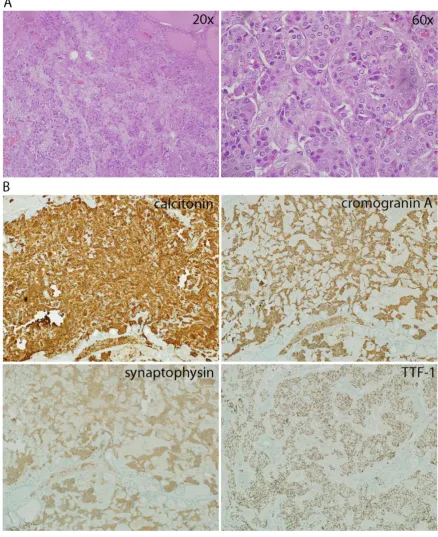
patient was discharged. Immunohistochemistry performed on the nodule showed the 84
presence of a polilobulated medullary thyroid cancer (MTC) of 1 cm, composed of cells with 85
fused shape with eosynophilic cytoplasm, agglomerated in solid nests with a predominantly 86
expansive growth pattern (Figure 1A). Histological examination showed that tumor cells 87
were positive for Ct, Cromogranin A, Synaptofisin, and TTF-1 (Figure 1B), and negative for 88
the presence of amyloid (not shown). Focal foci of C-cells hyperplasia were spread in the 89
entire gland. In none of the lymph nodes of the central compartment were found metastases. 90
92
Figure 1. A. Hematoxylin-Eosin (H&E) staining of MTC nodule at 20x and 60x magnification. B.
93
Immunohistochemistry for calcitonin, cromogramin A, synaptophysin and TTF-1 of the MTC nodule.
94
Magnification 10x.
95
To perform a more detailed morphological analysis, formalin-fixed paraffin-embedded 96
sections (3 μm thick) were subjected to antigen retrieval with citrate buffer at high pH and 97
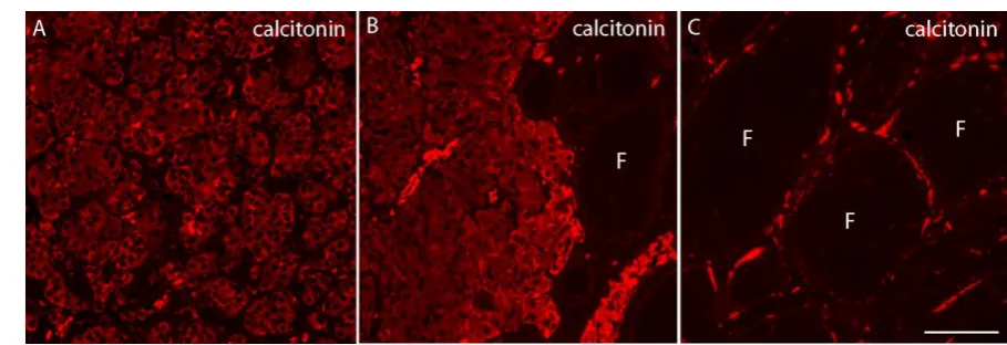
immunolabeled with rabbit monoclonal anti-calcitonin (SP17, Cell Marque) (Figure 2) and 98
were then incubated with the appropriate fluorescent secondary antibodies (anti-rabbit 99
strong intracellular positivity for Ct (Figure 2 A-B), as well as C-cells surrounding found in 101
follicolar parenchima (F) (Figure 2C). 102
103
104
Figure 2.Immunofluorescence labeling for calcitonin on semi-thick sections of the MTC nodule.
105
A-B represents tumor C cells positive for intracellular calcitonin. In C are depicted thyroid follicles (F)
106
surrounding the MTC nodule showing the presence of C cells positive for calcitonin. Images were taken at 40x
107
magnification, scale bar 10µm.
108
We next performed immunofluorescence labeling for RTK receptors ERBB1 (EGFR) using 109
a polyclonal antibody anti-EGFR (Sigma-Aldrich), and for ERBB2 using the monoclonal 110
humanized anti-ERBB2 antibody trastuzumab-alexa488 conjugated (10µg/ml), 111
(Genetech-Roche). The slides were examined and imaged with an inverted Olympus 112
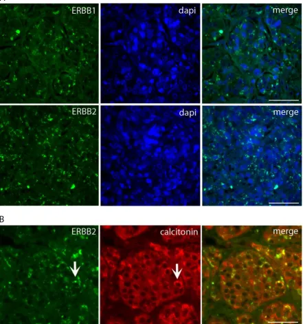
microscope (Olympus Italia, Segrate, Italy) at 40x and 63x magnification. As shown in 113
Figure 3A-B, tumor cells display strong punctate positivity for both ERBB1 and ERBB2 114
receptors, consistent with localization in intracellular compartments, likely the 115
endolysosomal system. Indeed, double immunofluorescence for ERBB2 and Ct showed a 116
limited overlap of the two proteins, indicating that Ct and ERBB2 are segregated in distinct 117
119
Figure 3. Immunofluorescence labeling for ERBB1 and ERBB2 on semi-thick sections of the MTC nodule. A
120
represents tumor C cells positive for intracellular punctate staining for ERBB1 and ERBB2. Magnification 40x,
121
scale bar 10µm. In B is depicted a double immunolabeling for ERBB2 and calcitonin showing only a partial
122
co-localization of the two proteins (arrows). Magnification 63x, scale bar 30µm.
123
124
For electron microscopy, small pieces of fresh tissue were immediately fixed after surgical 125
resection in 2.5% glutaraldehyde, post-fixed with 1% osmium tetroxide and embedded in 126
epoxy resin. Ultrathin sections were double stained with uranyl acetate and lead citrate and 127
examined under a transmission electron microscope (TEM, CM10 Philips, The Netherlands). 128
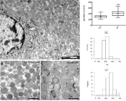
Ultrastructural examination of the tumor nodule displayed heterogeneous spindle-shaped 129
cells containing two distinct classes of tightly packed secretory granules uniformly 130
132
Figure 4.Representative TEM micrographs of MTC. A. Two adiacent cells containing small dense cored
133
granules (SG) and large pale-cored granules (LG). (N) nucleus, (mit) mithocondria. B-C. Higher magnification
134
insets of A showing granules morphology. Scale bars: 3 µm (A), 500 nm (B-C). In the right side of the panel are
135
shown a box plot and histograms representing the frequency distribution of granules size. LG granules are
136
significantly larger than SG (*** p<0.0001, t-test).
137
Round-shaped mitochondria (mit) with disorganized cristae were frequently observed 138
(Figure 4A). Tumor cells containing large, pale-cored secretory granules (LG) were the 139
majority with respect to cells containing small and dense-cored granules (SG). The tumor 140
nodule also contained blood and connective tissue in which cancer cells were scattered. 141
Consistently with other morphological studies [11, 12], morphometric analysis of granule 142
size and distribution showed two main classes of secretory granules based on their size and 143
morphology (Figure 4B-C). The small granules (SG) had an electron dense core and a size 144
ranging from 200 to 400nm, while the large granules (LG, 300-500nm in size) were 145
pale-cored. Statistical significant differences in granules size (*** p<0.0001) and 146
morphology might reflect distinct stages of hormonal production [11, 12]. In particular, 147
mature granules appear characterized by their uniform and moderate electron density and by 148
their generalized distribution throughout the cytoplasm, while small electron dense granules 149
would represent pre-secretory immature granules [13]. 150
151
3. Discussion 153
Medullary thyroid carcinoma (MTC) is a rare calcitonin-secreting neoplasm derived from C 154
cells that occurs in both a familial and sporadic form [14]. It is the most aggressive among 155
well-differentiated thyroid cancers, with survival rates of 40–80 % at 10 years [15]. 156
Because of the relative aggressiveness of MTC, routine measurement of serum calcitonin is 157
suggested as an integral part of the diagnostic evaluation of thyroid nodules to unmask its 158
diagnosis [2]. When an elevated calcitonin level is found, a calcitonin stimulation test with 159
calcium is often indicated to allow an early diagnosis and ultimately decrease morbidity and 160
mortality of this tumor [2, 3, 16]. Serum Ct is the most specific and sensitive marker of MTC, 161
not only for its diagnosis, but also during the postsurgical follow-up [17], since it is produced 162
in high concentrations by almost 100% of primary and metastatic MTCs. However, in a few 163
cases serum calcitonin can be negative or only slightly increased [18]. 164
Primary MTC is efficiently controlled by surgery, but only when the tumor is confined to the 165
thyroid [3]. Patients with progressive or symptomatic metastatic disease who cannot be 166
treated by surgery should be considered candidates for systemic and/or targeted therapy. 167
Current and experimental targeted therapies for advanced medullary thyroid carcinomas 168
include pan tyrosine kinase (TK) inhibitors such as sorafenib, sunitinib, and vandetanib 169
[19-22]. 170
The ERBB family of receptor tyrosine kinases has been implicated in carcinogenesis with 171
special attention to EGFR (ERBB1) and ERBB2. In particular, the expression or activation of 172
EGFR and ERBB2 and related signaling circuitries are altered in many epithelial tumors, 173
especially breast, gastric, lung and ovarian cancers [10]. Both pre-clinical and clinical studies 174
indicate that ERBB receptors have important roles in tumor progression and distant 175
metastasis [7]. Accordingly, these receptors have been intensely studied to understand their 176
importance in cancer biology and as therapeutic targets, and many ERBB inhibitors are now 177
used in the clinic [23, 24]. We report the case of a female patient with no familiar history of 178
MTC presenting a single thyroid nodule of 1 cm, incidentally discovered. The nodule was 179
classified as sporadic MTC and the patient was admitted for total thyroidectomy. After a 180
successful post-surgery recovery, the patient was discharged at hospital day 3 with 181
recommendation for 1-year follow up. We examined structural and ultrastructural features of 182
this case of MTC by immunofluorescence and electron microscopy and found that almost all 183
tumor cells showed strong granular staining for Ct, as expected. In addition, tumor C cells 184
displayed strong positivity for ERBB2 and ERBB1, but in distinct subcellular compartments, 185
most likely the endolysosomal compartment. As the presence of EGFR and ERBB2 within 186
the endocytic system is related to propagation of pro-survival signaling originating from 187
endosomes [13], our finding supports the hypothesis of a role for these receptors in 188
oncogenic C cells proliferation. Ultrastructural morphology of MTC showed the presence of 189
uniformly distributed secretory granules of different size and morphology. Tumor C cells 190
containing large, pale-cored, secretory granules were more represented with respect to cells 191
containing small and dense-cored granules. These characteristics suggest that MTC cells 192
might store and elaborate precursor forms of Ct and/or produce different hormones. As this 193
observation of predominant C cells containing mature, pale-cored, granules is in agreement. 195
To our knowledge, the biological and prognostic significance of ERBB expression in 196
sporadic MTCs and in endocrine organs is still largely unknown [6]. However, ERBB1 and 197
ERBB2 expression in MTCs have been recently correlated with more aggressive disease and 198
extrathyroid growth [8] suggesting a role for ERBB1 and ERBB2 as important prognostic 199
markers for tumor invasiveness. Thus, deregulation of these receptors and their signaling 200
pathways might be connected with essential MTC properties such as tumor cell proliferation 201
and invasive behavior [25]. 202
4. Conclusions 203
MTCs are rare but aggressive thyroid carcinomas that produce calcitonin. The therapeutic 204
option for patients with confined MTCs is total thyroidectomy, while options for metastatic 205
medullary thyroid carcinoma are still limited. Overexpression of ERBB receptors has been 206
related to metastasis in various cancers, including MTCs. This case suggests that further 207
preclinical and clinical studies are required to establish the role for ERBB1 and 2 receptors in 208
C cells proliferation, as novel biomarkers for tumor invasiveness and potential candidates for 209
additional targeted therapies. 210
211
Acknowledgments: Written consent was obtained from the patient for publication of the 212
study. We are grateful to Dr. Paola Baccini for providing reagents and to Maria Cristina 213
Gagliani for support in electron microscopy analysis. 214
215
Author Contributions: MM, EV and GM were involved in the direct patient care. KC, MM, 216
EC conceived and designed the experiments. ADS, GM and KC performed histology, 217
immunofluorescence and electron microscopy analysis. KC and MM wrote the manuscript. 218
All the authors have critically discussed, read and approved the final manuscript. 219
220
Conflicts of Interest: The authors declare no conflict of interest.
221
References 222
1. Johansson E, Andersson L, Ornros J, Carlsson T, Ingeson-Carlsson C, Liang S, 223
Dahlberg J, Jansson S, Parrillo L, Zoppoli P et al: Revising the embryonic origin of 224
thyroid C cells in mice and humans. Development 2015, 142(20):3519-3528. 225
2. Elisei R, Bottici V, Luchetti F, Di Coscio G, Romei C, Grasso L, Miccoli P, Iacconi 226
P, Basolo F, Pinchera A et al: Impact of routine measurement of serum calcitonin 227
on the diagnosis and outcome of medullary thyroid cancer: experience in 10,864 228
patients with nodular thyroid disorders. The Journal of clinical endocrinology and 229
metabolism 2004, 89(1):163-168. 230
3. Miccoli P, Minuto MN, Ugolini C, Molinaro E, Basolo F, Berti P, Pinchera A, Elisei 231
R: Clinically unpredictable prognostic factors in the outcome of medullary 232
4. Khurana R, Agarwal A, Bajpai VK, Verma N, Sharma AK, Gupta RP, Madhusudan 234
KP: Unraveling the amyloid associated with human medullary thyroid 235
carcinoma. Endocrinology 2004, 145(12):5465-5470. 236
5. Priya SR, Dravid CS, Digumarti R, Dandekar M: Targeted Therapy for Medullary 237
Thyroid Cancer: A Review. Frontiers in oncology 2017, 7:238. 238
6. Zhao WJ: Expression and Localization of Neuregulin-1 (Nrg1) and 239
ErbB2/ErbB4 Receptors in Main Endocrine Organs of the Rhesus Monkey. 240
International journal of endocrinology and metabolism 2013, 11(3):162-166. 241
7. Hynes NE, MacDonald G: ErbB receptors and signaling pathways in cancer. Curr 242
Opin Cell Biol 2009, 21(2):177-184. 243
8. Utrilla JC, Martin-Lacave I, San Martin MV, Fernandez-Santos JM, 244
Galera-Davidson H: Expression of c-erbB-2 oncoprotein in human thyroid 245
tumours. Histopathology 1999, 34(1):60-65. 246
9. Rodriguez-Antona C, Pallares J, Montero-Conde C, Inglada-Perez L, Castelblanco E, 247
Landa I, Leskela S, Leandro-Garcia LJ, Lopez-Jimenez E, Leton R et al: 248
Overexpression and activation of EGFR and VEGFR2 in medullary thyroid 249
carcinomas is related to metastasis. Endocrine-related cancer 2010, 17(1):7-16. 250
10. Yarden Y, Sliwkowski MX: Untangling the ErbB signalling network. Nat Rev Mol 251
Cell Biol 2001, 2(2):127-137. 252
11. Kameya T, Shimosato Y, Adachi I, Abe K, Kasai N, Kimura K, Baba K: 253
Immunohistochemical and ultrastructural analysis of medullary carcinoma of 254
the thyroid in relation to hormone production. The American journal of pathology 255
1977, 89(3):555-574. 256
12. Huang SN, Goltzman D: Electron and immunoelectron microscopic study of 257
thyroidal medullary carcinoma. Cancer 1978, 41(6):2226-2235. 258
13. Engedal N, Mills IG: Endosomal signaling and oncogenesis. Methods in 259
enzymology 2014, 535:179-200. 260
14. Gosnell JE, Duh QY: Medullary Thyroid Carcinoma-We Should Do Better. 261
JAMA surgery 2018, 153(1):59. 262
15. Jayakody S, Reagh J, Bullock M, Aniss A, Clifton-Bligh R, Learoyd D, Robinson B, 263
Delbridge L, Sidhu S, Gill AJ et al: Medullary Thyroid Carcinoma: Survival 264
Analysis and Evaluation of Mutation-Specific Immunohistochemistry in 265
Detection of Sporadic Disease. World journal of surgery 2018. 266
16. Rendl G, Manzl M, Hitzl W, Sungler P, Pirich C: Long-term prognosis of 267
medullary thyroid carcinoma. Clinical endocrinology 2008, 69(3):497-505. 268
17. Tashijan AH, Jr., Howland BG, Melvin KE, Hill CS, Jr.: Immunoassay of human 269
calcitonin. The New England journal of medicine 1970, 283(17):890-895. 270
18. Sand M, Gelos M, Sand D, Bechara FG, Bonhag G, Welsing E, Mann B: Serum 271
calcitonin negative medullary thyroid carcinoma. World journal of surgical 272
oncology 2006, 4:97. 273
19. Schlumberger M, Massicotte MH, Nascimento CL, Chougnet C, Baudin E, 274
Leboulleux S: Kinase inhibitors for advanced medullary thyroid carcinoma. 275
20. Nelkin B: Recent advances in the biology and therapy of medullary thyroid 277
carcinoma. F1000Research 2017, 6:2184. 278
21. Tampaki EC, Tampakis A, Droeser R, Patsouris E, Kouraklis G: Cabozantinib and 279
Vandetanib in medullary thyroid carcinoma: mitochondrial function and its 280
potential as a therapeutic target towards novel strategies to design anti-CSCs 281
drugs. Cancer biology & therapy 2018:0. 282
22. Antonelli A, Fallahi P, Ferrari SM, Ruffilli I, Santini F, Minuto M, Galleri D, Miccoli 283
P: New targeted therapies for thyroid cancer. Current genomics 2011, 284
12(8):626-631. 285
23. Anampa J, Makower D, Sparano JA: Progress in adjuvant chemotherapy for 286
breast cancer: an overview. BMC Med 2015, 13:195. 287
24. Albanell J, Codony J, Rovira A, Mellado B, Gascon P: Mechanism of action of 288
anti-HER2 monoclonal antibodies: scientific update on trastuzumab and 2C4. 289
Adv Exp Med Biol 2003, 532:253-268. 290
25. Martin-Lacave I, Utrilla JC: Expression of a neu/c-erbB-2-like product in 291
neuroendocrine cells of mammals. Histology and histopathology 2000, 292