REVIEW PAPER
Selenium nanoparticles role in organ systems functionality and
disorder
Seyed Mohammad Amini1,2*, Vahid Pirhajati Mahabadi3
1 Radiation Biology Research Center, Iran University of Medical Sciences, Tehran, Iran
2 Medical Nanotechnology Department, School of Advanced Technologies in Medicine, Iran University of Medical Sciences, Tehran, Iran
3 Neuroscience Research Center, Iran University of Medical Sciences, Tehran, Iran
* Corresponding Author Email: [email protected]
Extensive research on the nutritional and medical application of selenium nanoparticles (SeNPs) was performed in past decades. Besides nutritional values, new characteristics such as antibacterial and anticancer properties depict a bright future for high Selenium (Se) consumption in the coming years. Se is essential for the proper functioning of most of the major body organ systems meanwhile it could be highly toxic and even cancerous. The current knowledge of Se interaction with major organ systems functionality such as the central nervous system isn’t well studied and many physiological aspects aren’t clear to the science community. Meanwhile, various results were published on increasing organ system functionality through administrated SeNPs. So with the rapid entrance of SeNPs in the medical and nutritional industry, it may cause unintended complications. The intent of this review is to investigate current knowledge of SeNPs interaction with major body organ systems functionality. Investigated pharmacokinetic parameters of SeNPs was also reviewed.
ARTICLE INFO
Article History:
Received 2 April 2018 Accepted 06 June 2018 Published 01 July 2018
Keywords:
Antioxidant
Nanopharmacokinetic Nanotoxicity
Organ Function Selenium Nanoparticles
ABSTRACT
How to cite this article
Amini SM, Pirhajati Mahabadi V. Selenium nanoparticles role in organ systems functionality and disorder. Nanomed Res J, 2018; 3(3): 117-124. DOI: 10.22034/nmrj.2018.03.001
This work is licensed under the Creative Commons Attribution 4.0 International License. To view a copy of this license, visit http://creativecommons.org/licenses/by/4.0/.
INTRODUCTION
Se is an essential mineral with a high nutritional
value and very important role for majority of
physiological interactions in our body [1]. Our
body intake of selenium is provided from food or
water as a selenite, selenate, selenocysteine and
selenomethionine and was carried by different
human selenoproteins in the body. Humans with
Selenium deficiency are amenable to many Health
problems in a way that abnormal
selenoprotein
function or selenium deficiency could lead to
various neurological [2], thyroid [1], muscle [3]
and many other physiological disorders [4].
So the
need for selenium starts from an embryonic age
and continues until old age.
Nanoparticles are gaining lots of attention
and concerns in biomedical and industrial
application [5, 6]. Especially metal nanoparticles
with extraordinary characteristics are capable of
many diagnostic[7, 8], therapeutic [9, 10], health
[11, 12] and nutrition [13] application. Recently
many reports about a different biomedical
application of Se nanoparticles were published.
With antimicrobial, antioxidant and anticancer
properties [14] and lower toxicity in comparison to
the selenite (SeO
4−2) or selenite (SeO
3−2
) counterpart
[15, 16], these nanoparticles have attracted much
attention.
Similar to livestock, agriculture industry applied
Se nanoparticles for better plants growth and
productivity [22]. Furthermore, the extensive use
of Se as an antimicrobial agents in hygiene products
was recommended by many researchers [23, 24].
Future extensive use of Se nanoparticles may
lead to possible harmful effects on human health
and the environment.
Therefore the present review
provides possible pros and cons of Se nanoparticle
role in human organ functionality and disorder.
Importance of Se for organ functionality
The discovery of a congestive cardiomyopathy
known as Keshan disease and Kashin-Beck disease
in certain part of the People’s Republic of China,
which is deforming arthritis reviled the important
role of Se in dietary [25]. But Keshan disease is
not the only disorder of Se deficiency. Altogether,
many experimental bioassays, epidemiologic
studies, and even human clinical intervention trials
support that a tiny amount of Se is vital for proper
function of many human organs and systems such
as Reproductive system [26], Thyroid [27], brain
[28], cardiovascular system [29], Jacobson’s organ
function [30] and many others.
There is much evidence that indicates the direct
relation between Se deficiency and increasing the
risk of cancer [31, 32]. Molecular scientific bases
of Se intake and cancer prevention are not clear
but Glutathione peroxidase activity for hydrogen
peroxidase breakdown strongly depends on
available selenium [33]. Also, mitochondrial
electron transport was altered in Se deficiency
conditions [34] that could lead to more oxidative
stress [35].
Malnutrition may also be associated with
microbial infections. In case of Se deficiency, this has
been reported frequently by researchers. Different
research groups demonstrate that harmless viruses
could be virulent in Se-deficient hosts [36, 37].
Boyne
et al
. reported that Se deficiency may have
a negative effect on the mice immune system to
eliminate Candida albicans [38]. Same results
were reported for mice infected with Diplococcus
pneumoniae [39] and Listeria monocytogenes [40].
Also, Viral infection leads to high ROS generation
[41] and in-fact viral gene expression controls the
cellular gene expression via ROS generation which
leads to the appearance of cancer cells [42]. It’s
clear that under low Se condition, general oxidative
stress increased and the mentioned process lead
to higher viral infection. Also, Se deficiency may
cause a decrease in immune system efficiency that
provides a chance for opportunistic pathogens [43,
44].
Se toxicity and organ disorder
Se toxicity was well-known in livestock that
grazed in high Se soils. These animals developed
disorders such as “alkai disease” and “blind
staggers“ [45]. Alkai disease is characterized
by thickening hair and degenerative changes
in hooves and blind staggers is characterized
by impairment of vision and unsteady steps. In
human, acute selenium toxicity usually leads to
gastrointestinal disorders such as nausea, vomiting
or diarrhoea alongside hair losses and headache
[46]. Other acute toxicological symptoms such as
bronchitis [47] and hypochromic anemia [48] have
been reported. Long-term Se toxicity resembles in
acute discolouration of skin and nail [49]. Except
for the blood level of selenium, other biochemical
indicators of body Se would increase which
include: prothrombin time [50], alanine serum
concentrations [51] and alanine aminotransferase
enzyme activity [52]. Liver histopathological
studies reveal sinusoidal damage and nodular
regenerative hyperplasia in a rat model with high
Se intake [53].
Numerous experimental studies demonstrate the
carcinogenic effect of inorganic or organic Se form
[54, 55] which is contrary to the cancer-protective
activity of Se that was discussed. The same
contradictive reports happen to the neuroprotective
or neurodegenerative activity of Se. Amyotrophic
lateral sclerosis (ALS) is a rare neurodegenerative
disease that may be related to high Se intake [56].
Other nervous system abnormality may also be
related to Se toxicity [57, 58]. Also, the limited
evidence demonstrates Se toxicity in endocrine
[48], reproductive [59], immune [60] systems. A
direct link between high Se exposure and dental
caries is clarified by Hadjimarkos
et al
[61].
Pharmacokinetic parameters of Se NPs
Nanomed Res J 3(3): 117-124, Summer 2018
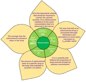
under the time-concentration curve (AUC) (Fig. 1).
In general metallic nanoparticles pharmacokinetic
is different from inorganic or organic metallic ion
forms [62].
In the human metabolic cycle, Se exists in
various organic or inorganic oxidation states
including -2, +2, +4 and +6 [63]. Unlike mentioned
oxidative states of Se, pharmacokinetic data
of elemental Se (Se
0) is scarce. In nature, some
microorganisms reduced selenite (Se
+4), or selenate
(Se
+6) into nanoparticles formed Se
0[64]. Moderate
absorption of Se
0NPs reduced by bacteria in the
chicken model was reported [65]. Bioavailability
and bioactivity of chemically synthesized Se
0NPs
are reported in Rats [66]. The retention of SeNPs
and Se-methylselenocysteine (MeSeCys) at the
nutritional level in mice blood and other tissues
was compared and no significant differences were
observed [67]. Similar results were observed for
kidney and liver accumulation for Se0NP and
selenite [68]. Same results were reported for
selenomethionine (SeMet) [69] and selenite [68].
The clearance of SeNPs from the body can be either
by renal and hepato-biliary excretion SeNPs or
excretion of Se metabolites. In a pharmacokinetic
study by Loeschner
et al
. the entrance of Se
0from
Se
0NPs into the metabolic cycle and its subsequent
elimination was confirmed by the apprehension of
the two urinary metabolites of Se alongside a high
amount of stool excretion of Se [68]. The authors
don’t investigate hair excretion.
Studies that indicate organ functionality with Se
Nanoparticles Treatments
Organ functionality strongly depends on
Antioxidant defensive capability. The antioxidant
defence system is consisting of enzymatic and
non-enzymatic antioxidants which are expanded
not only inside the cells but also in extracellular
environments. The first line of the antioxidant
defence system is to restrain the production of
free radicals. Reduction of disintegrated hydrogen
peroxide and hydroperoxides by water and alcohol
is a prime ROS generation mechanism which
was suppressed through some selenoproteins and
selenoenzymes such as glutathione peroxidase,
thioredoxin reductases and etc. In many studies
that applied SeNPs, increasing selenoproteins and
selenoenzymes activity was reported with lower
toxicity in comparison to an organic or inorganic
form of Se [62]. SeNPs provides not only higher
bioavailability but also they can scavenge free
Figure 1: Pharmacokinetic values
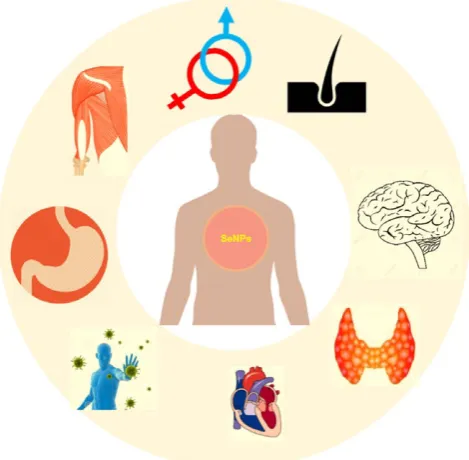
radicals directly [63]. The organ systems in which
the effect of selenium nanoparticles on their
function is examined is shown schematically in
Fig. 2.
Musculoskeletal system:
Beside direct
interaction with oxidative stress SeNPs could be
integrated with other metabolic cycles that are
involved with the musculoskeletal system oxidative
stress response. For example, increasing oxidative
stress can lead to a higher expression of heat shock
proteins (HSPs) [64], which is an adjusting process
corresponding to the interruption of cellular
integrity [65]. The nutritional supplement of SeNPs
could increase the expression level of HSP90 in a
rest period after extensive exercise. A higher level
of HSP expression could increase cells tolerability
in stressful condition [66]. The nutritional
supplement of SeNPs can significantly decrease the
blood urea nitrogen (BUN) and creatinine serum
concentration after intense exercise [67]. Both
of these compounds can damage cells through
oxidative stress [68].
Reproductive system:
The main cause of
sperm dysfunction is oxidative stress [18]. Some
researchers applied SeNPs as a male fertility
preservation substance. Increase semen quality
and reproductive functionality of male goats was
reported by Shi
et al
. through increase activity
of testicular GPx and improvement in testicular
microstructure and testicular spermatozoa.
Moreover, SeNPs treatment leads to higher Se
accumulation in testes, and testicular tissue [19].
Fertility preservation in those who have been treated
with chemotherapy is major concerns. Rezvanfar
et al
. observed the chemoprotective activity of
SeNPs against cisplatin-induced gonadotoxicity.
They demonstrate significant improvement in
spermatogenesis and semen quality and lessened
spermatic DNA destruction alongside
cisplatin-induced free radical stress [69].
Endocrine system:
Se is necessary for
efficient endocrine glands functionality. Many
selenoenzymes and selenoproteins are adjusting
redox status of endocrine cells and in some cases
involved in endocrine hormone metabolism
[70]. But few investigations were performed for
investigation of SeNPs role in endocrine glands
functionality. Hassanin
et al
. demonstrated that
accumulated SeNPs in thyroid cell can modify
thyroid chromium toxicity in rats [71]. Rezaeian
and Sadeghi report investigate the thyroid and
sex hormones levels after SeNPs administration
in rats. SeNPs administration can increase both
thyroids and sex hormones levels not only in
Figure 2: Schematic representation of SeNPs possible interaction with body organ systems: In this
manuscript, current knowledge about SeNPs interaction with musculoskeletal, reproductive, endocrine,
nervous, immune, digestive, circulation and the integumentary system was discussed.
Fig. 2: Schematic representation of SeNPs possible interaction with body organ systems: In this manuscript, current knowledge about SeNPs interaction with musculoskeletal, reproductive, endocrine, nervous, immune, digestive,
Nanomed Res J 3(3): 117-124, Summer 2018
normal condition but also in oxidative stress
condition [72]. Oral administration of SeNPs
can not only minimize diabetic complication
but also could enhance pancreatic efficiency.
Al-Quraishy
et al
report not only a significant
augment of blood serum insulin level was
observed but also histopathological studied show
a SeNPs protective role in β-cells in the islet of
Langerhans [73]. Similar results were provided
by a liposomal formulation of SeNPs by Ahmed
et al
[74].
Nervous system:
For normal nervous system
function selenoproteins are essential. Nervous
system damage due to nanoparticle was reported
before [75]. A link between selenoproteins
functionality and several nervous system disorder
was reviewed by Schweizer
et al
[28]. But SeNPs
were merely applied for investigation of central
nervous system functionality. Nazıroğlu
et al
. review
state that SeNPs could be a potential remedial
compound for Alzheimer disease treatment [76]. In
contrast, Yuan
et al
. studied the impacts of SeNPs
on sodium currents on dorsal root ganglion (DRG)
neurons activity, applying the whole-cell patch
clamp technique. In their report, SeNPs were able
to decrease sodium current in a concentration and
time-dependent manners which consider possible
neurotoxic characteristic for SeNPs [77].
Immune system:
It has been demonstrated
that Se is generally accumulated in immune
response organs such as liver, lymph nodes
and spleen [78].
To deal with immunogens, Se
treatment can increased antibodies production and
expand complement responses through different
mechanisms which include various selenoproteins
[78].
It has been demonstrated that
biogenic SeNPs
could be a stimulator for immune system [80]. In
particular, expanded neutrophils chemotactic and
respiratory burst activities were observed for SeNPs
in comparison with sodium selenite [81]. The
authors consider the different pharmacokinetics
parameters of SeNPs in comparison to selenite for
assessment of immune stimulator functionality of
SeNPs.
Digestive system:
Sarkar
et al
. claimed that
SeNPs could be an effective treatment for fatty
liver disease [82]
.
In a separate study induced
fatty liver male rats were treated with SeNP, and
lower level of free radical and inflammation
were reported [83]. SeNPs can improve rumen
fermentation in livestock. A diet which contains
SeNPs could increase total fiber digestion in sheep
[19].
Lactobacillus
species
could be applied for the
preparation of selenium nanoparticle-enriched
probiotics that represent an
antifungal activity
against Candida albicans [84].
Circulation system:
Selenoproteins and
selenoenzymes are essential for blood cells
protections against oxidative damages [85]. It has
been demonstrated that feeding of SeNPs has a
better impact in comparison to sodium selenite
upon peroxidative damage in blood cells [21].
SeNPs is also representing a better iron homeostasis
capability in compression to other Se forms. In
in-vivo compression investigation between SeNPs
and sodium selenite, the level of transferrin and
transferrin receptor was monitored. Total iron
binding capacity (TIBC) in groups that treated
by SeNPs is higher than a group that treated with
sodium selenite [85]. Se has an essential role in
inhibiting cardiovascular problems by augmenting
the oxidation stress defence system to combat
the oxidative alteration of lipids and subsequent
decrease platelets aggregation [29]. SeNPs Ability
to protect the cardiac cells from ischemia was
demonstrated in an in-vitro experiment by Soumya
et al
[87].
Integumentary system:
Se
compensates
skin-damaging compounds destructive role. It has been
used for skin and hair care through cosmetics
and health products. It has been demonstrated
that SeNPs are able to penetrate in skin tissue
and subsequently prevent the lipofusin formation
caused by UV-exposure and protect glutathione
peroxidase (GPx) activity in mice [88]. Anti-fungal
effect of SeNPs was as well as classical antidandruff
selenium sulfide with lower cytotoxicity [89].
CONCLUSION
supply could happen very soon and subsequently
human or environmental safety concerns such as
long-term toxic and possible carcinogenic effects
must be considered through further studies.
CONFLICTS OF INTEREST
The authors declare that there are no conflicts
of interest regarding the publication of this
manuscript.
REFERENCES
1. Ventura M, Melo M, Carrilho F. Selenium and Thyroid
Disease: From Pathophysiology to Treatment. International Journal of Endocrinology. 2017;2017:1-9.
2. Pillai R, Uyehara-Lock JH, Bellinger FP. Selenium and
selenoprotein function in brain disorders. IUBMB Life. 2014;66(4):229-39.
3. Lescure A, Rederstorff M, Krol A, Guicheney P, Allamand
V. Selenoprotein function and muscle disease. Biochimica et Biophysica Acta (BBA) - General Subjects. 2009;1790(11):1569-74.
4. Schweizer U, Dehina N, Schomburg L. Disorders of selenium
metabolism and selenoprotein function. Current Opinion in Pediatrics. 2011;23(4):429-35.
5. Zarchi AAK, Amini SM, Farsangi ZJ, Mohammadi E, Moosavi Z, Harati PG. A study on the possibility of drug delivery approach through ultrasonic sensitive nanocarriers. Nanomedicine Journal, 2018;5 (3):127-137.
6. Shirkhanloo H, Safari M, Amini SM, Rashidi M. Novel Semisolid Design Based on Bismuth Oxide (Bi2O3) nanoparticles for radiation protection. Nanomedicine Research Journal, 2017;2 (4):230-238.
7. Emami T, Madani R, Golchinfar F, Shoushtary A, Amini SM.
Comparison of Gold Nanoparticle Conjugated Secondary Antibody with Non-Gold Secondary Antibody in an ELISA Kit Model. Monoclonal Antibodies in Immunodiagnosis and Immunotherapy. 2015;34(5):366-70.
8. Fatemi F, Amini SM, Kharrazi S, Rasaee MJ, Mazlomi MA,
Asadi-Ghalehni M, et al. Construction of genetically engineered M13K07 helper phage for simultaneous phage display of gold binding peptide 1 and nuclear matrix protein 22 ScFv antibody. Colloids and Surfaces B: Biointerfaces. 2017;159:770-80.
9. Amini SM, Kharrazi S, Jaafari MR. Radio frequency
hyperthermia of cancerous cells with gold nanoclusters: an in vitro investigation. Gold Bulletin. 2017;50(1):43-50.
10. Karimi Zarchi AA, Amini SM, Salimi A, Kharazi S.
Synthesis and characterisation of liposomal doxorubicin with loaded gold nanoparticles. IET Nanobiotechnology. 2018;12(6):846-9.
11. Darabpour E, Kashef N, Amini SM, Kharrazi S, Djavid GE.
Fast and effective photodynamic inactivation of 4-day-old biofilm of methicillin-resistant Staphylococcus aureus using methylene blue-conjugated gold nanoparticles. Journal of Drug Delivery Science and Technology. 2017;37:134-40.
12. Shaabani E, Amini SM, Kharrazi S, Tajerian R. Curcumin coated gold nanoparticles: synthesis, characterization, cytotoxicity, antioxidant activity and its comparison with citrate coated gold nanoparticles. Nanomedicine Journal, 2017;4 (2):115-125.
13. Amini SM, Gilaki M, Karchani M. Safety of nanotechnology in food industries. Electronic physician, 2014;6 (4):962.
14. Hosnedlova B, Kepinska M, Skalickova S, Fernandez C,
Ruttkay-Nedecky B, Peng Q, et al. Nano-selenium and its nanomedicine applications: a critical review. International Journal of Nanomedicine. 2018;Volume 13:2107-28.
15. Benko I, Nagy G, Tanczos B, Ungvari E, Sztrik A, Eszenyi P,
et al. Subacute toxicity of nano-selenium compared to other selenium species in mice. Environmental Toxicology and Chemistry. 2012;31(12):2812-20.
16. Shakibaie M, Shahverdi AR, Faramarzi MA, Hassanzadeh
GR, Rahimi HR, Sabzevari O. Acute and subacute toxicity of novel biogenic selenium nanoparticles in mice. Pharmaceutical Biology. 2012;51(1):58-63.
17. Shi L-g, Yang R-j, Yue W-b, Xun W-j, Zhang C-x, Ren Y-s,
et al. Effect of elemental nano-selenium on semen quality, glutathione peroxidase activity, and testis ultrastructure in male Boer goats. Animal Reproduction Science. 2010;118(2-4):248-54.
18. Aitken RJ, Baker MA. Oxidative stress and male reproductive biology. Reproduction, Fertility and development, 2004;16 (5):581-588.
19. Shi L, Xun W, Yue W, Zhang C, Ren Y, Liu Q, et al. Effect
of elemental nano-selenium on feed digestibility, rumen fermentation, and purine derivatives in sheep. Animal Feed Science and Technology. 2011;163(2-4):136-42.
20. Hu CH, Li YL, Xiong L, Zhang HM, Song J, Xia MS.
Comparative effects of nano elemental selenium and sodium selenite on selenium retention in broiler chickens. Animal Feed Science and Technology. 2012;177(3-4):204-10.
21. Sadeghian S, Kojouri GA, Mohebbi A. Nanoparticles of
Selenium as Species with Stronger Physiological Effects in Sheep in Comparison with Sodium Selenite. Biological Trace Element Research. 2011;146(3):302-8.
22. El-Ramady H, Abdalla N, Taha HS, Alshaal T, El-Henawy
A, Faizy SEDA, et al. Selenium and nano-selenium in plant nutrition. Environmental Chemistry Letters. 2015;14(1):123-47.
23. Huang X, Chen X, Chen Q, Yu Q, Sun D, Liu J. Investigation of
functional selenium nanoparticles as potent antimicrobial agents against superbugs. Acta Biomaterialia. 2016;30:397-407.
24. Tran PA, Webster TJ. Antimicrobial selenium nanoparticle
coatings on polymeric medical devices. Nanotechnology. 2013;24(15):155101.
25. Chen X, Yang G, Chen J, Chen X, Wen Z, Ge K. Studies on
the relations of selenium and Keshan disease. Biological Trace Element Research. 1980;2(2):91-107.
26. Barrington JW, Lindsay P, James D, Smith S, Roberts A.
Selenium deficiency and miscarriage: a possible link? BJOG: An International Journal of Obstetrics and Gynaecology. 1996;103(2):130-2.
27. Olivieri O, Girelli D, Azzini M, Stanzial AM, Russo C, Ferroni
M, et al. Low Selenium Status in the Elderly Influences Thyroid Hormones. Clinical Science. 1995;89(6):637-42.
28. Schweizer U, Bräuer AU, Köhrle J, Nitsch R, Savaskan NE.
Selenium and brain function: a poorly recognized liaison. Brain Research Reviews. 2004;45(3):164-78.
29. N??ve J. Selenium as a risk factor for cardiovascular diseases.
Journal of Cardiovascular Risk. 1996;3(1):42-7.
30. Berliner DL, Monti-Bloch L, Jennings-White C,
Nanomed Res J 3(3): 117-124, Summer 2018
organ (VNO): Evidence for steroid receptors. The Journal of Steroid Biochemistry and Molecular Biology. 1996;58(3):259-65.
31. Willett W, Steven Morris J, Pressel S, Taylor J, Frank Polk B,
Stampfer M, et al. PREDIAGNOSTIC SERUM SELENIUM AND RISK OF CANCER. The Lancet. 1983;322(8342):130-4.
32. Shamberger RJ. Relationship of selenium to cancer. I. Inhibitory effect of selenium on carcinogenesis. Journal of the National Cancer Institute, 1970;44 (4):931-936.
33. Lawrence RA, Burk RF. Glutathione peroxidase activity in
selenium-deficient rat liver. Biochemical and Biophysical Research Communications. 1976;71(4):952-8.
34. Rani P, Lalitha K. Evidence for altered structure and
impaired mitochondrial electron transport function in selenium deficiency. Biological Trace Element Research. 1996;51(3):225-34.
35. Liu Y, Fiskum G, Schubert D. Generation of reactive oxygen
species by the mitochondrial electron transport chain. Journal of Neurochemistry. 2002;80(5):780-7.
36. Beck MA, Shi Q, Morris VC, Levander OA. Rapid genomic
evolution of a non-virulent Coxsackievirus B3 in selenium-deficient mice results in selection of identical virulent isolates. Nature Medicine. 1995;1(5):433-6.
37. Beck MA, Levander OA, Handy J. Selenium Deficiency and
Viral Infection. The Journal of Nutrition. 2003;133(5):1463S-7S.
38. Boyne R, Arthur JR. The Response of Selenium-Deficient
Mice to Candida albicans Infection. The Journal of Nutrition. 1986;116(5):816-22.
39. Hoffmann PR, Berry MJ. The influence of selenium on
immune responses. Molecular Nutrition & Food Research. 2008;52(11):1273-80.
40. Wang C, Wang H, Luo J, Hu Y, Wei L, Duan M, et al.
Selenium deficiency impairs host innate immune response and induces susceptibility to Listeria monocytogenes infection. BMC Immunology. 2009;10(1):55.
41. Love AJ. Cauliflower mosaic virus, a Compatible Pathogen
of Arabidopsis, Engages Three Distinct Defense-Signaling Pathways and Activates Rapid Systemic Generation of Reactive Oxygen Species. PLANT PHYSIOLOGY. 2005;139(2):935-48.
42. Waris G, Ahsan H. Reactive oxygen species: role in the development of cancer and various chronic conditions. Journal of carcinogenesis, 2006;5:14.
43. Descamps-Latscha B, Jungers P, Witko-Sarsat V. Immune
System Dysregulation in Uremia: Role of Oxidative Stress. Blood Purification. 2002;20(5):481-4.
44. Laddha NC, Dwivedi M, Mansuri MS, Gani AR, Ansarullah
M, Ramachandran AV, et al. Vitiligo: interplay between oxidative stress and immune system. Experimental Dermatology. 2013;22(4):245-50.
45. Kim YY, Mahan DC. Biological Aspects of Selenium in Farm
Animals. Asian-Australasian Journal of Animal Sciences. 2003;16(3):435-44.
46. Schuh B, Jappe U. Selenium intoxication: undesirable
effect of a fasting cure. British Journal of Dermatology. 2007;156(1):177-8.
47. Nemery B. Metal toxicity and the respiratory tract. European Respiratory Journal, 1990;3 (2):202-219.
48. Nagai I. An Experimental Study of SeleniumPoisoning. Igaku Kenkyu, 1959;29 (5):1505-1532.
49. Smith MI, Westfall BB. Further Field Studies on the
Selenium Problem in Relation to Public Health. Public Health Reports (1896-1970). 1937;52(40):1375.
50. Yang G, Yin S, Zhou R, Gu L, Yan B, Liu Y. Studies of safe maximal daily dietary Se-intake in a seleniferous area in China. Part II: Relation between Se-intake and the manifestation of clinical signs and certain biochemical alterations in blood and urine. Journal of trace elements and electrolytes in health and disease, 1989;3 (3):123-130.
51. Longnecker MP, Taylor PR, Levander OA, Howe M, Veillon
C, McAdam PA, et al. Selenium in diet, blood, and toenails in relation to human health in a seleniferous area. The American Journal of Clinical Nutrition. 1991;53(5):1288-94.
52. Coudray C, Haida H, Boucher F, Tirard V, De Leiris J,
Favier A. Effect of selenium supplementation on biological constants and antioxidant status in rats. Journal of Trace Elements in Medicine and Biology. 1996;10(1):12-9.
53. Dubuisson L, Boussarie L, Bedin C-A, Balabaud C,
Bioulac-Sage P. Transformation of sinusoids into capillaries in a rat model of selenium-induced nodular regenerative hyperplasia: An immunolight and immunoelectron microscopic study. Hepatology. 1995;21(3):805-14.
54. Birt DF, Pour PM, Pelling JC. The Influence of Dietary
Selenium on Colon, Pancreas, and Skin Tumorigenesis. Selenium in Biology and Medicine: Springer Berlin Heidelberg; 1989. p. 297-304.
55. Woutersen RA, Appel MJ, van Garderen-Hoetmer A.
Modulation of Pancreatic Carcinogenesis by Antioxidants. Food and Chemical Toxicology. 1999;37(9-10):981-4.
56. Vinceti M, Guidetti D, Pinotti M, Rovesti S, Merlin M,
Vescovi L, et al. Amyotrophic Lateral Sclerosis after Long-Term Exposure to Drinking Water with High Selenium Content. Epidemiology. 1996;7(5):529-32.
57. Pamphlett R, McQuilty R, Zarkos K. Blood Levels of
Toxic and Essential Metals in Motor Neuron Disease. NeuroToxicology. 2001;22(3):401-10.
58. Yang GQ, Wang SZ, Zhou RH, Sun SZ. Endemic selenium
intoxication of humans in China. The American Journal of Clinical Nutrition. 1983;37(5):872-81.
59. Vinceti M, Cann CI, Calzolari E, Vivoli R, Garavelli L,
Bergomi M. Reproductive outcomes in a population exposed long-term to inorganic selenium via drinking water. Science of The Total Environment. 2000;250(1-3):1-7.
60. Ferenčík M, Ebringer L. Modulatory effects of selenium
and zinc on the immune system. Folia Microbiologica. 2003;48(3):417-26.
61. Hadjimarkos TlDM. Selenium in Relation to Dental Caries.
Food and Cosmetics Toxicology. 1973;11(4):1083-95.
62. Zhang J, Wang X, Xu T. Elemental Selenium at Nano Size
(Nano-Se) as a Potential Chemopreventive Agent with Reduced Risk of Selenium Toxicity: Comparison with Se-Methylselenocysteine in Mice. Toxicological Sciences. 2007;101(1):22-31.
63. Peng D, Zhang J, Liu Q, Taylor EW. Size effect of elemental
selenium nanoparticles (Nano-Se) at supranutritional levels on selenium accumulation and glutathione S-transferase activity. Journal of Inorganic Biochemistry. 2007;101(10):1457-63.
64. Khassaf M, McArdle A, Esanu C, Vasilaki A, McArdle
Physiology. 2003;549(2):645-52.
65. Kinnunen S, Hyyppä S, Oksala N, Laaksonen DE, Hannila
ML, Sen CK, et al. α-Lipoic acid supplementation enhances heat shock protein production and decreases post exercise lactic acid concentrations in exercised standardbred trotters. Research in Veterinary Science. 2009;87(3):462-7.
66. Kregel KC. Invited Review: Heat shock proteins: modifying
factors in physiological stress responses and acquired thermotolerance. Journal of Applied Physiology. 2002;92(5):2177-86.
67. Kojouri GA, Sharifi S. Preventing Effects of
Nano-Selenium Particles on Serum Concentration of Blood Urea Nitrogen, Creatinine, and Total Protein During Intense Exercise in Donkey. Journal of Equine Veterinary Science. 2013;33(8):597-600.
68. Zhang Z, Dmitrieva NI, Park JH, Levine RL, Burg MB.
From The Cover: High urea and NaCl carbonylate proteins in renal cells in culture and in vivo, and high urea causes 8-oxoguanine lesions in their DNA. Proceedings of the National Academy of Sciences. 2004;101(25):9491-6.
69. Rezvanfar MA, Rezvanfar MA, Shahverdi AR, Ahmadi A,
Baeeri M, Mohammadirad A, et al. Protection of cisplatin-induced spermatotoxicity, DNA damage and chromatin abnormality by selenium nano-particles. Toxicology and Applied Pharmacology. 2013;266(3):356-65.
70. Beckett GJ, Arthur JR. Selenium and endocrine systems.
Journal of Endocrinology. 2005;184(3):455-65.
71. Hashem K, Hassnin KMA, AbdEl-Kawi SH. The prospective
protective effect of selenium nanoparticles against chromium-induced oxidative and cellular damage in rat thyroid. International Journal of Nanomedicine. 2013:1713.
72. Rezaeian-Tabrizi M, Sadeghi AA. Plasma antioxidant
capacity, sexual and thyroid hormones levels, sperm quantity and quality parameters in stressed male rats received nano-particle of selenium. Asian Pacific Journal of Reproduction. 2017;6(1):29-34.
73. Abdel Moneim A, Al-Quraishy S, Dkhil MA.
Anti-hyperglycemic activity of selenium nanoparticles in streptozotocin-induced diabetic rats. International Journal of Nanomedicine. 2015:6741.
74. Ahmed HH, Abd El-Maksoud MD, Abdel Moneim AE,
Aglan HA. Pre-Clinical Study for the Antidiabetic Potential of Selenium Nanoparticles. Biological Trace Element Research. 2016;177(2):267-80.
75. Amiri S, Yousefi-Ahmadipour A, Hosseini M-J,
Haj-Mirzaian A, Momeny M, Hosseini-Chegeni H, et al. Maternal exposure to silver nanoparticles are associated with behavioral abnormalities in adulthood: Role of mitochondria and innate immunity in developmental toxicity. NeuroToxicology. 2018;66:66-77.
76. Nazıroğlu M, Muhamad S, Pecze L. Nanoparticles as
potential clinical therapeutic agents in Alzheimer’s disease: focus on selenium nanoparticles. Expert Review of Clinical
Pharmacology. 2017;10(7):773-82.
77. Yuan H, Lin J, Lan T. Effects of Nano Red Elemental Selenium
on Sodium Currents in Rat Dorsal Root Ganglion Neurons. 2006 International Conference of the IEEE Engineering in Medicine and Biology Society; 2006/08: IEEE; 2006.
78. Dickson RC, Tomlinson RH. Selenium in blood and human
tissues. Clinica Chimica Acta. 1967;16(2):311-21.
79. McKenzie RC, Rafferty TS, Beckett GJ, Arthur JR. Effects of
selenium on immunity and aging. Selenium: Springer US; 2001. p. 257-72.
80. Yazdi M, Mahdavi M, Setayesh N, Esfandyar M, Shahverdi A.
Selenium nanoparticle-enriched Lactobacillus brevis causes more efficient immune responses in vivo and reduces the liver metastasis in metastatic form of mouse breast cancer. DARU Journal of Pharmaceutical Sciences. 2013;21(1):33.
81. Kojouri GA, Sadeghian S, Mohebbi A, Mokhber Dezfouli
MR. The Effects of Oral Consumption of Selenium Nanoparticles on Chemotactic and Respiratory Burst Activities of Neutrophils in Comparison with Sodium Selenite in Sheep. Biological Trace Element Research. 2011;146(2):160-6.
82. Sarkar B, Bhattacharjee S, Daware A, Tribedi P, Krishnani
KK, Minhas PS. Selenium Nanoparticles for Stress-Resilient Fish and Livestock. Nanoscale Research Letters. 2015;10(1).
83. Hegedüs V, Prokisch J, Fébel H, Kleiner D, Ditrói K, Szijártó
A, et al. Nanoselenium treatment in fatty liver. Zeitschrift für Gastroenterologie. 2012;50(05).
84. Kheradmand E, Rafii F, Yazdi M, Sepahi A, Shahverdi
A, Oveisi M. The antimicrobial effects of selenium nanoparticle-enriched probiotics and their fermented broth against Candida albicans. DARU Journal of Pharmaceutical Sciences. 2014;22(1):48.
85. Murthy NLN. Investigation Of Biochemical And Histopathological Modifications Induced By Selenium Toxicity On Albino Mice. 2013.
86. Kojouri GA, Jahanabadi S, Shakibaie M, Ahadi AM,
Shahverdi AR. Effect of selenium supplementation with sodium selenite and selenium nanoparticles on iron homeostasis and transferrin gene expression in sheep: A preliminary study. Research in Veterinary Science. 2012;93(1):275-8.
87. Soumya RS, Vineetha VP, Salin Raj P, Raghu KG. Beneficial
properties of selenium incorporated guar gum nanoparticles against ischemia/reperfusion in cardiomyoblasts (H9c2). Metallomics. 2014;6(11):2134-47.
88. Zhai X, Zhang C, Zhao G, Stoll S, Ren F, Leng X. Antioxidant
capacities of the selenium nanoparticles stabilized by chitosan. Journal of Nanobiotechnology. 2017;15(1).
89. Parsamehr N, Rezaie S, Khodavaisy S, Salari S, Hadizadeh