Kranthi and Sravani et al. World Journal of Pharmaceutical and Medical Research
VENOUS THROMBOEMBOLISM
*Ch. Kranthi, *A. Sravani, Dr. G. Susmitha, Dr. V. Tejaswi, Dr. G. Ramesh, Dr. P. Srinivasa Babu
Vignan Pharmacy College, Vadlamudi.
Article Received on 19/01/2018 Article Revised on 09/02/2018 Article Accepted on 01/03/2018
INTRODUCTION
Venous thromboembolism is a blood clot that starts inside a vein. Venous thromboembolism (VTE) is a major health and financial burden that affects the
community. A common type of venous
thromboembolism is a deep vein thrombosis (DVT), usually initiates in the calf area of the leg. If the thrombus breaks off (embolizes) and flows towards the lungs, it can become a pulmonary embolism (PE), a blood clot in the lungs.[1] About 60–70% of patient with symptomatic VTE develop DVT. Pulmonary Embolism (PE) symptoms, such as new or worsening dyspnoea, chest pain, or sustained hypotension with no alternative causeoccur in about 30–40% of patients with VTE. Both acquired and hereditary factors play essential roles in development of VTE. Clinical features are nonspecific. The initial diagnostic step for determination of VTE is the clinical probability assessment. For suspected DVT, the Wells score has well-established criteria. To prevent thrombus extension, decrease of the risk of recurrent thrombosis and subsequent death in patient with VTE pharmacological and/or mechanical approaches can be
administered. The initial treatment for venous
thromboembolism is typically with either low molecular weight heparin (LMWH) or unfractionated heparin.[3] The delayed recognition and treatment of DVT can lead to several early and late complications including PE,
venous gangrene, post-phlebitic syndrome and chronic venous insufficiency.
CLASSIFICATION
Venous thromboembolism is of two types:
1. Deep vein thrombosis (DVT)
2. Pulmonary embolism (PE)
1. Deep vein thrombosis (DVT)
Calf area of leg is the site where DVT usually initiates. The majority of thrombi in deep veins below the popliteal trifurcation are most likely to resolve with no symptoms spontaneously. Most of the patients with venous thromboembolism are likely to develop DVT.[2] When distal DVT extends to popliteal and femoral and other proximal veins, most of the patients develop symptoms. DVT can lead to complications like postphlebitic syndrome, pulmonary embolism and death. Patients with who are untreated with symptomatic proximal DVT develop pulmonary embolism within 3 months. Important complication of DVT is post thrombotic syndrome which results in lifelong limb pain, swelling, heaviness, oedema and leg ulcers.[5]
ISSN 2455-3301
WJPMR
WORLD JOURNAL OF PHARMACEUTICAL AND MEDICAL RESEARCH
www.wjpmr.com
*Corresponding Author:Ch. Kranthi and A. Sravani
Vignan Pharmacy College, Vadlamudi.
ABSTRACT
Venous thromboembolism is a blood clot that starts inside a vein. Venous thromboembolism (VTE) is a major health and financial burden that affects the community. A common type of venous thromboembolism is a deep vein thrombosis (DVT), usually initiates in the calf area of the leg. If the thrombus breaks off (embolizes) and flows towards the lungs, it can become a pulmonary embolism (PE), a blood clot in the lungs. About 60–70% of patient with symptomatic VTE develop DVT. Pulmonary Embolism (PE) symptoms, such as new or worsening dyspnoea, chest pain, or sustained hypotension with no alternative causeoccur in about 30–40% of patients with VTE. Both acquired and hereditary factors play essential roles in development of VTE. Clinical features are nonspecific. The initial diagnostic step for determination of VTE is the clinical probability assessment. For suspected DVT, the Wells score has well-established criteria. To prevent thrombus extension, decrease of the risk of recurrent thrombosis and subsequent death in patient with VTE pharmacological and/or mechanical approaches can be administered. The initial treatment for venous thromboembolism is typically with either low molecular weight heparin (LMWH) or unfractionated heparin.
Figure 1: Deep Vein Thrombosis.
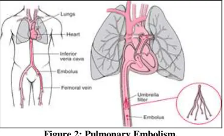
2. Pulmonary embolism (PE)
PE occurs when a DVT clot breaks free from a vein wall, travels to the lungs and blocks some or all of the blood supply.[7]
Blood clots in the thigh are more likely to break off and travel to the lungs when compared to blood clots in the lower leg or other parts of body.[8]
Figure 2: Pulmonary Embolism.
ETIOLOGY
Venous thromboembolic disease occurs when one or more of the elements of Virchow’s triad are present which results in deep vein thrombosis (DVT) and/or pulmonary embolism (PE).[9] Virchow’s triad include the following:
1. Endothelial factors
2. Hypercoagulability
3. Stasis
EPIDEMIOLOGY
The current incidence of venous thromboembolism is approximately 1 per 1000 adults annually .out of that one third of the patients are with pulmonary embolism and remaining are with deep vein thrombosis.[12] The one month of the mortality shows 6% DVT cases and 10% PEs. Autopsy results estimated the mortality rate to be 30%.it is predicted based on the observations that many PEs are not diagnosed at the time of death. But the hypercoagulable states such as malignancy increase the rate of mortality with PE and DVT when compared with idiopathic causes.[10] In patients with DVTs, despite of the anticoagulant therapy the risk of the reoccurrence is 7%.Eventhough the anticoagulant therapy is started at correct time, DVTs can lead to the persistent chronic disease that can be severely disabling .the impaired venous return may cause chronic symptoms which is called postthrombotic syndrome and it occurs in upto 20-50% of patients who are having an acute DVT.[3] PE can also have a devastating chronic condition termed as chronic thromboembolic pulmonary hypertension. DVT is most common in males than in females. The incidence of venous thromboembolism is low in children. Pregnant women have a much higher risk of VTE than nonpregnant women of similar age.[12]
PATHOGENESIS
There are three factors that contribute for the development of venous thromboembolism, which include stasis, vessel damage, and hypercoagulable state. Beyond all the factors stasis play an important role in development of venous thromboembolism. Valves or
venous sinuses are the places where venous
thromboembolism begins.[12] Venous thrombosis
originates as a small fibrin deposits in these areas where blood flow is low. The deposits then grow and occlude the vessels and eventually trigger coagulation cascade. Antithrombotic proteins such as thrombomodulin and endothelial protein C receptor (EPCR) which are sensitive to hypoxia and inflammation are regionally expressed on the valves. Stasis at the valvular sinus leads to hypoxia and decreased hematocrit forming a
hypercoagulable microenvironment. Conditions
red blood cells, and platelets form intravascular deposit known as the venous thrombus as the coagulation cascade unfolds.
RISK FACTORS
Risk factors include
a. Acquired
b. Inherited and
c. Mixed
a. Acquired
Older age.
Major surgery.
Cancers – all cancers increases the risk, especially if it has spread widely and if the cancer is of lungs, brain, lymphoma, gynecologic system (ovary or uterus), or gastrointestinal tract (pancreas or stomach). Chemotherapy and surgery for cancer increases the risk in cancer patients.[15]
Immobilization for longer time.
Pregnancy and postpartum period.
Antiphospholipid syndrome like lupus
anticoagulant.
Trauma.
Previous VTE.
Oral contraceptives and Hormone replacement
therapy.
Central venous catheters.
Autoimmune diseases
Obesity.
Infections.
Polycythemia.
Heart failure.
Major orthopedic surgery.
Lower extremity paralysis due to spinal cord injury.
b. Inherited
The factor V protein is mutated in carriers of factor V Leiden, which is most common inherited DVT risk factor.
Antithrombin deficiency.
Protein C deficiency.
Protein S deficiency.
Prothrombin G20210A.
Dysfibrinogenemia.
Non O- blood type.
c. Mixed
Low free protein S.
Activated protein C resistance.
High factor VIII levels.
Hyperhomocysteinemia.
High fibrinogen levels.
High factor IX levels.
High factor XI levels.
Warning signs
Large veins in lower leg and thigh are mainly effected. The clot blocks the blood flow and can cause:
Leg pain, tenderness of the thigh or calf, Leg swelling.
Affected area feels warm to touch.
Red streaks.
Pulmonary embolism which is fatal and occurs when DVT breaks from a vein wall and blocks some or all of the blood supply to the lungs can cause:[16]
Unexplained shortness of breath.
Cough, chest pain.
Dizziness or even fainting.
Rapid breathing, Tachycardia.
DIAGNOSIS
The commonly accepted evidence-based approach in diagnosis of VTE is the use of a clinical model that results in best clinical assessment. The most commonly recommended model is developed by Wells and colleagues.[2] Based on the clinical model, a score greater than or equal to 2 is likely to be DVT and a score less than 2 indicates that the DVT is unlikely. Whereas a score of >4 is likely to be PE and a score of < 4 indicates that the PE is unlikely.[3]
Wells score
Clinical characteristics Score
Active cancer +1
Paralysis or plaster immobilization +1
Bed rest >3 days or major surgery <4 weeks +1 Localized tenderness along the distribution
of the deep venous system +1
Entire leg swollen +1
Calf swelling >3 cm when compared with
asymptomatic leg +1
Pitting oedema +1
Collateral superficial veins (nonvaricose) +1
Previously documented deep vein
thrombosis +1
Alternative diagnosis at least as likely as
deep vein thrombosis -2
Clinical probability Score
Unlikely <2
Likely >2
Some other diagnostic tests include:
D – dimer assay
Venous ultrasonography
Contrast venography
Impendance plethysmography
Magnetic resonance imaging
D – Dimer assay
exclude VTE in 25% of patients who have the symptoms of VTE and it does not need any other investigation. Levels of D-dimer can be assessed by using any of the following assays:
Enzyme linked immunosorbent assay (ELISA)
Latex agglutination assay
Red blood cell whole blood agglutination assay
The three assays show different specificity, sensitivity, and variability among patients with suspected VTE. Of all the three, ELISA is the best assay.
Though D-dimer assays are highly sensitive, they have poor specificity. Negative predictive values for patient with a negative D-dimer blood test is nearly 100%. So, a negative value of D-dimer may safely ruleout DVT and PE. False positive results can be seen in case of inflammation, pregnancy, malignancy, and elderly patients.[22] Using age dependent cut-off values is still a matter of controversy. It is recommended that D-dimer assay should be done prior to administering heparin because false negative results have been noted after heparin use.
Venous ultrasonography
Venous ultrasonography is a noninvasive, safe, available, and relatively inexpensive investigation of choice in
patients stratified as DVT likely. Venous
ultrasonography is of three types: compression ultrasound, duplex ultrasound, and colour Doppler imaging alone.[21] Blood flow in normal veins is spontaneous, phasic with respiration, and can be
augmented by manual pressure in duplex
ultrasonography. Pulsed Doppler signal is used to produce images in colour flow sonography. Compression ultrasound is performed on the proximal deep veins, which includes common femoral, femoral, and popliteal veins. To investigate calf and iliac veins a combination of both duplex ultrasound and colour duplex is used.[3]
The major advantage of venous ultrasound is its ability to diagnose other pathologies like Baker’s cysts, superficial or intramuscular hematomas, lymphadenopathy, femoral aneurysm, thrombophlebitis, and abscess.[3] Other advantage is that there is no risk of exposure to irradiation, where as the main limitation is its reduced ability to diagnose distal thrombus.
Contrast venography
Venography is one of the definitive diagnostic test for DVT but is rarely done because noninvasive tests are most accurate and appropriate. It is carried out by injecting a contrast medium, usually noniodinated into
pedal vein through cannulation.[3] A constant
intraluminal filling defect evident in two or more views is the most reliable cardinal sign for diagnosis of phlebothrombosis using venogram. But being invasive and painful are the major drawbacks.
Impedance plethysmography
It is a portable, safe and noninvasive technique which measures the rate of change in impedance between two electrodes on calf when venous occlusion cuff is deflated.[1] Rapid changes in impedance is observed when the blood flow is free in the veins, where as in case of any delay in flow or in presence of DVT more gradual changes are seen.[21] But the main drawback is that the test is insensitive to calf thrombi and small, nonobstructing proximal vein thrombi.
Magnetic resonance imaging (MRI)
It is more sensitive and is mainly used in detecting calf and pelvic DVT and upper extremity venous thrombosis. It helps in ruling out differential diagnosis in patients suspected with DVT. When computed tomography, venography is contraindicated or technically inadequate, MRI is used for diagnosis of suspected iliac vein or inferior vena caval thrombosis.[3] Advantage is that there is no risk of ionizing radiation but the major disadvantages include cost, scarce and require expertise.
TREATMENT
Pharmacological and mechanical approaches can be administered to prevent thrombus extension, decrease of recurrent thrombosis and subsequent death in patient with VET.[2]
PHARMACOLOGICAL INTERVENTIONS
This includes wide range of traditional and new generation anticoagulants.
A. Conventional Anticoagulants for treatment of VTE
1) Unfractionated Heparin
Unfractionated heparin is the initial and standard pharmacological approach in patients with VTE and is followed by long term warfarin.
MOA
UFH plays anticoagulation action by binding to
antithrombin by pentasachharide, catalyzing the
inactivation of thrombin and other clotting factors as it is a heterogenous mixture of glycosaminoglycans.[2]
SIDE EFFECTS
Bleeding complications, immune thrombocytopenia and osteoporosis.
2) Low molecular- weight Heparin
3) Vitamin k Antagonists
Vitamin k antagonists like warfarin are the most common oral anticoagulants for prevention and treatment of VTE. But it has some disadvantages like slow onset of action, narrow therapeutic window, food and drug
interactions and wide interindividual dosing
differences.so they require intensive monitoring. The major encountered difficulties in this case are warfarin resistance and optimal warfarin initiation dose.[28]
B. New generation of anticoagulants for treatment of VTE
The new generation of oral anticoagulants have been developed to increase the safety and efficacy and which are more convenient. By using the recombinant DNA technology or structure based design these anticoagulants have been developed from the hematophaogus organisms.[28] These anticoagulants target specific steps in the coagulation cascade such as factor VIIa/tissue factor, factor Xa, activated protein C and soluble thrombodulin and thrombin. Phase II and Phase III trials had shown these anticoagulants to be effective in the long-term treatment of VTE.
Nematode Anticoagulant Protein c2 (NAPc2)
Nematode anticoagulant protein c2 (NAPc2) is an inhibitor of factor VIIa/tissue factor complex pathway. It is a natural protein initially isolated from canine hookworm and has currently also been produced in a recombinant from (rNAPc2). RNAPc2 directly inhibits factor VIIa/tissue factor complex by forming a complex by binding to a noncatalytic site on both factors x and/or Xa.[25] It has been shown that the rNAPc2 is as safe and effective as LMWH for prevention of VTE in patients after the elective, unilateral total knee replacement surgery.
Fondaparinux
Fondaparinux indirectly inhibits factor Xa by binding to AT. Fondaparinux binds to AT noncovalently and reversibly in form of UFH and LMWH structure .This induces a conformational change that enhances the affinity of AT for factor Xa resulting in 300-fold increase in its inhibitory effect.[29] The efficacy of fondaparinux will depend on the circulating level of AT and cannot be administered orally. It has been shown that Fondaparinux administrated once daily subcutaneously (S.C.) can be as effective and safe as adjusted dose of UFH which is given intravenously and body weight-adjusted LMWH which is given subcutaneously in the initial treatment of PE and DVT, respectively. Therefore, Fondaparinux can be a good alternative for LMWH.
Rivaroxaban
Rivaroxaban is an oral direct inhibitor of factor Xa. By a rapid and reversible binding rivoroxaban inhibits factor Xa in a concentration-dependent manner.[27] When compared with LMWH it has been reported that rivaroxaban reduces the rate of development of VTE in patients after total hip or knee arthroplasty with no
significant differences in risk of bleeding. Rivaroxaban as compared with enoxaparin has also shown to reduce costs associated with drug administration for prophylaxis and treatment of VTE events in this population.
Apixaban
Apixaban is a reversible active direct inhibitor of factor Xa that can also be administered orally. Benefit of apixaban is it does not have the risk of bleeding. It can be prescribed after knee and hip replacement for prevention of VTE. Along with this fixed-dose orally administered apixaban may replace LMWH combined with vitamin K antagonists in treatment of DVT.[26]
Dabigatran Etexilate
Dabigatran Etexilate which is a competitive reversible anticoagulant by converting into its active form dabigatran inhibits the thrombin directly.[24] It has the potential to replace traditional anticoagulants for prevention of VTE in patient’s undergone elective total hip or knee replacement surgery.
MECHANICAL APPROACHES
Nonpharmacological interventions[29] with advantage of management of VTE with no risk of bleeding include graduated compression stockings, intermittent pneumatic compression devices, and inferior vena cava filters.
Graduated Compression Stockings
Graduated compression stockings reduce pooling of blood in the deep veins which can enhance the velocity of blood outflow toward the heart as they apply greater pressure at the ankle than higher up the leg.[30] It is advised that patients with acute proximal DVT treated with LMWH should walk with compression bandage or medical compression stockings in order to assist the
recovery from pain and swelling. Below-knee
compression elastic stockings has been proven to reduce the risk of post thrombotic syndromes by approximately 50% in patients with proximal DVT. So, it has been recommended that graduated elastic compression stockings with pressure of 30–40 mm Hg at the ankle for 2 years after DVT diagnosis may prevent post thrombotic syndrome. But, there are controversial reports in regards to the efficiency of graduated compression stocking on prevention of VTE in patients.[29] Thigh-level stockings have shown not to be very effective in preventing VTE and below-knee stockings might even enhance thrombosis in patients with acute stroke. Future investigation is required to determine the role of these mechanical methods in prevention of VTE.[2]
Intermitted Pneumatic Compression Devices
been used prior to, during and following surgery to prevent DVT.[30] It has also been reported that combination of fondaparinux and intermitted pneumatic compression device was preferred than pneumatic compression alone in reducing the rate of VTE in patients who are undergoing abdominal surgery.
Inferior Vena Cava Filters
Inferior vena cava filters may be inserted through the infrarenal, jugular, femoral, or antecubital veins to relieve pulmonary vascular obstruction in patients with proximal DVT. However, these filters do not prevent VTE in the lower extremities, so they should be combined with pharmacological interventions to reduce the risk of further development of thrombosis.[30]
VTE and Pregnancy
Women who are pregnant, or have just had a baby are at greater risk of developing a blood clot.[17] The risk is greater in the presence of the following other factors:
Previous VTE.
A genetic predisposition to VTE or a family history
of VTE (especially in a first degree relative – parent, sibling)
Obesity.
Immobilization, such as bed rest and long distance travel.
Twin gestation.
Older maternal age.
Other medical illness during pregnancy, like cancer,
serious infection or toxemia/pre-eclampsia.[18]
CONCLUSION
VTE is a multifactorial disease with both environmental and genetic related risk factors. It is been a significant threat to individuals that causes various complications that lead to death as well as financial burden for people. Even though this condition is risky it can be diagnosed
by various noninvasive cost-effective diagnostic
algorithms in combination with noninvasive imaging techniques. Pharmacological and Nonpharmacological techniques, either alone or together can be used to prevent or manage this condition. The new generation of anticoagulants that target a specific step in coagulation cascade and with the possibility to replace the common pharmacological treatments due to their enhanced safety and reduced side effects are having the greatest importance in current investigations. On the other hand, the role of nonpharmacological approaches that are being easier to use without the risk of bleeding should not be neglected. The combination of both interventions may make the process of prevention and treatment of VTE easy. However, further more investigations are required to fulfil these goals.
REFERENCES
1. Sasan Behravesh, Peter Hoang, Alisha Nanda,
Pathogenesis of Thromboembolism and
Endovascular Management, 2017; Article ID 3039713: Page no: 1 to 13.
2. Fatemeh Moheimani and Denise E.Jackson, Venous
Thromboembolism: Classification, Risk Factors, Diagnosis, and Management, 2011; Article ID 124610: Page no: 1 to 7.
3. Emeka Kesieme, Chinenye Kesieme, Nze Jebbin,
Eshiobo Irekpita and Andrew Dongo, Deep Vein thrombosis: a clinical review, Page no: 1 to 11.
4. Morris TA, Natural history of venous
thromboembolism. Crit Care Clin, 2011; 27: 869-884, VI.
5. C. Kearon, “Natural history of venous
thromboembolism,” Circulation, 2003; 107(23)
supplement 1: 122-130.
6. Khan SR. The post thrombotic syndrome.
Thrombosis research, 2011; 127 (supplement 3): S89-S92. (Pub Med)
7. Angelli G, Becattini C. Acute pulmonary embolism.
The New England Journal of Medicine, 2010; 363(3): 210-274. (Pub Med).
8. S. McRae, “Pulmonary embolism,” Australian
Family Physician, 2011; 39(6): 462-466.
9. D. R. Kumar, E. R. Hanlin, I. Glurich, J. J. Mazza, and S. H. Yale, “Virchow’s contribution to the understanding of thrombosis and cellular biology,”
Clinical Medicine& Research, 2010; 8(3-4): 168-172.
10. Naess IA, Christiansen SC, Romundstad P, et al. Incidence and mortality of venous thrombosis: a population-based study. Journal of Thrombosis and Haemostasis, 2007; 5(4): 692-699. (Pub Med).
11. R. H. White, “The epidemiology of venous
thromboembolism,” Circulation, 2003; 107(23)
supplement 1: 14-18.
12. J. Hirsh, R. D. Hull, and G. E. Raskob,
“Epidemiology and pathogenesis of venous
thrombosis, “Journal of American College of
Cardiology, 1986; 8(6): 104B-113B.
13. E. F. Mammen, “Pathogenesis of venous
thrombosis,” Chest, 1992; 102(6): 640S-644S.
14. C. T. Esmon, “Basic mechanism and pathogenesis of
venous thrombosis,” Blood Review, 2009; 23(5): 225-229.
15. F.A.Anderson and F. A. Spencer, “Risk factors for
venous thromboembolism,” Circulation, 2003;
107(23): supplement 1: I 9-I 16.
16. W. K. Ho, “Deep vein thrombosis risks and
diagnosis,” Australian Family Physician, 2010;
39(7): 468-474.
17. https://en.wikipedia.org/wiki/Venous thrombosis.
18. http://www.heart.org/HEARTORG/Conditions/Vasc
ularHealth/VenousThromboembolism/RiskFactors-For-Venous-Thromboembolism-VTE UCM 479059 Article.jsp#.Wmb9wE-srcs.
19. Rosendaal FR, Reitsma PH. Genetics of venous
Haemostasis, 2009; 7(supplement 1): 301-304. (Pub Med).
20. Nicolaides AN, Kakkar VV, Field ES, Renny JT. The origin of deep vein thrombosis: a venographic study. Br J Radiol, 1971; 44(525): 653-663 (Pub Med).
21. Oudega R, Moons KGM, Hoes AW. Limited value
of patient history and physical examination in diagnosing deep vein thrombosis in primary care. Family practice, 2005; 22(1): 86-91. (Pub Med). 22. Tapson VF, Carroll BA, Davidson BL, et al. The
Diagnostic Approach to Acute Venous
Thromboembolism. Clinical Practice Guideline. American Thoracic Society. Am J Respir Crit Care Med, 1999; 160(3): 1043-1066. (Pub Med).
23. Kahn SR. The clinical diagnosis of deep vein thrombosis: integrating incidence, risk factors and symptoms and signs. Arch Inter Med, 1998; 158(21): 2315-2323. (Pub Med).
24. J. B. Segal, M. B. Streiff, L. V. Hofmann, K. Thornton, and E. B. Bass, “ Management of venous thromboembolism: a systemic review for a practice guideline,” Annals of Internal Medicine, 2007; 146(3): 211-222.
25. D.G.MacLellan and J.P.Fletcher, “Mechanical
compression in the prophylaxsis of venous thromboembolism,”ANZ Journal of surgery, 2007; 77(6): 418-423.
26. J.Hirsh, M.o’Donneill, and J.I.Weitz, “New
anticoagulants,” blood, 2005; 105(2): 453-463.
27. J.Hirsh and R.Raschke, “Heparin and
low-molecular-weight heparin: the seventh ACCP Conference on antithrombotic and thrombolytic therapy,” Chest, 2004; 126(3): 188s-203s.
28. B.I.Eriksson, L.C.Borris, O.E.Dahl et al., “Dose-escalation study of rivaroxaban (BAY 59-7939) – an oral,direct Factor Xa inhibitor – for the prevention of venous thromboembolism in patients undergoing total hip replacement,” Thrombosis Research, 2007; 120(5): 685-693.
29. J.B.Long, “Venous thromboembolism:
pharmacological and nonpharmacological
interventions,” Journal of Cardiovascular Nursing, 2009; 24(6): S8-S13.
30. J.A.Caprini,”Mechanical methods for thrombosis