R E S E A R C H
Open Access
Efficacy and safety of low-dose Sirolimus in
Lymphangioleiomyomatosis
Hee-Young Yoon
1, Jung Jin Hwang
2, Dong Soon Kim
1and Jin Woo Song
1*Abstract
Background:Lymphangioleiomyomatosis is a rare disease caused by unregulated activation of mammalian
target of rapamycin (mTOR) signalling pathway. Sirolimus showed efficacy in a phase 3 trial of patients with lymphangioleiomyomatosis, but the optimal dose remains unclear.
Methods: We investigated the efficacy and safety of low-dose compared with conventional-dose sirolimus. Clinical data of 39 patients with lymphangioleiomyomatosis (mean age, 34.8 years; median treatment period, 29.6 months) who received sirolimus were retrospectively reviewed. Low-dose sirolimus was defined as any dose that maintained mean blood trough levels lower than those maintained with conventional doses (5–15 ng/mL).
Results:Fifty-one percent of patients received low-dose therapy. The rate of decline in lung function decreased after treatment in the whole group (forced expiratory volume in 1 s [FEV1],−0.12 ± 0.47 [before] vs. 0.24 ± 0.48% predicted/ month [after],p= 0.027; diffusing capacity for carbon monoxide [DLco],−0.33 ± 0.61 vs. 0.03 ± 0.26% predicted/month, p= 0.006) compared with before treatment. In the low-dose group, the rate of decline in FEV1(−0.08 ± 0.38 [before] vs. 0.19 ± 0.51% predicted/month [after],p= 0.264) and DLco (-0.13 ± 0.62 vs. 0.02 ± 0.28% predicted/month,p= 0.679) showed a numeric trend towards improvement after treatment; however, the conventional-dose group showed significant improvement in FEV1 (−0.26 ± 0.54 [before] vs. 0.22 ± 0.38 [after] % predicted/month, p= 0.024) and DLco (−0.55 ± 0.58 vs. 0.04 ± 0.25% predicted/month,p= 0.002) after treatment. Adverse events (AEs) occurred in 89.7% of patients and the most common AEs was hypercholesterolaemia (43.6%), followed by stomatitis (35.9%). The occurrences of AE were similar between the low- and conventional-dose groups (85.0% vs. 94. 7%,p= 0.605).
Conclusions:Low-dose sirolimus may stabilise lung function decline in lymphangioleiomyomatosis patients, but its efficacy appears to be inferior to that of conventional-dose sirolimus.
Keywords:Lymphangioleiomyomatosis, Sirolimus, Low dose, Respiratory function tests, Treatment outcome
Background
Lymphangioleiomyomatosis (LAM) is a rare progressive lung disease that mainly affects women of child-bearing age [1, 2]. The disease occurs sporadically or in associ-ation with tuberous sclerosis complex (TSC), and both are characterised by smooth muscle cell infiltration, cys-tic lung destruction, systemic angiomyolipoma (AML)
and lymphangioleiomyoma formation [2, 3]. LAM is
caused by mutations in the tuberous sclerosis genes, resulting in activation of the mammalian target of the
rapamycin complex 1 (mTORC1) signalling network [4], a protein kinase that controls cell growth, proliferation and survival, and contributes to the uncontrolled proliferation of LAM cells [5]. Sirolimus, a highly specific inhibitor of mTORC1, can suppress the growth of spontaneously occur-ring renal tumours in aTsc2+/−Eker rat model [6] and in
Tsc1+/−andTsc2+/−mice [7], as well as in TSC2-deficient xenograft tumours in immune-deficient mice [8].
Based on early preclinical data, trials of sirolimus ther-apy in human tuberous sclerosis or LAM have been per-formed [9–11]. In a phase 3 trial of patients with LAM, sirolimus improved lung function, quality of life and
functional performance [10]. In that study, the blood
trough level of sirolimus was maintained between 5 and
* Correspondence:jwsong@amc.seoul.kr
1Departments of Pulmonary and Critical Care Medicine, Asan Medical Center,
University of Ulsan College of Medicine, 88, Olympic-Ro 43-Gil, Songpa-Gu, Seoul 05505, Republic of Korea
Full list of author information is available at the end of the article
15 ng/mL, based on a previous phase 1–2 trial [9]. How-ever, the optimal treatment dose was not given because a significant number of patients developed problematic side effects, such as stomatitis, and the potential risk of devel-oping a malignant tumour increased with long-term use
[12]. In a recent study, low-dose sirolimus treatment
(trough level < 5 ng/mL) was shown to improve lung func-tion in nine patients without chylous effusion and to re-solve chylothoraxes in seven patients with chylous effusions [13]. That study was limited by the small num-ber of patients and the absence of comparison with conventional-dose therapy. Therefore, we aimed to com-pare the efficacy and safety of low- and conventional-dose sirolimus in patients with LAM.
Materials and methods Study population
The present study included 39 patients with LAM (82.1% biopsy-proven cases) treated with sirolimus be-tween May 2011 and March 2016 at Asan Medical Cen-ter, Seoul, Republic of Korea (Fig. 1). All subjects met the diagnostic criteria of the American Thoracic Society/
Japanese Respiratory Society guideline [14]. Subjects
treated with sirolimus, who had mean blood trough levels maintained < 5 ng/mL, were classified as the low-dose sirolimus group. Informed consent was waived, and the study was approved by the Asan Medical Center Institutional Review Board (2016–0480).
Methods
Clinical and survival data for all patients were retro-spectively obtained from medical records, telephone
interviews and/or National Health Insurance records. All subjects were routinely follow-up at 3-month intervals, and pulmonary function tests and measurement of blood sirolimus levels were performed at each follow-up visit. Whole blood sirolimus levels were measured by liquid chromatography-tandem mass spectrometry (LC-MS). Spirometry and measurement of the diffusing capacity of the lung for carbon monoxide (DLco) were performed according to the recommendations of the American Thoracic Society (ATS) and European Respiratory Soci-ety, and the results were expressed as percentages of normal predicted values [15–17]. The six-minute walk test was performed according to ATS guidelines [18].
Evaluation of efficacy and safety
Efficacy was evaluated in patients who were treated with sirolimus for more than 12 weeks and who underwent pulmonary function tests more than three times before and after treatment (Fig. 1). Changes in lung function, specifically, forced expiratory volume in 1 s (FEV1), forced vital capacity (FVC) and DLco, from baseline to 12 or 24 months before and after treatment were evalu-ated. The rate of decline in lung function was estimated by linear regression modelling and compared before and after treatment. For categorical comparison, disease
pro-gression was defined as any decline in FEV1during the
observation period. The treatment response of extra-pul-monary manifestations was assessed in patients with AML or lymphangioma who had follow-up CT images using the Response Evaluation Criteria in Solid Tumours (RECIST) criteria (version 1.1) [19] and classified as fol-lows: complete response (completely disappeared tumor),
partial response (≥30% decrease in the sum of longest di-ameters of target lesions), progression (≥20% increase in the sum of longest diameters), and stability (all other changes). In this study, complete or partial responses were regarded as‘improvement’.
Safety was evaluated in all patients who received at least one dose of sirolimus (Fig. 1). AEs were identified from the initiation of treatment to 28 days after the last dose and were classified using the preferred terms in the Common Terminology Criteria for Adverse Events (Ver-sion 4.0). SAEs were defined as any AEs occurring with any dose that resulted in any of the following outcomes: death, hospitalisation for life-threatening causes, disabil-ity or permanent damage, intervention to prevent per-manent impairment or damage, or other serious medical events.
Statistical analysis
All values are reported as mean ± standard deviation (SD) for continuous variables or as percentages for cat-egorical variables. Student’st-test and the Mann– Whit-ney U test were used for continuous data, and Pearson’s chi-square test and Fisher’s exact test were used for cat-egorical data. Comparison of the rate of lung function decline and changes in lung function before and after
treatment were performed by unpaired t-tests with or
without Welch’s correction, as appropriate. All statistical analyses were performed using IBM SPSS, Version 21.0
(IBM Corp., Armonk, NY, USA). A two-tailed p-value
< 0.05 was considered to indicate statistical significance.
Results
Baseline characteristics of the subjects
Of the total of 39 patients, 51% were classified as receiv-ing low-dose treatment. The median treatment period was 29.6 months (29.2 months in the low-dose group vs.
30.0 months in the conventional-dose group, p= 0.261),
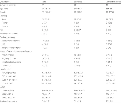
and mean blood sirolimus level was 5.5 ± 2.8 ng/ml (3.5 ± 1.3 ng/ml in the low-dose group vs. 7.7 ± 2.3 ng/ml in the conventional-dose group,p< 0.001). In the low-dose group, the mean trough level of sirolimus was maintained below 5 ng/ml during the treatment period (Additional file1: Figure S1). There were no differences between the low- and conventional-dose groups in age, gender, smoking history, prior treatment, extrapulmonary manifestations, lung func-tion or exercise capacity (Table1). However, more subjects in the low-dose group had TSC (30.0% vs. 0.0%,
p= 0.020). Most subjects in the low-dose group
main-tained low blood trough levels due to adverse events (AEs, 67.5%) or a stable disease course after initial treatment (25.0%) (Additional file2: Table S1). Among patients with
TSC-LAM (n= 6), four received low-dose sirolimus due
to azotaemia (n= 1) and treatment history of the renal
procedure due to AML (n= 3) and remaining two did due
to mucositis.
Changes in lung function
In the whole group, changes in FEV1 significantly
im-proved 12 and 24 months after treatment (ΔFEV1, 3.4 ±
9.3% predicted at 12 months, p= 0.004; 6.9 ± 11.5%
pre-dicted at 24 months,p= 0.007) compared with those
be-fore treatment (ΔFEV1, −4.2 ± 8.2% predicted; Fig. 2a). The changes in DLco also exhibited similar trends after
treatment (ΔDLco, 3.1 ± 7.7% predicted at 12 months,
p= 0.006; 2.4 ± 8.0% predicted at 24 months, p= 0.032;
Fig.2b). By contrast, FVC only showed a numerical
im-provement after treatment (ΔFVC, 3.1 ± 7.7% predicted
at 12 months, p= 0.250; 6.8 ± 14.8% predicted at
24 months,p= 0.582; Fig.2c).
In the low-dose group, FEV1 showed an improving
trend at 12 and 24 months after treatment (ΔFEV1, 4.2 ± 11.6% predicted at 12 months,p= 0.169; 7.2 ± 12.0% pre-dicted at 24 months,p= 0.212) without statistical signifi-cance (Fig. 2a). DLco (ΔDLco, 6.6 ± 14.0% predicted at 12 months,p= 0.145; 6.6 ± 14.0% predicted at 24 months,
p= 0.250) and FVC (ΔFVC, 3.4 ± 10.7% predicted at
12 months,p= 0.283; 2.7 ± 11.1% predicted at 24 months,
p= 0.891) also showed similar trends (Fig.2band c). On the other hands, the conventional-dose group showed
sig-nificant improvements in FEV1 (ΔFEV1, 2.7 ± 7.0%
pre-dicted at 12 months, p= 0.010; 6.6 ± 11.7% predicted at 24 months, p= 0.015; Fig. 2a) and DLco (ΔDLco, 2.8 ±
3.7% predicted at 12 months, p= 0.001; 2.1 ± 2.6%
pre-dicted at 24 months, p= 0.010; Fig. 2b) at 12 and
24 months after treatment; however, only numerical im-provements in FVC were observed after treatment (ΔFVC, 2.2 ± 5.9% predicted at 12 months, p= 0.608; 6.9 ± 6.5% predicted at 24 months,p= 0.233; Fig.2c). There were no differences between the two groups in changes in lung function (FEV1, FVC and DLco) before and after treatment.
Rate of decline in lung function
In the whole group, the rate of decline in FEV1was sig-nificantly reduced after treatment (−0.12 ± 0.47% pre-dicted/month [before] vs. 0.24 ± 0.48% prepre-dicted/month
[after], p= 0.027) compared with before treatment
(Additional file 3: Table S2). The rate of decline in
DLco (−0.33 ± 0.61% predicted/month [before] vs. 0.03
± 0.26% predicted/month [after],p= 0.006) was also re-duced after treatment, but that of FVC was not.
In the low-dose group, the rate of decline in FEV1
showed an improving trend after treatment (−0.08 ±
0.38% predicted/month [before] vs. 0.19 ± 0.51% pre-dicted/month [after], p= 0.264) without statistical
sig-nificance (Additional file 3: Table S2). There were
Table 1Comparison of baseline characteristics between the low-dose and conventional-dose groups
Characteristic Total Low-dose Conventional-dose
Number of patients 39 20 19
Age, years 34.8 ± 8.3 34.0 ± 8.7 35.6 ± 8.1
Female 39 (100.0) 20 (100.0) 19 (100.0)
Smoking
Never 36 (92.3) 19 (95.0) 17 (89.5)
Former 3 (7.7) 1 (5.0) 2 (10.5)
Current 0 (0.0) 0 (0.0) 0 (0.0)
TSC 6 (15.4) 6 (30.0)* 0 (0.0)
Postmenopausal state 2 (5.1) 1 (5.0) 1 (5.3)
Previous treatment
Medroxyprogesterone 14 (35.9) 7 (35.0) 7 (36.8)
LHRH 4 (10.3) 1 (5.0) 3 (15.8)
Bilateral oophorectomy 1 (2.6) 1 (5.0) 0 (0.0)
History of extrapulmonary manifestation
Pneumothorax 24 (61.5) 15 (75.0) 9 (47.4)
Angiomyolipoma 14 (35.9) 9 (45.0) 5 (26.3)
Lymphangioleiomyoma 5 (12.9) 1 (5.0) 4 (21.1)
Chylothorax 3 (7.7) 2 (10.0) 1 (5.3)
Lung function
FEV1, % predicted 67.7 ± 26.4 62.9 ± 27.4 72.3 ± 2.3
FVC, % predicted 86.2 ± 14.9 83.3 ± 13.8 88.9 ± 15.7
DLco, % predicted 51.7 ± 21.8 52.6 ± 23.1 50.9 ± 21.2
FEV1/FVC ratio 66.2 ± 20.8 64.1 ± 2 4.1 68.2 ± 17.6
6MWT
Distance, meter 456.9 ± 103.6 458.4 ± 103.5 455.1 ± 106.9
Initial SaO2, % 97.6 ± 1.7 97.7 ± 1.7 97.6 ± 1.7
Lowest SaO2, % 93.7 ± 4.7 94.5 ± 4.9 92.8 ± 4.5
Sirolimus level, ng/mL 5.5 ± 2.8 3.5 ± 1.3* 7.7 ± 2.3
Data are presented as mean ± SD or number (%)
Abbreviations:TSCtuberous sclerosis complex,LHRHluteinising hormone-releasing hormone,FEV1forced expiratory volume in 1 s,FVCforced vital capacity,DLco diffusing capacity of the lung for carbon monoxide,6MWTsix-minute walk test,SaO2oxygen saturation
*P < .05(compared with conventional-dose group)
Fig. 2Changes in lung function before and after treatment. Changes in FEV1(a), DLco (b) and FVC (c) before and after treatment. Bars and lines
show the mean ± standard error of changes in lung function. Abbreviations: Pre_12M, 12 months before treatment; Post_12M, 12 months after treatment; Post_24M, 24 months after treatment; FEV1, forced expiratory volume in 1 s; DLco, diffusing capacity of the lung for carbon monoxide;
after treatment. However, in the conventional-dose group, the rates of decline in FEV1 (−0.26 ± 0.54% predicted/
month vs. 0.22 ± 0.38% predicted/month, p= 0.024) and
DLco (−0.55 ± 0.58% predicted/month vs. 0.04 ± 0.25%
predicted/month, p= 0.002) were significantly reduced
after treatment (Additional file3: Table S2).
Disease progression
In the whole group, the rate of disease progression, which was defined as any decline in FEV1, decreased after
treat-ment (77% before vs. 33% at 12 months [p= 0.008] vs.
35% at 24 months [p= 0.024]) (Fig. 3a). In the low-dose group, the rate of disease progression showed a trend to-wards decrease after treatment (63% before vs. 43% at 12 months [p= 0.659] vs. 44% at 24 months [p= 0.637]) (Fig. 3b). In the conventional-dose group, however, the rate of disease progression decreased significantly after treatment (100% before vs. 25% at 12 months [p= 0.006] vs. 25% at 24 months [p= 0.021]) (Fig.3c).
Treatment response of extra-pulmonary manifestations Among 18 patients with extra-pulmonary manifesta-tions, 11 (61.1%) were assessed for treatment responses. The median observation time from the initiation of siro-limus to the last CT follow-up during treatment was 2.9 years (range: 1.4–5.8 years; 2.8 [low] vs. 3.1 [conven-tional] years,p= 0.631). In the whole group, five (45.5%) patients showed improvement and 6 (54.5%) showed sta-bility. Comparison of the results between the low and conventional groups were similar; in the low-dose group, improvement and stability were observed in 2 (28.6%)
and 5 patients (71.4%), respectively and in the
conventional-dose group, those were observed in 3 (75.0%) and 1 patients (16.7%), respectively (p= 0.242).
Adverse events
Of all patients, 89.7% experienced AEs, averaging 3.46 AEs per patient (Table2and Additional file4: Table S3).
The most common AE was hypercholesterolaemia (43.6%), followed by stomatitis (35.9%). The rate of AEs in the
low-dose group did not differ from that in the
conventional-dose group (85.0% vs. 94.7%, p= 0.605). Al-though there were no significant differences in AEs between the groups, the most common AE in the low-dose group was stomatitis (50.0%), whereas hypercholesterolaemia was the most common in the conventional-dose group (52.6%). The rate of AEs per patient was also comparable in the two groups (3.70 events per patient in the low-dose group and 3.21 events per patient in the conventional-dose group,
p= 0.406) (Additional file4: Table S3).
Serious adverse events (SAEs) occurred in 17.9% of all subjects, and there was no significant difference in the rate of SAEs between the low- and conventional-dose groups (15.0% vs. 21.0%, p= 0.695) (Table 2). Although there were no significant differences in the rate of AEs between the groups, the most common SAEs were infec-tion (15.0%) in the low-dose group and pneumothorax (10.5%) in the conventional-dose group. There were no deaths during follow-up.
Discontinuation of treatment
Seven patients (17.9%) permanently discontinued treatment due to planned pregnancy (7.7%), AEs (5.1%), or stable dis-ease status (1.1%). Although the overall discontinuation rate was lower in the low dose group (5.0% [low] vs. 31.6% [con-ventional],p= 0.044) than in the conventional dose group, the discontinuation rate due to AEs were not different be-tween two groups (5.0% vs. 5.3%,p= 1.000; Additional file5: Table S4). Of the two patients who discontinued sirolimus due to AEs, one in the low-dose group discontinued be-cause of stomatitis and one in the conventional-dose group discontinued due to stomatitis and urticaria.
Discussion
In our current study, low-dose sirolimus may stabilise lung function decline in patients with LAM. The rates of
Fig. 3Categorical changes in lung function before and after treatment. Changes in disease progression (any decline in FEV1) before and after
treatment in all subjectsa, in the low-dose groupband in the conventional-dose groupc. Bars show the proportion of patients in each group. Abbreviations: Pre_12M, 12 months before treatment; Post_12M, 12 months after treatment; Post_24M, 24 months after treatment; FEV1, forced
decline in lung function and disease progression showed a trend towards improvement after treatment, similar to the conventional-dose group. The frequency of AEs and the rate of treatment discontinuation due to AEs were also similar in the both low and conventional-dose groups.
Ando et al. showed that low-dose sirolimus, achieving trough levels < 5 ng/mL, effectively resolved chylous ef-fusion in seven patients with LAM and improved lung
function (FVC, −101.0 [before] vs. + 190.0 mL/year
[after], p= 0.046; FEV1,−115.4 vs. + 127.8 mL/year, p= 0.015) in nine LAM patients without chylous effusion
[13]. Our findings also showed trends in improvement
of FEV1 and disease progression after sirolimus
treat-ment in the low-dose group, although they were not sta-tistically significant. This is consistent with the results of recent studies [20–22]. Bee et al. demonstrated that lower serum sirolimus level was associated with fewer AEs but not with lower efficacy in FEV1decline in a
pro-spective LAM national cohort [20]. Another mTOR
in-hibitor, everolimus, has recently been used for treatment of LAM in open-label studies; this agent could also sta-bilise lung function and reduce AML and lymphangio-leiomyomas with tolerable safety when administered in low doses [21,22].
Although low-dose sirolimus may be cost-effective and capable of reducing AEs during chronic suppressive treat-ment, this treatment has been thought to be at the expense of inferior efficacy compared with conventional-dose ther-apy [23]. Our results suggesting the efficacy of low dose sir-olimus are consistent with those of previous report [13],
but the improvement in lung function was greater in the conventional-dose group than in the low-dose group. Al-though we did not observe significant improvement in lung function changes after treatment in the low-dose sirolimus group, the rates of disease progression and lung function decline were improved. The absence of statistical signifi-cance in the results from the low-dose group might be due to the small number of subjects or the lower efficacy com-pared with the conventional-dose group.
The proportion of patients with TSC-LAM was greater in the low-dose group than in the conventional-dose group. These findings might be attributed from frequent renal involvement in patients with TSC-LAM, such as AML, renal cyst and renal-cell carcinoma. Because of renal complications, these patients tend to undergo
nephrectomy or embolisation more frequently [24]. In
our cohort, four of six patients with TSC-LAM under-went renal procedures including embolisation and
neph-rectomy. Because sirolimus has the potential to
exacerbate pre-existing or newly occurring renal lesions by causing massive proteinuria, glomerulonephritis or
thrombotic microangiopathy [25], patients with TSC
who already have impaired renal function might prefer low-dose sirolimus therapy. Actually, most patients with TSC-LAM in our cohort received low-dose sirolimus due to a potential risk of renal impairment and showed the stable course of lung function changes without dis-continuation after treatment.
In the present study, the rates of infection, including upper respiratory infection (23.0% vs. 44.0%), urinary tract infection (10.3% vs. 16.0%) and cellulitis (2.5% vs. 12.0%), were lower than those in a study by Bissler et al. [9]. In particular, the incidence of pneumonia requiring hospital-isation in our total cohort was also lower than that in an-other study (7.7% vs. 30.0%) [26]. Although sirolimus did not increase the risk of infection compared with the pla-cebo in phase 3 clinical trial involving patients with LAM [10], considering the increased risk of infection with siroli-mus treatment in transplant patients [27,28], these results were probably due to the effect of low-dose sirolimus. The rates of stomatitis (35.9% vs. 68.0%) and diarrhoea (17.9% vs. 28.0%) were also lower in our study than in a conventional-dose study [9]; however, the rate of hyper-cholesterolaemia in our total subjects was similar to that in patients receiving conventional doses of sirolimus in other studies (43.6% vs. 42.1–52.0%) [9,29].
The AE rates were comparable between the two treat-ment groups in our study, although the rate of stomatitis showed a trend towards being higher in the low-dose group. However, it should be noted that this was a retro-spective study, and most patients who were included in the low-dose group had maintained low doses due to AEs, in-cluding mostly stomatitis. Nevertheless, the majority of pa-tients with AEs continued sirolimus with conservative
Table 2Comparison of adverse events and serious adverse events between the low-dose and conventional-dose groups
Type of event Total Low-dose Conventional-dose
Number of patients 39 20 19
Adverse events 35 (89.7) 17 (85.0) 18 (94.7)
Hypercholesterolaemia 17 (43.6) 7 (35.0) 10 (52.6)
Stomatitis 14 (35.9) 10 (50.0)* 4 (21.1)
URI 9 (23.0) 3 (15.0) 6 (31.6)
Diarrhoea 7 (17.9) 4 (20.0) 3 (15.8)
Headache 5 (12.8) 4 (20.0) 1 (5.3)
Vaginal bleeding 5 (12.8) 1 (5.0) 4 (21.1)
Acneiform lesions 4 (10.3) 3 (15.0) 1 (5.3)
UTI 4 (10.3) 2 (10.0) 2 (10.5)
Serious adverse events 7 (17.9) 3 (15.0) 4 (21.1)
Infectiona 4 (10.3) 3 (15.0) 1 (5.3)
Pneumothorax 3 (7.7) 1 (5.0) 2 (10.5)
Malignancyb 1 (2.3) 0 (0.0) 1 (5.3)
Data are presented as number (%)
Abbreviations:URIupper respiratory infection,UTIurinary tract infection *P < .1(compared with conventional-dose group)
a
Infection included pneumonia (n= 3) and cellulitis (n= 1)
b
therapy, resulting in comparable AE-related discontinu-ation rates between the two groups (5% [low dose] vs. 5.3% [conventional dose],p= 1.000). These findings indicate that the AEs in the low-dose group were not severe enough to cause discontinuation of treatment. Thus, low-dose siroli-mus can be considered tolerable for patients who experi-ence AEs while taking conventional doses.
Our study has some important limitations. First, the number of patients included was relatively small, espe-cially for analysis of efficacy in each group. This is re-lated to the lack of statistical significance in subgroup analysis. However, our results showed that the reduction in lung function before treatment was changed to an in-crease in lung function after treatment in both groups. Second, this was a retrospective study conducted in a single tertiary referral centre; however, the demographic features and lung function of our patients were compar-able to those in other studies. Finally, the proportion of patients with TSC-LAM was higher in the low-dose group than in the conventional-dose group. Neverthe-less, baseline lung function did not differ between the two groups, and a recent study showed that there were no differences in changes of FEV1, DLco and cyst scores between patients with TSC-LAM and those with spor-adic LAM [30]. Therefore, the difference in the propor-tion of patients with TSC-LAM between two groups would not affect the results of lung function analysis.
Conclusions
In conclusion, our data suggest that low-dose sirolimus may stabilise lung function decline in patients with LAM who have moderately impaired lung function, and that low-dose sirolimus may be less effective than conventional-dose ther-apy in preventing lung function decline and disease progres-sion. Based on our results, low-dose sirolimus could be considered a treatment option in patients with LAM, espe-cially those who suffer from intolerable AEs or who have a long-term stable course. Further prospective studies are war-ranted to confirm these findings.
Additional files
Additional file 1:Figure S1.The mean blood trough level of sirolimus during the first two years of treatment. Dots and error bars show the mean ± 95% confidential interval (DOCX 13 kb)
Additional file 2:Table S1.Reasons to maintain low-dose sirolimus treatment. (DOCX 18 kb)
Additional file 3:Table S2.Comparison of the rates of decline in lung function between the low-dose and conventional-dose groups. (DOCX 19 kb)
Additional file 4:Table S3.Comparison of adverse events and serious adverse events between the low-dose and conventional-dose groups. (DOCX 19 kb)
Additional file 5:Table S4.Comparison of reasons for treatment discontinuation between the low-dose and conventional-dose groups (DOCX 13 kb)
Abbreviations
AEs:Adverse events; AML: Angiomyolipoma; DLco: The diffusing capacity of the lung for carbon monoxide; FEV1: Forced expiratory volume in 1 s;
FVC: Forced vital capacity; LAM: Lymphangioleiomyomatosis; mTORC1: Mammalian target of the rapamycin complex 1; SAEs: Serious adverse events; SD: Standard deviation; TSC: Tuberous sclerosis complex
Acknowledgements
We would like to express our deep gratitude to Minkyu Han, a statistician of Department of Clinical Epidemiology and Biostatistics, Asan Medical Center, University of Ulsan College of Medicine, for his valuable advice in the statistical analysis.
Funding
This study was supported by a grant (16–495) from the Asan Institute for Life Sciences, Asan Medical Center, Seoul, Republic of Korea. The funders had no involvement in study design, analysis, interpretation of data or the decision to submit this manuscript.
Availability of data and materials
Any data generated and/or analysed during the current study are available from the corresponding author on reasonable request.
Authors’contributions
HYY and JWS take full responsibility for the content of this manuscript, including its data and analysis. JWS made substantial contributions to the conception and design of the study. HYY and JWS made substantial contributions to the analysis and interpretation of data. HYY and JWS drafted the initial manuscript. All authors read and approved the final manuscript.
Ethics approval and consent to participate
The study was approved by the Asan Medical Center Institutional Review Board (2016–0480).
Consent for publication
Not applicable.
Competing interests
The authors declare that they have no competing interests.
Publisher’s Note
Springer Nature remains neutral with regard to jurisdictional claims in published maps and institutional affiliations.
Author details
1Departments of Pulmonary and Critical Care Medicine, Asan Medical Center,
University of Ulsan College of Medicine, 88, Olympic-Ro 43-Gil, Songpa-Gu, Seoul 05505, Republic of Korea.2Department of Convergence Medicine, Asan Medical Center, University of Ulsan College of Medicine, 88, Olympic-Ro 43-Gil, Songpa-Gu, Seoul 05505, Republic of Korea.
Received: 2 August 2018 Accepted: 30 October 2018
References
1. Henske EP, McCormack FX. Lymphangioleiomyomatosis - a wolf in sheep's clothing. J Clin Invest. 2012;122(11):3807–16.
2. Harari S, Torre O, Cassandro R, Moss J. The changing face of a rare disease: lymphangioleiomyomatosis. Eur Respir J. 2015;46(5):1471–85.
3. Harari S, Torre O, Moss J. Lymphangioleiomyomatosis: what do we know and what are we looking for? Eur Respir Rev. 2011;20(119):34–44. 4. McCormack FX. Lymphangioleiomyomatosis: a clinical update. Chest. 2008;
133(2):507–16.
5. Ballou LM, Lin RZ. Rapamycin and mTOR kinase inhibitors. J Chem Biol. 2008;1(1–4):27–36.
6. Kenerson HL, Aicher LD, True LD, Yeung RS. Activated mammalian target of rapamycin pathway in the pathogenesis of tuberous sclerosis complex renal tumors. Cancer Res. 2002;62(20):5645–50.
8. Goncharova EA, Goncharov DA, Li H, Pimtong W, Lu S, Khavin I, et al. mTORC2 is required for proliferation and survival of TSC2-null cells. Mol Cell Biol. 2011;31(12):2484–98.
9. Bissler JJ, McCormack FX, Young LR, Elwing JM, Chuck G, Leonard JM, et al. Sirolimus for angiomyolipoma in tuberous sclerosis complex or
lymphangioleiomyomatosis. N Engl J Med. 2008;358(2):140–51. 10. McCormack FX, Inoue Y, Moss J, Singer LG, Strange C, Nakata K, et al.
Efficacy and safety of sirolimus in lymphangioleiomyomatosis. N Engl J Med. 2011;364(17):1595–606.
11. Davies DM, de Vries PJ, Johnson SR, McCartney DL, Cox JA, Serra AL, et al. Sirolimus therapy for angiomyolipoma in tuberous sclerosis and sporadic lymphangioleiomyomatosis: a phase 2 trial. Clin. Cancer Res. 2011;17(12): 4071–81.
12. Branco F, Cavadas V, Osorio L, Carvalho F, Martins L, Dias L, et al. The incidence of cancer and potential role of sirolimus immunosuppression conversion on mortality among a single-center renal transplantation cohort of 1,816 patients. Transplant Proc. 2011;43(1):137–41.
13. Ando K, Kurihara M, Kataoka H, Ueyama M, Togo S, Sato T, et al. The efficacy and safety of low-dose sirolimus for treatment of
lymphangioleiomyomatosis. Respir Investig. 2013;51(3):175–83. 14. Gupta N, Finlay GA, Kotloff RM, Strange C, Wilson KC, Young LR,
Taveira-DaSilva AM, Johnson SR, Cottin V, Sahn SA, et al.
Lymphangioleiomyomatosis diagnosis and management: high-resolution chest computed tomography, Transbronchial lung biopsy, and pleural disease management. An official American Thoracic Society/Japanese respiratory society clinical practice guideline. Am J Respir Crit Care Med. 2017;196:1337–48.
15. Wanger J, Clausen J, Coates A, Pedersen O, Brusasco V, Burgos F, Casaburi R, Crapo R, Enright P, Van Der Grinten C. Standardisation of the measurement of lung volumes. Eur Respir J. 2005;26:511.
16. Miller MR, Hankinson J, Brusasco V, Burgos F, Casaburi R, Coates A, Crapo R, Enright P, van der Grinten CP, Gustafsson P, et al. Standardisation of spirometry. Eur Respir J. 2005;26:319–38.
17. Macintyre N, Crapo RO, Viegi G, Johnson DC, van der Grinten CP, Brusasco V, Burgos F, Casaburi R, Coates A, Enright P, et al. Standardisation of the single-breath determination of carbon monoxide uptake in the lung. Eur Respir J. 2005;26:720–35.
18. ATS Committee on Proficiency Standards for Clinical Pulmonary Function Laboratories. ATS statement: guidelines for the six-minute walk test. Am J Respir Crit Care Med. 2002;166:111–7.
19. Therasse P, Arbuck SG, Eisenhauer EA, Wanders J, Kaplan RS, Rubinstein L, et al. New guidelines to evaluate the response to treatment in solid tumors. European Organization for Research and Treatment of Cancer, National Cancer Institute of the United States, National Cancer Institute of Canada. J Natl Cancer Inst. 2000;92(3):205–16.
20. Bee J, Fuller S, Miller S, Johnson SR. Lung function response and side effects to rapamycin for lymphangioleiomyomatosis: a prospective national cohort study. Thorax. 2018;73:369–75.
21. Avdeev S, Makarova M, Merzhoeva Z, Chuchalin A. Treatment with low-dose everolimus in patients with sporadic lymphangioleiomyomatosis (LAM). Eur Respir J. 2014;44:P1519.
22. Mohammadieh AM, Bowler SD, Silverstone EJ, Glanville AR, Yates DH. Everolimus treatment of abdominal lymphangioleiomyoma in five women with sporadic lymphangioleiomyomatosis. Med J Aust. 2013;199:121–3. 23. McCormack FX. Chronic sirolimus therapy for lymphangioleiomyomatosis.
Am J Respir Crit Care Med. 2014;190:1332–3.
24. O'Callaghan FJ, Noakes MJ, Martyn CN, Osborne JP. An epidemiological study of renal pathology in tuberous sclerosis complex. BJU Int. 2004;94: 853–7.
25. Marti HP, Frey FJ. Nephrotoxicity of rapamycin: an emerging problem in clinical medicine. Nephrol Dial Transplant. 2005;20:13–5.
26. Neurohr C, Hoffmann AL, Huppmann P, Herrera VA, Ihle F, Leuschner S, von Wulffen W, Meis T, Baezner C, Leuchte H, et al. Is sirolimus a therapeutic option for patients with progressive pulmonary
lymphangioleiomyomatosis? Respir Res. 2011;12:66.
27. Fisher A, Seguel JM, de la Torre AN, Wilson D, Merchant A, Arora RK, Koneru B. Effect of sirolimus on infection incidence in liver transplant recipients. Liver Transpl. 2004;10:193–8.
28. Alsuwaidi AR, George JA, Almarzooqi S, Hartwig SM, Varga SM, Souid A-K. Sirolimus alters lung pathology and viral load following influenza a virus infection. Respir Res. 2017;18:136.
29. Taveira-DaSilva AM, Hathaway O, Stylianou M, Moss J. Changes in lung function and chylous effusions in patients with lymphangioleiomyomatosis treated with sirolimus. Ann Intern Med. 2011;154:797–805.