R E V I E W
Open Access
Mandibular molar uprighting using
orthodontic miniscrew implants: a
systematic review
Panagiota Magkavali-Trikka
1, Georgios Emmanouilidis
2and Moschos A. Papadopoulos
2*Abstract
The purpose of this systematic review was to identify studies and present the use of miniscrew implants (MIs) as an alternative treatment to mandibular molar uprighting. An electronic search and handsearching were conducted by two independent reviewers to identify relevant articles, published up to January 27, 2017. In order to
methodologically assess the eligible studies, a pilot checklist consisting of 22 items was also implemented. After exclusion of all the irrelevant papers, only 17 studies were included, presenting 27 cases of mandibular molar uprighting in all planes using both direct and indirect force traction by MIs. Regarding the quality evaluation, the mean score of the included studies was 13.2, indicating a rather poor methodology implemented in the majority of the included cases. Due to many advantages, MIs provide a unique treatment alternative and constitute a reliable solution for treating tipped or impacted molars. Regarding the force application, a direct method is simpler, as it requires one MI and a single bracket or button, minimizing the patient’s discomfort and also reducing chair time compared to more complex indirect anchorage. It also eliminates the possibility of unwanted movement of the anchorage unit, which can occur even with indirect anchorage as a result of technical errors. However, direct anchorage has limitations in cases of lingually tipped or rotated molars because a single force may be insufficient to upright the tooth.
Background
A tipped mandibular molar is a frequent situation among orthodontic patients, which usually occurs after premature loss of adjacent teeth leading to the inclin-ation of the molars [1, 2]. Inadequate mandibular arch length, excessive teeth size, loss of the adjacent first molar, premature eruption of the mandibular third molar, and unusually mesial eruption pathway of the sec-ond molar can also cause its partial or total impaction with a reported incidence of 0.03–0.3% of the general population and 2–3% of orthodontic patients [3–5].
Tilted molars can cause numerous problems in a pa-tient’s mouth, especially if a prosthetic rehabilitation is planned. According to Zachrisson, periodontal status can be aggravated, with signs of inflammation, angular bone loss, and an apparent pocket at the mesial surface
of a tipped mandibular molar [6]. In excessive
inclination, overeruption of the antagonist molar, prema-ture contacts, and occlusal interferences impede pros-thetic restoration [7]. However, molar uprighting into its correct position leads to the normalization of the func-tional and periodontal condition [7]. Finally, an impacted mandibular second molar can lead to caries, periodontal disease, or external root resorption of the adjacent first molar [4].
Conventional methods for molar uprighting
Several orthodontic approaches are suggested for man-dibular molar uprighting, such as Australian uprighting spring, cantilever spring, prefabricated Sander spring, helical uprighting spring, NiTi coil spring, push spring appliance, and traction from removable appliances are few of the currently available options [6, 8, 9]. Molar uprighting requires good anchorage control, and subse-quently, a full-arch fixed appliance is necessary. Further-more, ankylosed teeth, dental implants, and extraoral * Correspondence:[email protected]
2Department of Orthodontics, School of Health Sciences, Faculty of Dentistry,
Aristotle University of Thessaloniki, Thessaloniki, Greece Full list of author information is available at the end of the article
appliances could also be effective, enhancing anchorage, and protecting from undesirable tooth movements [8, 10]. Among others, the Uprighter Jet developed by Carano provides a complete control of molar uprighting, minim-izing extrusion, requiring no brackets, and no special pa-tient cooperation [1]. In another uprighting case of an impacted molar, in combination with rapid maxillary ex-pansion (RME), vertical elastic forces were directed from a hook on the RME device to an orthodontic attachment bonded on the tooth to be uprighted. This method saves time, requires no additional anchorage preparation, and appears more physiologic as the force vector is in the direction of normal eruption path [9]. According to Pogrel [11], surgical uprighting of lower second molars is a quick procedure with minimal morbidity and long-term prognosis. Most of the uprighted teeth remained firm with excellent bone formation and periodontal sta-tus after 18 months of follow up [11].
However, conventional treatment methods for molar uprighting have some disadvantages, including extru-sion of the target molar, unwanted reciprocal
move-ment of the anchorage units, need for bulky
appliances, and longer treatment time [1, 2, 12–15]. In order to minimize the first two side-effects, intra-arch stabilization is usually needed [2, 12, 13], which is undertaken through the use of osseointegrated im-plants. Yet, an osseointegrated dental implant is costly, needs sufficient bone space, limiting our choices, and is very difficult to be removed after the treatment. It also requires osseointegration before orthodontic force application, increasing the treatment time [5, 10, 12].
In addition, surgical uprighting should not be consid-ered as a routine method due to the possible pulp necro-sis, ankylonecro-sis, external root resorption, or even rupture during the procedure. After treatment, occlusal equili-bration may be needed, and the post-surgical stability of the tooth may be questionable [4]. Further, the possibil-ity of pulpal calcification and vitalpossibil-ity loss is high [11].
Use of orthodontic miniscrew implants for molar uprighting
The development of orthodontic miniscrew implants (MIs) provided solutions to most of the aforementioned problems. MIs are fabricated from pure titanium or ti-tanium alloy with a diameter of 1–2 mm and length of 8–20 mm [16]. They remain stable during orthodontic treatment with minimum anchorage loss and are more effective than conventional anchorage means [17–19]. Their success rate ranges from 59 to 100% with an aver-age of 86.5% [18–20].
Their attachment to the bone is mechanical with no intent to establish any form of osseointegration [21, 22]. Therefore, after the treatment, when they are no longer
needed, they can be removed through a simple proced-ure, with negligible risks for the patient [23].
This new type of skeletal anchorage is simpler, smaller, less-invasive, and more economical than conventional osseointegrated implants [16]. Moreover, MIs do not re-quire a long interval between placement and force appli-cation since loading can occur immediately after placement [10, 16, 24].
Their main advantage though is their ability to move specific teeth or even the entire quadrants directly, with-out involving other teeth or using inter-arch mechanics. Thus, they eliminate the reaction forces usually applied on the anchor teeth, leading to unwanted tooth move-ment and anchorage loss [5, 14, 15]. Patients are also more satisfied with the more invisible treatment com-pared to conventional methods [10].
MI anchorage is preferable to conventional mechanics when a third molar is in direct contact with the second molar root [4]. In some cases, it is better from a bio-mechanical perspective not to extract the third molar bud, since its extraction can change the center of resist-ance of the second molar and uprighting can be realized with distal crown tipping. This is undesirable when the second molar is planned to be uprighted mostly with mesial root tipping [24].
Materials and methods
In order to find the appropriate articles to be included in this systematic review, Medline was electronically searched via PubMed on January 27, 2017, using the fol-lowing search strategy:
(mini implants OR mini-implants OR screw implants OR miniscrew implants OR mini-screw implants OR mini screw implants OR microscrew implants OR micro-screw implants OR micro screw implants OR microimplants OR micro-implants OR micro implants) AND (orthodont*) AND ((molar) OR (“preprosthetic”) OR (upright) OR (upright*) OR (tipped) OR (tipping))
Random searching on Google Scholar and other data-bases was also held. All types of human studies includ-ing case reports were selected for inclusion. The reference lists of each article eligible for inclusion were also manually reviewed.
case. Finally, a judgment of the total quality of the re-spective case reports was made, based on the following interpretations: low quality when the total score was 0– 11, lower medium quality when the total score was 12– 22, upper medium quality when the total score was 23– 33, and high quality when the total score was 34–44.
Results
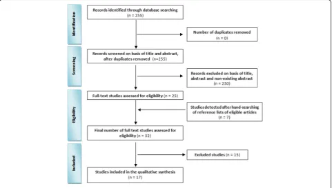
Initially, 255 citations were found in total. Two hundred thirty citations were excluded as non-relevant, and 25 articles remained for further evaluation using their full-text. Through handsearching, 7 more articles were added. After excluding 15 non-relevant articles, only 17 papers remained for inclusion and their findings are re-ported in this systematic review (Fig. 1).
There are two methods of application of orthodontic forces when utilizing MIs: (a) using direct anchorage and (b) using indirect anchorage. Direct anchorage de-scribes situations where the teeth desired to be moved are directly pulled or pushed towards or against the MIs. In contrast, indirect anchorage refers to the stabilization of certain teeth via a rigid connection with the MI and subsequent use of these stabilized anchors to move other teeth in the dental arch.
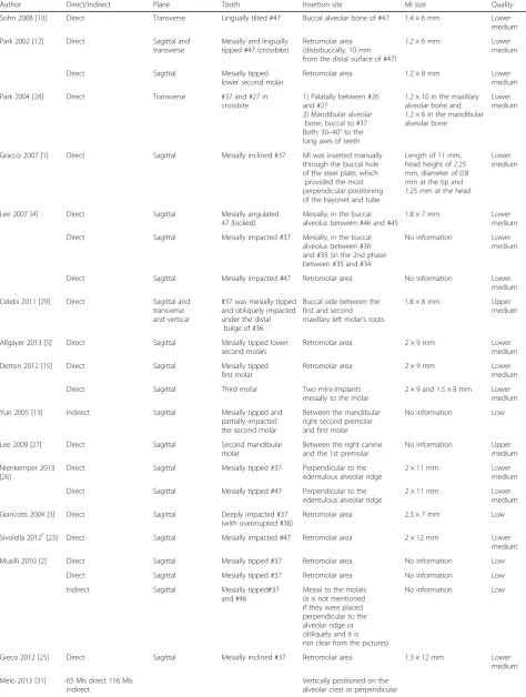
According to the current literature search, 27 cases were documented with both direct and indirect force traction by MIs, although most of them used the direct method (Table 1).
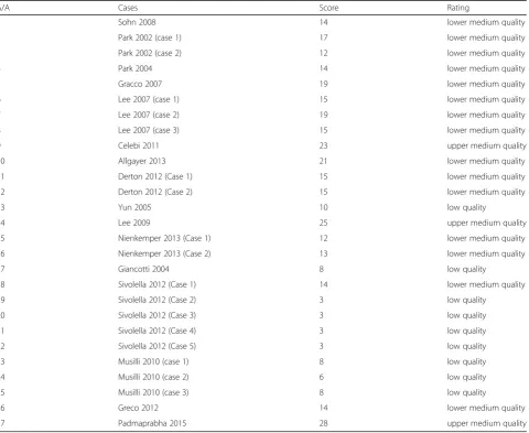
As far as the quality evaluation is concerned, the mean score of the included studies was 13.2, indicating a ra-ther poor methodology implemented in the majority of the included cases. In detail, 9 cases were judged with low quality, 15 with lower medium quality, 3 with upper medium quality while none of the investigated case re-ports was found to present high quality (Table 2).
Molar uprighting using MIs with direct anchorage
Molar uprighting using MIs with direct anchorage was the sole or partial subject of the 15 included papers. One of these 15 papers [2] describes two cases treated with direct use of MIs and one case with indirect use, and this is why it will be discussed again in the following paragraph.
Regarding the use of direct application of forces on the MIs for the correction of molars on thesagittal plane, cases of uprighting all three mandibular molars were found, which were initially either mesially tilted or impacted. In order to treat these cases, several options regarding the in-sertion sites of the MIs were used: (a) in the retromolar area [2–5, 12, 15, 23, 25] (Figs. 2 and 3), (b) vertically in the al-veolar ridge of a mesial edentulous molar site [2, 26] (Fig 4), or (c) mesial to the mandibular molar and between the roots of the adjacent teeth [4, 15, 27] (Fig 5).
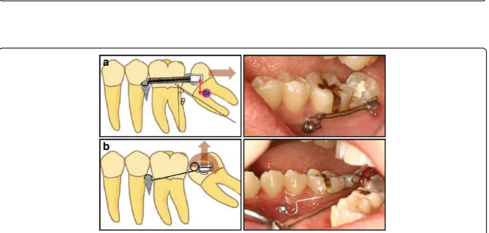
The origin of force differed also, as in several cases uprighting was achieved (a) by using an open or closed coil spring [3, 4] (Fig 6) or (b) by using buttons and elastomeric chains [2, 4, 5, 25, 27]. In a case of a mesially
Table 1Summary of cases included in this review
Author Direct/indirect Plane Tooth Insertion site MI size Quality
Sohn 2008 [10] Direct Transverse Lingually tilted #47 Buccal alveolar bone of #47 1.4 × 6 mm Lower medium
Park 2002 [12] Direct Sagittal and transverse
Mesially and lingually tipped #47 (crossbite)
Retromolar area (distobuccally, 10 mm from the distal surface of #47)
1.2 × 6 mm Lower medium
Direct Sagittal Mesially tipped lower second molar
Retromolar area 1.2 × 8 mm Lower medium
Park 2004 [28] Direct Transverse #37 and #27 in crossbite
1) Palatally between #26 and #27
2) Mandibular alveolar bone, buccal to #37 Both: 30–40° to the long axes of teeth
1.2 × 10 in the maxillary alveolar bone and 1.2 × 6 in the mandibular alveolar bone
Lower medium
Gracco 2007 [1] Direct Sagittal Mesially inclined #37 MI was inserted manually through the buccal hole of the steel plate, which
provided the most perpendicular positioning of the bayonet and tube
Length of 11 mm, head height of 2.25 mm, diameter of 0.8 mm at the tip and 1.25 mm at the head
Lower medium
Lee 2007 [4] Direct Sagittal Mesially angulated 47 (locked)
Mesially, in the buccal alveolus between #46 and #45
1.8 × 7 mm Lower medium
Direct Sagittal Mesially impacted #37 Mesially, in the buccal alveolus between #36 and #35 (in the 2nd phase: between #35 and #34
No information Lower medium
Direct Sagittal Mesially impacted #47 Retromolar area No information Lower medium
Celebi 2011 [29] Direct Sagittal and transverse and vertical
#37 was mesially tipped and obliquely impacted under the distal
bulge of #36
Buccal side between the first and second maxillary left molar’s roots
1.8 × 8 mm Upper medium
Allgayer 2013 [5] Direct Sagittal Mesially tipped lower second molars
Retromolar area 2 × 9 mm Lower medium
Derton 2012 [15] Direct Sagittal Mesially tipped first molar
Retromolar area 2 × 9 mm Lower medium
Direct Sagittal Third molar Two mini-implants mesially to the molar
2 × 9 and 1.5 × 8 mm Lower medium
Yun 2005 [13] Indirect Sagittal Mesially tipped and partially impacted the second molar
Between the mandibular right second premolar and first molar
No information Low
Lee 2009 [27] Direct Sagittal Second mandibular molar
Between the right canine and the 1st premolar
No information Upper medium
Nienkemper 2013 [26]
Direct Sagittal Mesially tipped #37 Perpendicular to the edentulous alveolar ridge
2 × 11 mm Lower medium
Direct Sagittal Mesially tipped #47 Perpendicular to the edentulous alveolar ridge
2 × 11 mm Lower medium
Giancotti 2004 [3] Direct Sagittal Deeply impacted #37 (with overerupted #38)
Retromolar area 2.3 × 7 mm Low
Sivolella 2012*
[23] Direct Sagittal Mesially impacted #47 Retromolar area 2 × 12 mm Lower medium
Musilli 2010 [2] Direct Sagittal Mesially tipped #37 Retromolar area No information Low Direct Sagittal Mesially tipped #37 Retromolar area No information Low
Indirect Sagittal Mesially tipped#37 and #46
Mesial to the molars (it is not mentioned if they were placed perpendicular to the alveolar ridge or obliquely and it is not clear from the pictures)
No information Low
Greco 2012 [25] Direct Sagittal Mesially inclined #37 Retromolar area 1.3 × 12 mm Lower medium
Melo 2013 [31] 65 MIs direct 116 MIs indirect
tilted second molar, buttons were placed on its labial, lingual, and mesial surface, and uprighting force was ap-plied through three elastic chains, which were connected to a MI in the retromolar area [25]. A distalizing and uprighting movement was produced, which avoided un-desirable rotation of the molar [25] (Fig 7). Other treat-ment options included uprighting springs [26], a small cantilever [2], a sequence of archwires and a running
loop [15], and a modified version of the Uprighter Jet called Uprighter Screw [1] (Fig 8).
As regards the direct force application on the MIs for the correction of molars on the transverse plane, two cases were reported [10, 28], where there was a lingually tilted second molar and a lingual crossbite of a second mandibular molar, respectively. In the first case, de-scribed by Sohn, the MI was inserted in the buccal
Table 2Quality assessment of the included case reports-series using the pilot assessment scale
A/A Cases Score Rating
1 Sohn 2008 14 lower medium quality
2 Park 2002 (case 1) 17 lower medium quality
3 Park 2002 (case 2) 12 lower medium quality
4 Park 2004 14 lower medium quality
5 Gracco 2007 19 lower medium quality
6 Lee 2007 (case 1) 15 lower medium quality
7 Lee 2007 (case 2) 19 lower medium quality
8 Lee 2007 (case 3) 15 lower medium quality
9 Celebi 2011 23 upper medium quality
10 Allgayer 2013 21 lower medium quality
11 Derton 2012 (Case 1) 15 lower medium quality
12 Derton 2012 (Case 2) 15 lower medium quality
13 Yun 2005 10 low quality
14 Lee 2009 25 upper medium quality
15 Nienkemper 2013 (Case 1) 12 lower medium quality
16 Nienkemper 2013 (Case 2) 13 lower medium quality
17 Giancotti 2004 8 low quality
18 Sivolella 2012 (Case 1) 14 lower medium quality
19 Sivolella 2012 (Case 2) 3 low quality
20 Sivolella 2012 (Case 3) 3 low quality
21 Sivolella 2012 (Case 4) 3 low quality
22 Sivolella 2012 (Case 5) 3 low quality
23 Musilli 2010 (case 1) 8 low quality
24 Musilli 2010 (case 2) 6 low quality
25 Musilli 2010 (case 3) 8 low quality
26 Greco 2012 14 lower medium quality
27 Padmaprabha 2015 28 upper medium quality
Rating was done according to the following: low quality when the total score was 0–11, lower medium quality for scores 12–22, upper medium quality for scores 23–33 and high quality for scores 34–44
Table 1Summary of cases included in this review(Continued)
Author Direct/indirect Plane Tooth Insertion site MI size Quality
to the buccal face of the alveolar bone
7, 9, or 11 mm in length and 1.3 or 1.6 mm in diameter
Padmaprabha 2015 [30]
Direct Transverse and vertical
Lingually tipped and supraerupted #46
In the interradicular region of #46
1.2 × 6 mm Upper medium
*
alveolar bone and the force was applied through an elastomeric chain to a buccal button for 3 months [10]. In the second case, two MIs were placed in both the mandible and the maxilla in order to treat the crossbite with elastomeric threads attached to buttons, by produ-cing a buccal and intrusive force on the lower molar and a palatal and intrusive force on the upper one, respect-ively [28] (Fig 9).
Direct force application was also used in three cases, where there was a correction of molars in more than one
planes of space. In the first case report [12], a second molar was mesially and lingually tipped and was simultan-eously in crossbite with the maxillary molar. In this spe-cific case, the MI was placed distobuccally in the retromolar region, and for the following 3 months, an uprighting force was applied to a lingual button on the mesiolingual surface of the second mandibular molar, through an elastomeric thread. In the second case [29], after the distal uprighting of a mesially tipped and ob-liquely impacted molar via a removable appliance and an Fig. 2Uprighting of #37 with a miniscrew implant and elastomeric chain.aA miniscrew positioned in the retromolar area with an elastomeric chain between the screw and the molar; uncontrolled tipping.bInitial radiograph at the area of #37.cFinal radiograph after the uprighting of #37, with the miniscrew placed distally and an implant in the site of #36.dOcclusal view of uprighted #37. (From Musilli et al. [2], with kind permission of Progress in Orthodontics)
Fig. 4Preprosthetic uprighting of #37 with a miniscrew implant and an uprighting spring.aActivation of the uprighting spring with a mesial eccentric V-bend.b,cA mini-implant with an incorporated bracket abutment placed in the alveolar ridge as an attachment of the uprighting spring.d,eAfter 5 months of molar-uprighting treatment, no extrusion has taken place. (From Nienkemper et al. [26], with kind permission of Journal of Clinical Orthodontics)
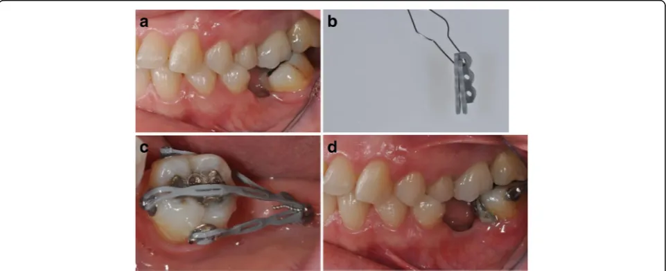
uprighting spring, a MI was inserted buccally between the roots of the maxillary molars. Force was applied via an elastic chain placed between the buccal MI and a wire on the occlusal surface of the mandibular molar in order to upright the tooth buccally and vertically (Fig 10). The third case [30] describes the simultaneous uprighting and intrusion of a lingually tipped and supraerupted lower first molar. The clinicians used elastomeric chains connecting a lingual attachment on the molar and a MI placed in the interradicular region. Buccolingual uprighting of 2.3 mm and intrusion of 1.8 mm was achieved in 45 days.
Molar uprighting using MIs with indirect anchorage Two out of the 17 included papers, which reported only two cases, assessed molar uprighting using MIs with indirect anchorage. As mentioned above, the paper of Musilli et al. [2] presents cases treated with both types of anchorage and will be described again in the next paragraph.
The first case of a second mandibular molar tilted on the sagittal plane was presented by Yun et al. [13]. A MI placed between the second premolar and the first molar was connected to the tooth by a rigid Fig. 6Uprighting of an impacted #37 with miniscrew implant and a closed coil spring tied to an orthodontic bracket.aA 7-mm titanium miniscrew inserted in the retromolar area and loaded with 50-g force on nickel-titanium closed coil spring. After the extrusion of the crown, the tooth was uprighted with fixed sectional appliance.bProgress radiographs during the treatment (from Giancotti et al. [3], with kind permission of American Journal of Orthodontics and Dentofacial Orthopedics)
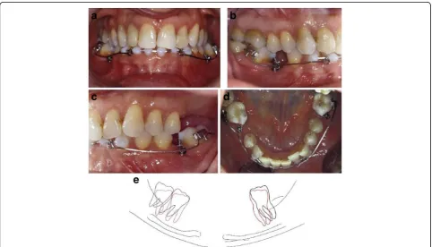
stainless steel wire serving as indirect anchorage. In addition, a peerless single tube was bonded on the first molar, and a metal button was attached on the second molar, in order to achieve appropriate traction with an uprighting spring. In the second case pre-sented by Musilli [2], two mesially tilted molars (teeth #37 and #46) were simultaneously uprighted in 9 weeks using a long cantilever on each side. Two MIs were used indirectly, locking the molars with
steel ligature in order to prevent molar’s extrusion and distal tipping. Thus, the uprighting is mainly achieved by mesial root tipping (Fig 11).
Molar uprighting using MIs both with direct and indirect anchorage
Finally, molar uprighting using MIs both with direct and indirect anchorage was the subject of the one remaining paper.
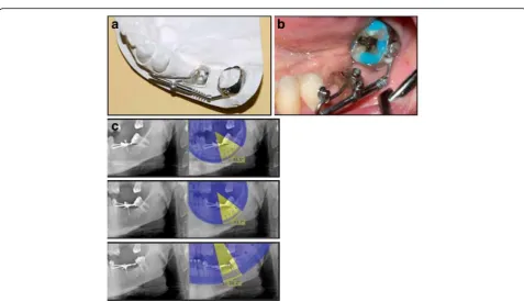
Fig. 8Uprighting of mesially inclined #37 with a modified Uprighter Jet.aUprighter Screw consists of a molar band with a welded lingual button that allows crown rotation when a force is applied; a .036 in. tube positioned parallel to the occlusal plane; a wire with a bayonet bend on the mesial end, curving back distally from the tube, and a loop on the distal end that is screwed to the molar band; an adjustable screw clamp; and a 150-g nickel titanium open coil spring.bAfter 2 months of uprighting.cRadiographs with Uprighter Screw in place.aAt the time of appliance insertion, showing 46.5° inclination of the second molarʼs long axis relative to miniscrew.bAfter 2 months of uprighting, showing 43.7° inclination.cAfter 5 months of uprighting, showing 16.2° inclination (from Gracco et al. [1], with kind permission of Journal of Clinical Orthodontics)
Fig. 9Uprighting #27 and #37 with miniscrew implants, elastomeric threads, and a temporary bite plane.aForce system used for molar uprighting with mini implants.bElastomeric thread which was attached from mini implants to buttons bonded to occlusal surfaces of left upper
There was one clinical trial included in the current re-view in which 181 MIs were inserted in 102 patients and were used both as a direct and indirect anchorage for lower molar uprighting [31]. With regard to the direct anchorage, 65 MIs were inserted in 31 patients using
two different options. The first option included two ver-tically inserted MIs in the edentulous alveolar crest area and a bracket that was bonded on a resin uniting the two MIs. A segment of wire from this bracket to the molar was activated to realize the uprighting. The Fig. 10Uprighting of a mesially tipped #37 with miniscrew implants, a modified removable appliance with an uprighting spring, and an
elastomeric power chain.aA removable appliance with an incorporated uprighting spring (0.8 mm of SS wire) that provoked the distal uprighting of the #37.bThe buccal mini screw was inserted in the maxilla in order to initiate buccal and vertical uprighting of the lower left second molar (from Celebi et al. [29], with kind permission of Journal of Medical Cases)
Fig. 11Uprighting of mesially tipped #37 and #46 with miniscrew implant and long cantilevers. The cantilevers generate molar uprighting, molar extrusion, and intrusion of the anterior teeth. Ligature between miniscrew and molar is used to control the extrusive force and the distal crown movement of each molar. Thus, the uprighting is mainly achieved by mesial root tipping.aThe beginning of uprighting of #37 and #46 with long cantilevers, from the molar to the anterior teeth, and a screw mesial to the molar.bLateral view at the initiation stage of uprighting of #46.
second option included the use of a cantilever directly activated on a MI that was inserted perpendicular to the buccal surface of the alveolar bone. With re-gard to the indirect anchorage, 116 MIs were inserted in the buccal surface of the alveolar bone of 71 patients and were connected via a segment of stainless steel wire with the canine and premolars. Then, a single or a double cantilever system was used to upright the molar. According to the au-thors, MIs showed high success rates in both an-chorage methods with a slight superiority of the direct anchorage. Only 18 MIs failed, including 15 that were used as indirect anchorage and 3 that were used as direct anchorage.
Direct vs. indirect anchorage
According to Lee et al. [4], direct MI for molar upright-ing is simpler, as it requires one MI and a supright-ingle bracket or button attachment, minimizing patient’s discomfort and also reducing chair time compared to more complex indirect anchorage. Furthermore, direct MI connection with the target tooth eliminates the possibility of un-wanted movement of the anchorage unit, which can occur even with indirect MI anchorage as a result of technical errors. However, direct anchorage has some limitations and especially in cases of lingually tipped or rotated molars because a single force may be insufficient to upright the tooth. Usually, such cases require a se-quential application of different force systems and re-peated changes of appliances. Lee et al. also report that direct MI application is not indicated in cases of ex-truded molars since the force system lacks an intrusive component [4].
Discussion
This systematic review presents evidence from 17 stud-ies that included a total of 27 cases of mandibular molar uprighting. From these 17 studies, 16 were case reports/ series and only 1 was a clinical trial.
A common point of these studies is that mandibular molar uprighting is a frequent and complicated proced-ure, which requires good anchorage control. Even a small amount of anchorage loss can result in adverse ef-fects on other tooth units, extrusion of the molar, or a compromised outcome. The introduction of MIs as orthodontic anchorage auxiliaries provided orthodontists with a very significant tool that will help them among others to upright molars easier, faster, with less side-effects, and less inconvenience for the patient. Moreover, in patients with many missing teeth or with periodontal compromised teeth, when conventional full-arch anchor-age cannot be applied, MIs provide a unique treatment alternative to molar uprighting.
The present review incorporated an innovative pilot-formed checklist in an effort to evaluate the method-ology followed in each of the included case reports/ series. Although the use of the aforementioned tool is not evidence-based, it seems to provide a brief yet ad-equate quality analysis in matters of case reports. Ac-cording to the results of the corresponding checklist, most of the cases examined were judged with rather low quality.
Conclusions
This paper presents a variety of clinical applications of MIs in mandibular molar uprighting in all three planes of space, both with direct and indirect anchorage. Due to numerous advantages, MIs seem to constitute a reli-able solution for treating tipped or impacted molars. However, since the majority of the included studies were case reports/series, which were also judged with rather low quality, the outcomes of the respective studies should be interpreted with caution and probably cannot be generalized to the average patient with similar dental malocclusions.
Acknowledgements
We thank Mr. Vasileios F. Zymperdikas, Resident, Department of Orthodontics, Faculty of Dentistry, School of Health Sciences, Aristotle University of Thessaloniki, for his invaluable assistance regarding the quality assessment of the case reports/series.
Authors’contributions
MAP was responsible for the topic’s conception, correction of the draft, and guidance of the flow of the subject matter. PM and GE collected, interpreted, and analyzed the data; drafted the article; and critically revised them. All authors read and approved the final manuscript.
Competing interests
The authors declare that they have no competing interests.
Publisher’s Note
Springer Nature remains neutral with regard to jurisdictional claims in published maps and institutional affiliations.
Author details
1Hamdan Bin Mohammed College of Dental Medicine, Mohammed Bin
Rashid University of Medicine and Health Sciences, Dubai, United Arab Emirates.2Department of Orthodontics, School of Health Sciences, Faculty of Dentistry, Aristotle University of Thessaloniki, Thessaloniki, Greece.
Received: 22 May 2017 Accepted: 5 December 2017
References
1. Gracco A, Lombardo L, Cozzani M, Siciliani G. Uprighting mesially inclined mandibular second molars with a modified Uprighter Jet. J Clin Orthod. 2007;41:281–4.
2. Musilli M, Marsico M, Romanucci A, Grampone F. Molar uprighting with mini screws: comparison among different systems and relative biomechanical analysis. Prog Orthod. 2010;11:166–73.
3. Giancotti A, Arcuri C, Barlattani A. Treatment of ectopic mandibular second molar with titanium miniscrews. Am J Orthod Dentofac Orthop. 2004;126:113–7. 4. Lee KJ, Park YC, Hwang WS, Seong EH. Uprighting mandibular second
molars with direct miniscrew anchorage. J Clin Orthod. 2007;41:627–35. 5. Allgayer S, Platcheck D, Vargas I, Loro R. Mini-implants: mechanical resource
6. Zachrisson BU, Bantleon HP. Optimal mechanics for mandibular molar uprighting. World J Orthod. 2005;6:80–7.
7. Ruellas A, Pithon M, Santos R. Miniscrew-supported coil spring for molar uprighting: description. Dental Press J Orthod. 2013;18:45–9.
8. Shellhart WC, Moawad M, Lake P. Case report: implants as anchorage for molar uprighting and intrusion. Angle Orthod. 1996;66:169–72.
9. Prashanth CS, Dharma RM, Akshai Shetty KR. Molar uprighting simplified. Int J Cont Dent. 2010;1:43–6.
10. Sohn D, Lee J, An K. Minor tooth movements using microimplant anchorage: case reports. Implant Dent. 2008;17:32–9.
11. Pogrel MA. The surgical uprighting of mandibular second molars. Am J Orthod Dentofac Orthop. 1995;108:180–3.
12. Park HS, Kyung HM, Sung JH. A simple method of molar uprighting with micro-implant anchorage. J Clin Orthod. 2002;36:592–6.
13. Yun SW, Lim WH, Chun YS. Molar control using indirect miniscrew anchorage. J Clin Orthod. 2005;39:661–4.
14. Leung M, Lee T, Rabie A, Wong R. Use of miniscrews and miniplates in orthodontics. J Oral Maxillofac Surg. 2008;66:1461–6.
15. Derton N, Perini A, Mutinelli S. Mandibular molar uprighting using mini-implants: different approaches for different clinical cases—two case reports. Orthodontics (Chic). 2012;13:138–45.
16. Papadopoulos MA, Tarawneh F. The use of mini-screw implants for temporary skeletal anchorage in orthodontics: a comprehensive review. Oral Surg Oral Med Oral Pathol Oral Radiol Endod. 2007;103:e6–15.
17. Papadopoulos MA, Papageorgiou SN, Zogakis IP. Clinical effectiveness of orthodontic miniscrew implants: a meta-analysis. J Dent Res. 2011;90:969–76. 18. Papadopoulos MA, Papageorgiou SN. Current evidence on clinical
performance of miniscrew implants in orthodontic treatment. Semin Orthod. 2013;19:162–73.
19. Papadopoulos MA, Papageorgiou SN, Zogakis IP. Success rates and risk factors of orthodontic miniscrew implants used as temporary anchorage devices for orthodontic purposes. In: Papadopoulos MA, editor. Skeletal anchorage in orthodontic treatment of class II malocclusion: contemporary applications of orthodontic implants, miniscrew implants and mini plates. Edinburgh: Elsevier, Mosby; 2014. p. 258–73.
20. Papageorgiou SN, Zogakis IP, Papadopoulos MA. Failure rates and associated risk factors of orthodontic miniscrew implants: a meta-analysis. Am J Orthod Dentofac Orthop. 2012;142:577–95.
21. Mizrahi E, Mizrahi B. Mini-screw implants (temporary anchorage devices): orthodontic and pre-prosthetic applications. J Orthod. 2007;34:80–94. 22. Polyzois I, Leonard G, Synodinos P. Biological principles and biomechanical
considerations of implants, miniplates and miniscrew implants. In:
Papadopoulos MA, editor. Skeletal anchorage in orthodontic treatment of class II malocclusion: contemporary applications of orthodontic implants, miniscrew implants and mini plates. Edinburgh: Elsevier, Mosby; 2014. p. 29–33. 23. Sivolella S, Roberto M, Bressan P, Bressan E, Cernuschi S, Miotti F, Berengo
M. Uprighting of the impacted second mandibular molar with skeletal anchorage. In: Bourzgui F, editor. Orthodontics: basic aspects and clinical considerations. Rijeka: In Tech; 2012.
24. Muggiano F, Giannantoni I, Anastasi G, Giorgia C, Quaranta A. TADs versus traditional devices and tecniques in lower second molar uprighting procedures. Webmed Central ORTHODONTICS. 2014;5:WMC004494. https:// doi.org/10.9754/journal.wmc.2014.004494.
25. Greco M, Meddis V, Giancotti A. The G-chain and miniscrew anchorage: simple mechanics for molar uprighting. J Clin Orthod. 2012;46:24–5. 26. Nienkemper M, Pauls A, Ludwig B, Wilmes B, Drescher D. Preprosthetic
molar uprighting using skeletal anchorage. J Clin Orthod. 2013;47:433–7. 27. Lee KJ, Joo E, Yu HS, Park YC. Restoration of an alveolar bone defect caused by
an ankylosed mandibular molar by root movement of the adjacent tooth with miniscrew implants. Am J Orthod Dentofac Orthop. 2009;136:440–9. 28. Park HS, Kwon OW, Sung JH. Uprighting second molars with micro-implant
anchorage. J Clin Orthod. 2004;38:100–3.
29. Celebi A, Gelgor I, Catalbas B. Correction of mesially impacted lower second molar. Journal of Medical Cases. 2011;2:236–9.
30. Padmaprabha BP, Ponnambathayil SA, Aynipully H, Vinod M, Reghunathan DP. A precise method of measuring simultaneous intrusion and uprighting of mandibular molar using denta scan: a case report. J Clin Diagn Res. 2015; 9:ZD01–3.