R E S E A R C H A R T I C L E
Open Access
Reduction and functional outcome of open
reduction plate fixation versus minimally
invasive reduction with percutaneous screw
fixation for displaced calcaneus fracture: a
retrospective study
Ye Peng
†, Jianheng Liu
†, Gongzi Zhang, Xinran Ji, Wei Zhang, Lihai Zhang
*and Peifu Tang
*Abstract
Background:Minimally invasive surgery has become popular because of the lower incidence of wound complications. However, achieving an anatomic reduction that provides a satisfactory outcome is difficult using minimally invasive surgery. Our study aimed to evaluate the reduction and clinical outcomes of closed reduction and percutaneous fixation treatment using a closed reduction traction device for displaced intra-articular calcaneal fractures compared with traditional open reduction plate fixation using an extended lateral approach.
Methods:A total of 40 patients and 45 feet with calcaneus fractures from 2012 to 2016 were studied. The open reduction plate fixation group (24 feet) was compared to the closed reduction percutaneous fixation group (21 feet) with a traction device. The reduction assessments included length, width, height, Bohler’s angle, Gissane’s angle, and varus or valgus angle before and after surgery. The clinical outcomes included the American Orthopaedic Foot and Ankle Society hindfoot score and the visual analog score for pain, length of stay, and complication rate.
Results:The patients were followed up for an average of 16.53 ± 3.95 months. No significant differences in
reduction were observed between the open and closed groups (P> 0.05). The American Orthopaedic Foot and
Ankle Society scores of the two groups were 80.29 ± 6.15 and 83.62 ± 6.95 (open versus closed) (P= 0.0957). The visual analog scores of the open and closed groups were 1.50 ± 1.22 and 0.81 ± 0.87 (P= 0.0364). The lengths of stay in the open and closed groups were 9.63 ± 2.72 days and 6.71 ± 1.85 days (P= 0.0002). The complication rates of the open and closed groups were 20.8% (5/24) and 4.8% (1/21) (P< 0.0001).
Conclusions:The closed reduction percutaneous fixation with traction device method may provide equivalent reduction results and superior outcomes for the length of stay, VAS score, and complication rate for displaced intra-articular calcaneal fractures.
Keywords:Minimally invasive, Calcaneus fracture, Percutaneous fixation
© The Author(s). 2019Open AccessThis article is distributed under the terms of the Creative Commons Attribution 4.0 International License (http://creativecommons.org/licenses/by/4.0/), which permits unrestricted use, distribution, and reproduction in any medium, provided you give appropriate credit to the original author(s) and the source, provide a link to the Creative Commons license, and indicate if changes were made. The Creative Commons Public Domain Dedication waiver (http://creativecommons.org/publicdomain/zero/1.0/) applies to the data made available in this article, unless otherwise stated.
* Correspondence:zhanglihai74@qq.com;tangpf301@126.com †Ye Peng and Jianheng Liu contributed equally to this work.
Introduction
Calcaneus fracture is a common fracture with an incidence of approximately 2% of all fractures and is the most com-mon tarsal fracture (> 60%) [1]. Approximately 70% of cal-caneus fractures are intra-articular fractures [2]. The gold standard treatment for intra-articular fractures is open re-duction internal fixation using an extended lateral ap-proach. However, many calcaneus fractures are associated with severe soft tissue injuries, which increase the risks of skin necrosis and infections, particularly in patients who smoke or have diabetes [3–5]. Surgeons have opted for less invasive techniques to reduce risks associated with per-forming the lateral extensile approach. For recent years, minimally invasive percutaneous fixation has become in-creasingly popular [6–8] because the minimally invasive method was associated with lower complication rates, shorter hospital stays, and more rapid healing [8,9]. How-ever, minimally invasive surgical techniques include limited exposure, which makes achieving a satisfactory closed re-duction technically demanding and difficult to accomplish [10]. Many surgeons have questioned the quality of reduc-tions achieved with minimally invasive techniques. In con-trast, many open reduction surgeons have started to question the value of precise open reduction techniques be-cause of the high rates of posttraumatic arthritis, chronic pain, postop fibrosis, and loss of subtalar function.
Many scholars have been working on the minimally in-vasive treatment of calcaneus fixations. Cao et al. [11] re-ported that a small lateral wall incision and minimally invasive plate fixation showed a good-to-excellent rate of
93.94% but did not report the wound complication rates. Battaglia et al. [12] and Dayton et al. [13] reported that with external fixation for calcaneus fractures, 18 of the 50 ankles (36%) developed arthritic changes in the lower ankle and 3 with sinus tarsi syndrome. Those results might be associated with the unsatisfactory reduction. None of them reported the reduction details or the results of reduction. Very few comparative studies have reported on open reduction plate fixation versus closed reduction percutaneous fixation and their functional outcomes.
To address this problem, we need a minimally invasive method to achieve a precise reduction. Therefore, we fashioned two types of percutaneous closed reduction devices (Fig.1) to provide traction for the subtalar joints. On the one hand, we can achieve a satisfactory reduc-tion using a tracreduc-tion device and surgical technique. On the other hand, this reduction device is a closed reduc-tion tool that will not affect the wound or increase the complication rate. After satisfactory reduction, percutan-eous fixation can be accomplished according to standard procedures. In this study, we attempted to compare re-ductions via X-ray radiographs and AOFAS, VAS, com-plications of the short-term outcome and function of open reduction plate fixation versus closed reduction percutaneous fixation.
Materials and methods
We retrospectively reviewed 40 cases of displaced intra-articular calcaneal fractures affecting 45 feet from June 2012 to December 2016. This study was approved
by the People’s Liberation Army General Hospital Insti-tutional Review Board. The hospital is a level 1 trauma center. The inclusion criterion was patients who under-went surgical treatment of a unilateral calcaneal intra-articular closed fracture without other associated fractures, and the exclusion criteria were patients who were treated conservatively and the lack of surgical con-ditions. The ICD-10 was used to identify the patients. In total, 23 patients were male, and 17 patients were fe-male, with an average age of 43.7 years (range 18–67). The patients were separated into a closed reduction per-cutaneous group and an open reduction internal fixation group. The open reduction group operation was per-formed at 7–10 days after injury, after the swelling had subsided, and the closed reduction group operation was performed at 1–3 days after injury. The waiting days were accounted in the bed stay. All of the patients were discharged 2–5 days after surgery, depending on the situation of the wound. The surgery times of the two groups were recorded. A single senior orthopedic sur-geon performed the surgeries. Smoking and diabetes sta-tus were recorded. X-rays and CT scans were obtained before the surgery. The VAS score was recorded on post-operative day 1 by the surgeon. The open reduction plate fixation group included calcaneus fractures of 24 feet: 11 of these were Sanders type II, 10 of these were Sanders type III, and 3 of these were Sanders type IV. The closed reduction percutaneous fixation group in-cluded calcaneus fractures of 21 feet: 12 of these were Sanders type II, 7 of these were Sanders type III, and 2 of these were Sanders type IV. The patient characteris-tics are shown in Table1.
Lateral and axial X-ray views were obtained before the surgery, and the length, width, height, Bohler’s angle, Gissane’s angle, and varus or valgus angle of the calca-neus were recorded before and after the surgery accord-ing to these views. The measurement method used was in accordance with a previous study [14]. The varus angle was defined as a positive value, and the valgus angle as a negative value. The measurements obtained from the X-ray images were determined by a senior at-tending orthopedic surgeon. The AOFAS hindfoot score was utilized for clinical outcome assessment. The AOFAS hindfoot score was recorded at the final
follow-up, whereas the visual analog scores for pain were recorded after surgery. The length of stay was recorded during the hospitalization, and the complication rate was recorded separately. The complications included early complications (i.e., superficial infections, deep in-fections, and wound necrosis) and late complications (i.e., posttraumatic arthritis, stiffness, chronic pain, fix-ation failure, lateral impingement, and joint penetrfix-ation).
Implants
The calcaneus locking plate system was selected for open reduction plate fixation. A 4.0-mm-diameter can-nulated screw and a 7.0-mm-diameter cancan-nulated screw and a high-strength injectable calcium phosphate graft were selected for closed reduction percutaneous fixation. A 7.0-mm-diameter cannulated screw was also im-planted and removed to create a bone tunnel for reduc-tion (Figs.2and3).
Standard operative technique
The extensile lateral approach used was similar to the previously referenced technique [15]. For closed reduc-tion percutaneous fixareduc-tion, the patient was posireduc-tioned in the prone position. After surgical draping, a localization K-wire on the skin was positioned perpendicular to the subtalar joint on the lateral view. Two K-wires were sep-arately placed to penetrate perpendicularly to the calca-neus axis in the talus neck and into the calcacalca-neus tuberosity. In some more comminuted cases (such as Sanders type IV), the front traction K-wire should be im-planted in both the tibia and the navicular bone instead of the talus bone. Subsequently, we assembled the trac-tion device on both sides of the calcaneus and gradually initiated traction with the pair of devices simultaneously. The traction was monitored by lateral views to confirm the subtalar joint was distracted. The length of the calca-neus was reduced, and the presence of the subtalar joint gap to elevate the compressed articular surface and lat-eral wall was verified. Then, an axial view of the calca-neus was obtained to permit calcacalca-neus varus and valgus adjustment by retracting the unilateral or bilateral trac-tion device. Additrac-tionally, the width was reduced by com-pression on both sides of the calcaneus applied by the assistant’s fists as monitored on the axial view. Next, an
Table 1Patient characteristics
ORPF CRPF
Gender 4 females/20 males 2 females/19 males
Age, years 39.2 (range, 16–65) 40.8 (range, 23–60)
Smoker,n(%) 7/24 (29.2%) 5/21 (24%)
Diabetes,n(%) 2/24 (8.3%) 1/21 (4.7%)
Sanders classification 11 type II/10 type III/3 type IV 12 type II/7 type III/2 type IV
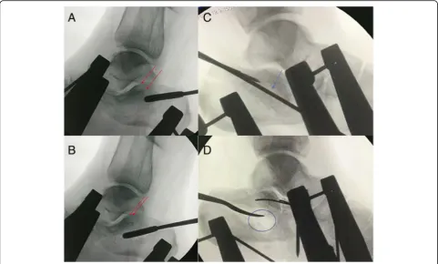
elevator bone tunnel could be established near the Achilles tendon attachment to inferior of the com-pressed subtalar joint by implanting and then removing the 7.0-mm cannulated screw from posterior to anterior. Through the bone tunnel, a smooth periosteal elevator may be used to elevate the central compressed articular surface under lateral view monitoring (Fig. 4). Through the established tunnel, the lateral and central surfaces of the subtalar joint may be reduced and temporarily fixed using a 4.0-mm cannulated screw guide wire percutan-eously. When subtalar joint reductions were completed and assessed by lateral and axial views, a 4.0-mm cannu-lated screw was implanted for subtalar articular fixation. Then, calcium phosphate cement was injected through the tunnel to the bone defect space, and a 7.0-mm head-less cannulated screw was fixed through the reduction tunnel along the calcaneal axis (Fig.5). Finally, the wound was closed with sutures, and a bandage was applied.
Postoperative management
Full exercise of the foot in bed was permitted the next day. Non-weight-bearing ambulation was begun from the second day until 4–6 weeks. After 4 weeks, lateral and axial view X-rays were obtained. Weight-bearing from a tolerated to full weight-bearing was permitted at 6–8 weeks after surgery. After 8 weeks, the patients were
allowed to return to light work (after 10 weeks for medium work and 12 weeks for heavy work). All of the patients were followed up for more than 1 year (average 16.53 ± 3.95 months (range, 12 to 27)).
Statistical analysis
Paired Student’s T tests were used to assess the reduc-tion, length of stay, and scores for the different
treat-ment groups. Chi-square tests were applied for
complication rates. Statistical significance was defined as
P< 0.05. The SPSS statistical software package for Win-dows (22.0) was used for the statistical analysis.
Results
Reduction assessment
Postoperatively, lateral and axial views were obtained to assess the reduction. We analyzed length, width, height, varus, and valgus angles for the calcaneus morphology reduction, and Bohler’s and Gissane’s angles for articular surface reduction.
to 9.79) cm preoperatively to 7.91 ± 0.69 (range, 6.79 to 8.93) cm postoperatively.
The open reduction plate fixation group width was re-stored from 4.60 ± 0.48 (range, 3.20 to 6.40) cm pre-operatively to 3.93 ± 0.46 (range, 3.11 to 4.92) cm postoperatively. The reduction percutaneous fixation group width was restored from 4.24 ± 0.52 (range, 3.24
to 4.99) cm preoperatively to 3.84 ± 0.48 (range, 3.11 to 4.57) cm postoperatively.
The open reduction plate fixation group height was re-stored from 4.05 ± 0.59 (range, 3.40 to 4.79) cm preopera-tively to 4.09 ± 0.51 (range, 3.19 to 4.72) cm postoperapreopera-tively. The reduction percutaneous fixation group height was re-stored from 4.05 ± 0.36 (range, 3.40 to 4.55) cm preopera-tively to 4.21 ± 0.56 (range, 3.35 to 4.83) cm postoperapreopera-tively.
The open reduction plate fixation group Bohler’s angle was restored from 1.60 ± 25.94° (range, −44.00 to 35.50°) preoperatively to 19.97 ± 9.48° (range, 5.90 to 41.00°) post-operatively. The reduction percutaneous fixation group Boh-ler’s angle was restored from 1.63 ± 13.55° (range,−13.11 to 38.70°) preoperatively to 21.70 ± 10.33° (range, 11.7 to 47.3°) postoperatively.
The open reduction plate fixation group Gissane’s angle was restored from 118.57 ± 12.21° (range, 100.20 to 144.20°) preoperatively to 116.51 ± 10.61° (range, 101.7 to 140.00°) postoperatively. The reduction percutaneous fix-ation group Gissane’s angle was restored from 122.10 ± 14.95° (range, 84.40 to 135.50°) preoperatively to 120.18 ± 16.76° (range, 91.40 to 148.10°) postoperatively.
The open reduction plate fixation group varus or val-gus angle was restored from 1.81 ± 4.20° (range, −8.00 to 11.00°) preoperatively to 1.52 ± 2.35° (range,−2.00 to 8.00°) postoperatively. The reduction percutaneous fix-ation group Gissane’s angle was restored from 3.30 ± 5.17° (range, −8.00 to 13.00°) preoperatively to 1.41 ± 1.59° (range,−1.00 to 4.00°) postoperatively.
The reduction measurements are shown in Table2. The average surgery times were 87.00 ± 9.78 min for the open reduction internal fixation group and 85.86 ± 11.17 min for the closed reduction percutaneous fixation group (P = 0.614). There was no significant difference between the two groups.
Clinical outcome results
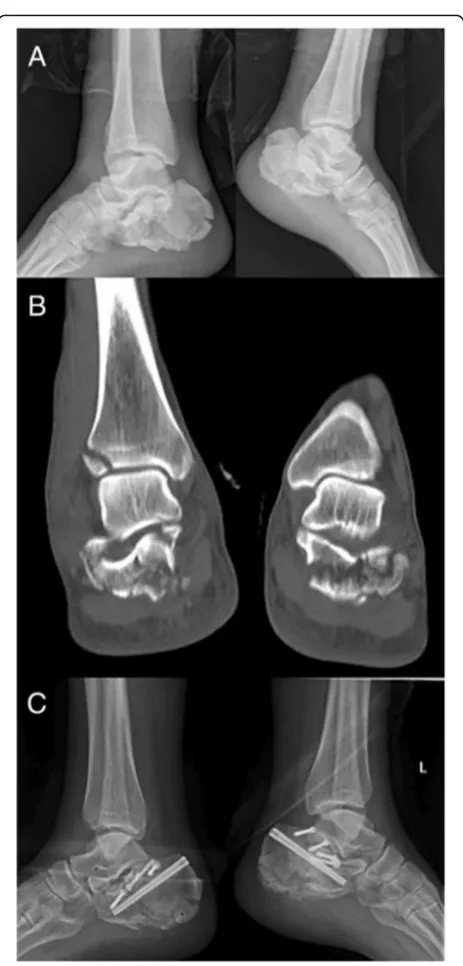
The patients were followed up for an average of 16.53 ± 3.95 months (range, 12 to 27). According to the AOFAS ankle-hindfoot score, the open reduction plate fixation group score was 80.29 ± 6.15 (range 67–91), and that of the closed reduction percutaneous group was 83.62 ± 6.95 (range 74–100) (t= 1.73, P = 0.0957 > 0.05). The visual analog score of the traditional open reduction plate fixation group was 1.50 ± 1.22 (range 0–4), whereas that of the percutaneous fixation group was 0.81 ± 0.87 (range 0–3) (t = 2.159, P = 0.0364 < 0.05). The length of stay of the traditional open reduction plate fixation group was 9.63 ± 2.72 days (range 6–16), whereas that of the percutaneous fixation group was 6.71 ± 1.85 days (range 4–10) (t = 4.141,P = 0.0002 < 0.05). The complication rate of the open reduction plate fixation group was 20.8% (5/24) (wound infection in 1 foot, skin necrosis in 1 foot, chronic pain in 3 feet), and that of the Fig. 3Case 2: A 38-year-old male who was a smoker with bilateral
closed reduction percutaneous fixation group was 4.8% (1/24) (chronic pain in 1 foot) (P = 0.0051). The clinical outcome results are shown in Table3.
During the 1-year follow-up, 1 patient developed trau-matic arthritis and received subtalar fusion in the open reduction group, which was Sanders type IV. No hard-ware was removed.
Discussion
The standard treatment of displaced intra-articular calca-neus fracture is open reduction plate fixation. Open reduc-tion plate fixareduc-tion can provide better fracture visualizareduc-tion
and facilitate direct restoration of the subtalar joint and the anatomic parameters, including length, width, height, and alignment [16]. Although open reduction plate fixation may offer superior reduction and rigid fixation, it has some dis-advantages, such as skin necrosis and infection complica-tions, which occur in approximately 20–37% of such cases [17–21], with higher rates among smokers and diabetics.
In recent years, many scholars have reported minim-ally invasive plate fixation with sinus tarsi incision or small lateral wall incisions and external fixation for cal-caneus fracture [11–13]. The minimally invasive treat-ments have become prevalent as a way to reduce tissue Fig. 4a–dReduction of the depressed articular surface through the created bone tunnel with a curved smooth periosteal elevator. Before reduction (a). After reduction (b). Subtalar joint articular surface (red arrow). Depressed subtalar joint articular surface (blue arrow). Large bone defect after reduction (blue circle). Before reduction (c). After reduction (d)
damage and the complication rate and to accelerate healing [8, 22–24]. Compared with open reduction in-ternal fixation, the timing of surgery is more liberal be-cause surgery may be performed soon after the injury. However, minimally invasive percutaneous fixation relies on indirect K-wire reduction techniques that require substantial use of fluoroscopy to ensure exact anatomic reduction, which remains an important issue [25]. Add-itionally, many surgeons have questioned the quality of reductions that surgeons are achieving with minimally invasive techniques, and very few comparative studies of open reduction plate fixation versus closed reduction percutaneous fixation have reported the reduction re-sults and functional outcomes.
To overcome the disadvantage of potentially subopti-mal reduction in minisubopti-mally invasive surgery, we devised two types of traction device based on that of Fröhlich et al. [26]. We analyzed 40 cases including 45 feet with cal-caneus fractures to determine whether the open reduc-tion plate fixareduc-tion and closed reducreduc-tion percutaneous fixation groups might sustain the same reduction effects and followed up with the patients to determine the clin-ical outcomes.
Many traction devices exist for calcaneus fracture re-duction [27]. We believe that our bilateral traction de-vice is superior to some types of unilateral traction. The traction device may retract the gap of the subtalar joint to restore the length and height of the calcaneus and to
prepare for the width reduction. The width may be re-duced by compression on both sides of the calcaneus after traction. Additionally, the alignment (varus and val-gus) of the calcaneus may be adjusted easily through bilat-eral traction in the axial view. Another crucial surgical technique is articular reduction through a bone tunnel. The bone tunnel is made through the cannulated screw from posterior to inferior of the depressed articular surface. Through the created bone tunnel, the central fragment may easily be elevated by a curved smooth periosteal elevator after traction. Bohler’s angle and the joint articular surface can be reduced clearly by the lateral view.
At the same time, our device can also be combined with sinus tarsi incision or small lateral wall incisions. It is reusable and costless and can achieve traction only through K-wires. The traction device is easily assembled and is adjustable for length and varus and valgus angles. This hands-free traction device exists as two types, i.e., 2- and 3-point traction devices. For Sanders type II or III fractures, 2-point traction might be used for direct traction on the subtalar joint. For some Sanders type IV or more comminuted fractures, which may be too com-minuted such that 2-point traction is not suitable, 3-point traction can be used for traction on the tibia-calcaneus and calcaneocuboid joint to restore the anatomical shape of the calcaneus. With the aid of the traction device, the proper length, height, and width reductions may be achieved. The articular surface and Bohler’s and Gissane’s angles may be restored under fluoroscopy.
Furthermore, we found that Gissane’s angle did not reflect the displacement of calcaneus fractures on the lateral view. Ultimately, we believe that a satisfactory reduction is crucial regardless of whether open or closed reduction is used to provide a superior func-tional outcome. Standards for reduction should not be reduced because minimally invasive surgery is chosen. The closed reduction with traction device
Table 2Reduction radiographic parameters
ORPF CRPF P
Length before surgery 7.96 ± 0.70 (cm) 8.52 ± 0.80 (cm) –
Length after surgery 7.48 ± 0.61 (cm) 7.91 ± 0.69 (cm) 0.8764
Width before surgery 4.60 ± 0.48 (cm) 4.24 ± 0.52 (cm) –
Width after surgery 3.93 ± 0.46 (cm) 3.84 ± 0.48 (cm) 0.5245
Height before surgery 4.05 ± 0.59 (cm) 4.05 ± 0.36 (cm) –
Height after surgery 4.09 ± 0.51 (cm) 4.21 ± 0.56 (cm) 0.4560
Bohler’s angle before surgery 1.60 ± 25.94 ° 1.63 ± 13.55° –
Bohler’s angle after surgery 19.97 ± 9.48° 21.70 ± 10.33° 0.5611
Gissane’s angle before surgery 118.57 ± 12.21° 122.10 ± 14.95° –
Gissane’s angle after surgery 116.51 ± 10.61° 120.18 ± 16.76° 0.3789
Varus (+) or valgus (−) before surgery 1.81 ± 4.20° 3.30 ± 5.17° –
Varus (+) or valgus (−) after surgery 1.52 ± 2.35° 1.41 ± 1.59° 0.8571
Table 3Outcome assessments
ORIF CRPF P
AOFAS hindfoot score 80.29 ± 6.15 83.62 ± 6.95 P= 0.0957
VAS postoperatively 1.50 ± 1.22 0.81 ± 0.87 P= 0.0364 *
Complication rate 20.8% 4.8% P= 0.0051 *
Length of hospital stay 9.63 ± 2.72 days 6.71 ± 1.85 days P< 0.001 *
method may provide a satisfactory reduction effect compared with open reduction.
Many minimally invasive methods of calcaneus fix-ation exist [11–13, 28]. Our percutaneous fixation does not violate the plantar surface, which may cause add-itional damage. We used 4.0-mm cannulated screws with shim fixation for the articular surface and 7.0-mm head-less screws with injectable calcium phosphate cement for the axis of the calcaneus. The percutaneous screw might restore the calcaneal biomechanics [29], and the injectable calcium phosphate cement might effectively fill the bone defect and increase the initial stability. In our percutaneous fixation group, the clinical outcome results were superior to those of the open reduction plate fixation group. The minimally invasive incision, shorter length of stay, lower VAS score, and a lower complication rate reflected the superior short-term clin-ical outcomes. However, in the long-term follow-up, the AOFAS scores did not differ significantly.
There were some limitations to this study. First, the number of cases was limited, particularly for Sanders IV fractures. This study describes the early experience and results of closed reduction percutaneous fixation. Sec-ond, the outcomes were only evaluated by simple X-rays; CT scan results would be expected to evaluate the qual-ity of fracture reduction in further studies. Third, the follow-up time was relatively brief; follow-up CT scans might provide additional study advantages. Finally, a multicenter randomized comparative study was not available, which might have led to selection bias.
Conclusion
This study was focused on the comparison of reduction and functional outcomes of minimally invasive surgery using a traction device for open versus closed reduction plate fixation. The traction devices were quite helpful for the closed reduction, and most of the displaced intra-ar-ticular calcaneus fractures were satisfactorily reduced. With the use of this device, there were no significant dif-ferences in the reduction regarding length, width, height, and Bohler’s, Gissane’s, and varus and valgus angles be-tween the open reduction plate fixation group and closed reduction percutaneous fixation group. The func-tional outcome assessments showed that the closed re-duction percutaneous fixation group had lower VAS scores after surgery, a lower complication rate, and a shorter hospital stay; however, no significant differences in the AOFAS hindfoot scores were observed. With the development of a reduction device, the closed reduction percutaneous fixation method will be not only a viable treatment option but will also have wider indications, which will produce better outcomes in the near future. However, multicenter controlled randomized clinical
trials are still required prior to widespread practical implementation.
Abbreviations
VAS:Visual analog score; AOFAS: American Orthopaedic Foot and Ankle Society
Acknowledgements Not applicable
Funding
This research did not receive any specific grant from funding agencies in the public, commercial, or not-for-profit sectors.
Availability of data and materials Please contact the author for data requests.
Authors’contributions
LHZ and PFT contributed to conception, design, and surgery. LHZ and YP contributed to the fracture surgery. YP contributed to the follow-up and drafting of the article. WZ contributed to the acquisition of data from gery. GZZ contributed to the analysis and interpretation of data before sur-gery, and XRJ and JHL contributed to the analysis and interpretation of data after surgery. All authors read and approved the final manuscript.
Ethics approval and consent to participate
The treatments were provided with authorization from the People’s Liberation Army General Hospital’s Ethics Committee. Informed consent, including publication, has been obtained from the patients.
Consent for publication
For manuscripts that include details, images, or videos relating to individual persons, written informed consent for the publication of these details has been obtained from the patients.
Competing interests
The authors declare that they have no competing interests.
Publisher’s Note
Springer Nature remains neutral with regard to jurisdictional claims in published maps and institutional affiliations.
Received: 12 December 2018 Accepted: 23 April 2019
References
1. Zwipp H, Rammelt S, Barthel S. Calcaneal fractures—the most frequent tarsal fractures. Ther Umsch. 2004;61(7):435–50.
2. Bajammal S, Tornetta PR, Sanders D, Bhandari M. Displaced intra-articular calcaneal fractures. J Orthop Trauma. 2005;19:360–4.
3. Al-Mudhaffar M, Prasad CV, Mofdi A. Wound complications following operative fixation of calcaneal fractures. Injury. 2000;31(6):461–4. 4. Shuler FD, Conti SF, Gruen GS, Abidi NA. Wound-healing risk factors after
open reduction and internal fixation of calcaneal fractures: does correction of Bohler’s angle alter outcomes? Orthop Clin North Am. 2001;32(1):187–92. 5. Assous M, Bharma MS. Should calcaneus fractures in smokers be fixed? A
review of 40 patients. Injury. 2001;32(8):631–2.
6. Biggi F, Di Fabio S, D’Antimo C, Isoni F, Salfi C, Trevisani S. Percutaneous calcaneoplasty in displaced intraarticular calcaneal fractures. J Orthop Traumatol. 2013;14(4):307–10.
7. Arastu M, Sheehan B, Buckley R. Minimally invasive reduction and fixation of displaced calcaneal fractures: surgical technique and radiographic analysis. Int Orthop. 2013;38:539–45.
8. Kikuchi C, Charlton TP, Thordarson DB. Limited sinus tarsi approach for intraarticular calcaneus fractures. Foot Ankle Int. 2013;34:1689–94. 9. Xia S, Lu Y, Wang H, Wu Z, Wang Z. Open reduction and internal fixation
10. Nosewicz T, Knupp M, Barg A, Maas M, Bolliger L, Goslings JC, Hintermann B. Mini-open sinus tarsi approach with percutaneous screw fixation of displaced calcaneal fractures. Foot Ankle Int. 2012;33(11):925–33. 11. Cao L, Weng W, Song S, Mao N, Li H, Cai Y, Zhou Q Jr, Su J. Surgical treatment
of calcaneal fractures of Sanders type II and III by a minimally invasive technique using a locking plate. J Foot Ankle Surg. 2015;54(1):76-81. 12. Battaglia A, Catania P, Gumina S, Carbone S. Early minimally invasive
percutaneous fixation of displaced intra-articular calcaneal fractures with a percutaneous angle stable device. J Foot Ankle Surg. 2015;54(1):51–6. 13. Dayton P, Feilmeier M, Hensley NL. Technique for minimally invasive
reduction of calcaneal fractures using small bilateral external fixation. J Foot Ankle Surg. 2014;53(3):376–82.
14. Schepers T, Ginai AZ, Mulder PG, Patka P. Radiographic evaluation of calcaneal fractures: to measure or not to measure. Skeletal Radiol. 2007; 36(9):847–52.
15. Benirschke SK, Sangeorzan B. Extensive intraarticular fractures of the foot: surgical management of calcaneal fractures. Clin Orthop. 1993;29:128–34. 16. Schepers T, van Lieshout EMM, van Ginhoven TM, Heetveld MJ, Patka P.
Current concepts in the treatment of intraarticular calcaneal fractures: results of a nationwide survey. Int Ortho. 2008;32(5):711–5.
17. Bezes H, Massart P, Delvaux D, Fourquet JP, Tazi F. The operative treatment of intraarticular calcaneal fractures: indications, technique, and results in 257 cases. ClinOrthop Relat Res. 1993;(290):55–59.
18. Zwipp H, Tscherne H, Thermann H, Weber T. Osteosynthesis of displaced intraarticular fractures of the calcaneus: results in 123 cases. Clin Orthop Relat Res. 1993;(290):76–86.
19. Abidi NA, Dhawan S, Gruen GS, Vogt MT, Conti SF. Wound-healing risk factors after open reduction and internal fixation of calcaneal fractures. Foot Ankle Int. 1998;19:856–61.
20. Harvey EJ, Grujic L, Early JS, Benirschke SK, Sangeorzan BJ. Morbidity associated with ORIF of intra-articular calcaneus fractures using a lateral approach. Foot Ankle Int. 2001;22:868–73.
21. Folk JW, Starr AJ, Early JS. Early wound complications of operative treatment of calcaneus fractures: analysis of 190 fractures. J Orthop Trauma. 1999;13: 369–72.
22. Zhang T, Su Y, Chen W, Zhang Q, Wu Z, Zhang Y. Displaced intra-articular calcaneal fractures treated in a minimally invasive fashion: Longitudinal approach versus sinus tarsi approach. J Bone Joint Surg Am. 2014;96(4):302–9. 23. Kline AJ, Anderson RB, Davis WH, Jones CP, Cohen BE. Minimally invasive
technique versus an extensile lateral approach for intra-articular calcaneal fractures. Foot Ankle Int. 2013;34(6):773–80.
24. De Vroome SW, van der Linden FM. Cohort study on the percutaneous treatment of displaced intra-articular fractures of the calcaneus. Foot Ankle Int. 2014;35(2):156–62.
25. Rammelt S1, Zwipp H. Fractures of the calcaneus: current treatment strategies. Acta Chir Orthop Traumatol Cech. 2014;81(3):177–96. 26. Fröhlich P, Zakupszky Z, Csomor L. Experience of closed screwing of
intraarticular fractures of the calcaneum. Operation technique and clinical outcome. Unfallchirurg. 1999;102:359–64.
27. T. Schepers · P. Patka. Treatment of displaced intra-articular calcaneal fractures by ligamentotaxis: current concepts’review. Arch Orthop Trauma Surg. 2009; 129:1677–1683.
28. Abdelgaid SM. Closed reduction and percutaneous cannulated screws fixation of displaced intra-articular calcaneus fractures. Foot Ankle Surg. 2012;18(3):164–79.