DEVELOPMENT AND VALIDATION OF RAPID AND SENSITIVE HPLC METHOD FOR THE DETERMINATION OF 5-FLUOROURACIL IN HUMAN SERUM
Nagulu Malothu*, Uday Kiran Veldandi1, Rama Krishna Devarakonda1.
1
Department of Pharmacology and Clinical Pharmacy, University College of Pharmaceutical
Sciences, Kakatiya University, Warangal – 506 009, Andhra Pradesh, INDIA.
Summary
This study describes a simple and fast high-performance liquid chromatography method for the determination of 5-Fluorouracil in serum. Samples were collected from adult cancer patients receiving high dose 5-Fluorouracil at Mahathma Gandhi Memorial hospital (Warangal, AP.India) at various time intervals after the end of each infusion. Serum was deproteinized with trichloroacetic acid and the supernatant was injected into a 250×4.6 mm octadecylsilane column. Mobile phase was methanol: water (10:90) with a flow rate of 1ml/min. Ultraviolet detection was done at 313 nm and at ambient temperature. Para aminoacetophenone was used as internal standard. 5-Fu and internal standard [thymine] retention times were 4.6 and 9.5 minutes, respectively. Results showed that reproducibility (precision) of method within a day was 98 to 99.8 percent and between days was 95.6 to 99.7 percent. The recovery of the method was between 88.6 and 99.7 percent. The quantitation limit of the method for Fu was 10µM. This method is suitable for quantitation of 5-Fu after infusion of high doses of this drug and has good accuracy, precision and quantitation limit.
Key Words: 5-Fu; HPLC; Serum Concentration.
*Correspondence Address: Nagulu.M
M.Pharm, PhD Associate Professor
Department of pharmacology and clinical pharmacy Vagdevi College of Pharmaceutical sciences
Waranagal; 506009 Andhrapradesh; India.
Introduction
5-Fluorouracil (5-Fu) is a commonly used antineoplastic agent and frequently used for the treatment
of Colon cancer [1, 2]. Several analytical methods have been previously reported for the assay of
5-Fu in biological fluids, including those methods that were based on solid phase extraction [3], gas
chromatography (GC) [4], gas chromatography combined with mass spectroscopy (GC–MS) [5],
liquid–liquid extraction using four serial columns [6], and LC–MS/MS [7]. These methods required
high sophisticated equipment and are not amenable to rapid and routine clinical assay [8]. The
described techniques needed a relatively large plasma volume (2.0 ml) which may not be always
available, and involving tedious extraction and derivatization steps. A successful analytical
technique should be cost saving and easily available in most laboratories with minimal tedious
work. Therefore, the aim of this present study was to describe a simple, fast, accurate and precise
method for the determination of 5-fu in serum for pharmacokinetic studies and routine therapeutic
drug monitoring in high-dose intravenous infusion of this drug.
Materials & Methods
Chemicals: 5-Fu (purity > 99%) was a kind gift sample of Dabur pharmaceuticals ltd [Delhi]. Thymine used as the internal standard purchased from SD Fine chemicals ltd [Mumbai]. Methanol
and ethyl acetate were of HPLC grade and were obtained from Merck Laboratory Supplies
(Germany). Distilled and deionized water was obtained by passage through ELGA® (a trade name
of Vivendi Water Systems Ltd.,Wycombe, Bucks, UK).Stock solutions were prepared by dissolving
the compounds in water. The standard solutions were prepared every day, stored in the dark and
refrigerated.
Chromatographic conditions: A Schimadzu liquid chromatography system equipped with a LT 10AT VP pump, a SPD 10A VP variable wavelength UVvisible spectrophotometric detector and a
Rheodyne 20 microliter loop injector system was used(Schimadzu, Kyoto, Japan). INERTSIL
ODS-3V C-18, 4.6x250mm [GL sciences Inc, Japan] chromatography column was used for analysis.
The mobile phase consisted methanol: water, with the ratio of 10:90, respectively. The flow rate
was 1ml/minute and the eluent was monitored spectrophotometrically at 260nm at room
temperature.
with drug free plasma to prepare different concentrations of 5-fu (10, 50,100,200,400 and
500µM/ml).
Sample collection and preparation: Received 5-fu at doses of 500mg/m2 as 4 or 6 hours small infusions, as part of protocols for the treatment of various cancer diseases at Mahathma Gandhi
memorial [MGM] Hospital (Kakatiya medical college, Warangal, India). Blood samples were
collected at various times after the end of each infusion. Different concentrations of 5-Fu were first
added to 50µl of thawed serum samples which were vortex-mixed for 2 min. Then, 50 µl of 10
µg/ml internal standard was added. A solution of 1ml of ethyl acetate, as an extracted solvent, was
added to the serum samples. Samples were vortex-mixed for 7 min and then centrifuged (4000 g, 10
min) (Jouan, GR 412, Saint Mazaire Cedex, France). The supernatant was collected and the organic
extraction process was repeated collecting organic supernatant into the same glass tube. Samples
were evaporated by heating on water bath and reconstituted in 200µl of water, vertex mixed. 10 or
20µL aliquots of the supernatant was directly injected into the chromatography column. Each
sample was analyzed in duplicate. All samples or standard solutions were stored at –80ºC until
analyzed.
Recovery and precision: The recovery was studied by preparation of the various amounts of 5-Fu in blank plasma (spiked blank). 5-Fu was determined according to the described method. The
recovery was calculated by comparison of the found amounts with the added ones.
Results
Under the conditions used for the chromatography, the retention times for 5-fu and the internal
standard were 5.4±0.03 and 7.5±0.98 minutes, respectively. Figure 1and 2 shows the
chromatograms of human blank serum used for preparation of different concentrations of 5-fu
standard solution (2) and patients samples (1 and 9) withdrawn from patients who received
500mg/sqm of 5-fu as a short infusion.. The chromatographic condition employed was quite
specific for 5-fu and thymine [Fig 3 & 4]. Other drugs that might be administered concomitantly
with 5-fu such as, cyclophosphamide, and methotrexate could not interfere with 5-fu Peaks because
either they have no significant absorption at 260 nm or their retention times are quite different. This
fact was proved in this study by adding each of these drugs to plasma samples containing
Fig 1 Human Blank serum Fig 2 Serum sample spiked with 50 µM/ml 5-Fu and Thymine [Internal standard]
-15 0 15 30 45 60 75 90 105
0 2 4 6 8 10 12 14 16
Retention time[Minutes] Vo lt a g e (m V ) 0 10 20 30 40 50 60 70 80
0 2 4 6 8 10 12
retention time [min]
vo lt ag e [ m V ]
Fig 3 5-Fu and Thymine in patient serum Fig 4 5-Fu and Thymine in patient serum sample, at 30 min after 4h infusion sample, at 10 min after 4h infusion
0 10 20 30 40 50
0 2 4 6 8 10 12
Retentin time [min]
Vo lt a g e [ m V] 0 10 20 30 40 50
0 2 4 6 8 10 12
Retention time [min]
V o la te g e [m V ]
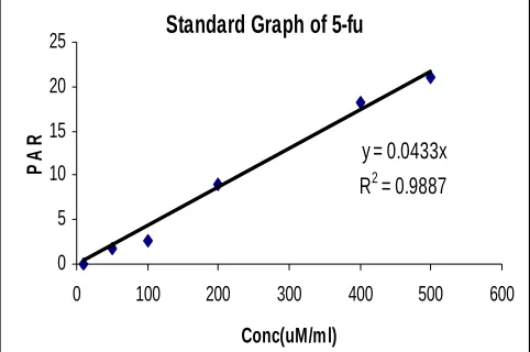
Fig 5 Linear standard curve determination of 5-Fu in serum (concentration range 5-70 µM/ml)
Standard Graph of 5-fu
y = 0.0433x R2 = 0.9887
In order to determine plasma concentration of 5-fu, internal standardization method was used. After
preparation of various concentrations of 5-fu and analyzing chromatography each standard solution,
two standard curves were prepared by plotting the ratio of peak area of 5-fu to internal standard
(thymine) versus concentration of 5-fu. A good linearity was seen in the standard curve of 5-fu
(Figure 5). To assess the accuracy of the method, recovery of 5-fu from serum samples with
known concentrations was compared with the solutions of 5-fu at the same concentrations as shown
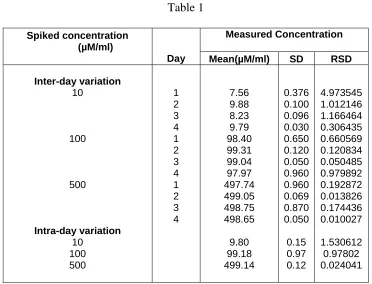
in Table 1. For the assessment of method precision, reproducibility of the results obtained for
different concentrations of 5-fu was determined at 5 different days and 5 times in one day. The
results of reproducibility study are shown in Table 2 as recovery and accuracy determination.
The limit of quantitation of 5-fu in serum with the above sample pretreatment method was 10µM.
The precision of method within a day was 98 to 99.8 percent and between days was 95.6 to 99.7
percent. The recovery of the method was between 88.6 and 99.7 percent.
Table 1
Measured Concentration Spiked concentration
(µM/ml)
Day Mean(µM/ml) SD RSD
Inter-day variation 10 100 500 Intra-day variation 10 100 500 1 2 3 4 1 2 3 4 1 2 3 4 7.56 9.88 8.23 9.79 98.40 99.31 99.04 97.97 497.74 499.05 498.75 498.65 9.80 99.18 499.14 0.376 0.100 0.096 0.030 0.650 0.120 0.050 0.960 0.960 0.069 0.870 0.050 0.15 0.97 0.12 4.973545 1.012146 1.166464 0.306435 0.660569 0.120834 0.050485 0.979892 0.192872 0.013826 0.174436 0.010027 1.530612 0.97802 0.024041
Table 2 Assessment of Recovery and Accuracy determination of 5-Fu in human serum
Substance
Concentration
Recovery (%) Mean±SD
Accuracy (%) Mean±SD
5-FU 5-FU 5-FU Internal Standard
10(µM) 100(µM) 500(µM) 10(µM/ml)
88.6±11.5 98.6±0.6 99.7±0.11 95.95±1.52
100.06±1.9 97.7±1.07
99.8±.77 96.05±1.09
SD: Standard deviation and mean value of 5 determinations
Discussion
Various methods of high-performance liquid chromatography for the determination of 5-fu in
biological fluids have been described so far which differ in chromatography type (reverse phase or
ion-pair chromatography) or detection system (UV or fluorescence) (6-8). Reverse-phase high
performance liquid chromatography with UV detection has been most recommended. (10, 11). But,
an important point is that most of them are tedious and expensive because they use more material
and have many stages of experiment. Besides, they are not suitable for a routine and quick
therapeutic drug monitoring (TDM) test which is necessary for a child cancer patient in a high dose
infusion therapy at hospital, or reference laboratories.
In this article a simple and fast method for the determination of 5-fu in serum is described that has
equal precision and accuracy to other similar methods (3-5,7-8). A full chromatography takes 10
minutes. To include sample preparation time it may need 25 minutes for the whole of each analysis,
which is comparatively a short time. The short duration of assay time is of quite importance in
routine monitoring of the drug in serum to predict and prevent future toxicity in high-dose 5-fu
intravenous infusion. On the other hand this method has a satisfactory quantitation limit that makes
it ideal for pharmacokinetic studies and therapeutic drug monitoring of 5-fu after the administration
of high doses of this drug. To improve the quantitation limit further we could use solid phase
extraction technique along with fluorescence detection after post column derivitization of the 5-fu
to fluorescent compounds so that the method become suitable for determination of 5-fu after the
administration of lower doses of the drug but such methods are more tedious, time consuming and
References
1. Fleisher M. Antifolate analogs, mechanism of action, analytical methodology and clinical
efficacy. Ther.Drug Monit. 1993; 15: 521-529
2. Boyd JR. (Ed) Drugs Facts and Comparisons, JP Lippincot Company, St. Louis 1992;653-654
3. Assadullahi TP, Dalgi E and Warner JO. Highperformance liquid chromatography method for
serum methotrexate levels in children with steroiddependent asthma. J. Chromatogr 1991;565:
349- 356
4. Schilsky RL. Clinical pharmacology of methotrexate. In: Ame AM, Powis G and Kovash IS
(Eds) Pharmacokinetics of Anticancer Agents in Humans. Elsevier Science Publishers, New
York 1983;187- 205
5. Breithaupt H, Kuenzelen E and Goebel G. Rapid high-performance liquid chromatographic
determination of methotrexate and its metabolites 7- hydroxymethtrexate and
2,4-diamino-N10-methyl pteroic acid in biological fluids. Anal. Biochem 1982;127: 103-113
6. Brimmed PA and Sams DJ. Rapid and simple assay for the measurement of methotrexate in
serum, urine and red blood cells by reversed-phase highperformance liquid chromatography. J.
Chromatogr. 1987; 413: 320-325
7. Cosolo W, Drummer OH and Christophidis N. Comparison of high- performance liquid
chromatography and the abbot fluorescence polarization radioimmunoassay in the measurement
of methotrexate. J. Chromatogr 1981; 223: 225-231
8. Farid YZ, Watson ID and Stewart MJ. An assay for methotrexate and its metabolites in serum
and urine by ion-pair high-performance liquid chromatography. J. Pharm. Biomed. Anal 1983;1:
55-63
9. Lawson GJ and Dixon PF. Rapid and simple method for measurement of methotrexate and 7-
hydroxymethotrexate in serum by high-performance liquid chromatography. J. Chromatogr
1981;223: 225-231
10.Najjar TA, Matar KM and Alfawaz IM. Comparison of a new high-performance
chromatography with fluorescence polarization immunoassay for analysis of methotrexate.
Ther. Drug Monit 1991;14: 142- 146
11.Salamoun J and Frantsiek J. Determination of methotrexate and its metabolites 7-
hydroxymethotrexate and 2,4-diamino-N10-methyl pteroic acid in biological fluid by liquid