UCL
TOWARDS A NEW
METHODOLOGY FOR THE
DETERMINATION OF
SEQUENCE SELECTIVITY IN
SMALL MOLECULE-DNA
BINDING
MICHAEL BRENNAN PICKUP BA (Hons)
LONDON
SEPTEMBER 1997
Submitted in partial fulfilment of the requirements of
the University of London for the degree of
ProQuest Number: U642969
All rights reserved
INFORMATION TO ALL USERS
The quality of this reproduction is dependent upon the quality of the copy submitted.
In the unlikely event that the author did not send a complete manuscript and there are missing pages, these will be noted. Also, if material had to be removed,
a note will indicate the deletion.
uest.
ProQuest U642969
Published by ProQuest LLC(2015). Copyright of the Dissertation is held by the Author.
All rights reserved.
This work is protected against unauthorized copying under Title 17, United States Code. Microform Edition © ProQuest LLC.
ProQuest LLC
789 East Eisenhower Parkway P.O. Box 1346
DECLARATION
I, Michael Pickup, hereby certify that this thesis has been composed by myself, that it is a record o f my own work, and that it has not been accepted in partial or complete fulfilment o f any other degree or professional qualification
ABSTRACT
Described in this thesis is work aimed towards the determination of the sequence selectivity o f aciacinomycin, an anthracycline, for dsDNA, by an in vitro
selection experiment. In particular the work was directed at the development of a technique to distinguish between pieces o f dsDNA which were, and were not, being bound by the drug.
The first methodology attempted was the attachment to aciacinomycin to a solid support, with the aim of performing an affinity chromatography experiment on a random dsDNA library, which had been synthesised. The successful synthesis of a biotinylated linker, for the immobilisation of aciacinomycin on a streptavidin based support, is described, with an overall yield o f 48%, starting from dodecan-l,12-diol, over a five step protocol. Attempts to attach this to aciacinomycin via its methyl ester group, however proved to be futile. A wide range o f methods were used, both chemical and enzymatic, but only breakdown products or starting material were observed in the reaction mixtures.
In order to study the hydrolysis o f the aciacinomycin methyl ester, attempts to synthesise a two-ring model compound of the aglycone portion o f the drug were made. Several potential routes were investigated starting from a- tetralone, however no viable method was found to produce the desired target compound. The most successful methodology was found only to result in a mixture of compounds which were unidentifiable by spectroscopic methods.
I f w e see light at the en d o f the tunnel it is
the light o f an on-coming train
ACKNOWLEDGEMENTS
I would like to thank everybody who has helped me throughout this work both inside and outside the lab. Firstly to Dr Alethea Tabor for allowing me to work for her and for her endless enthusiasm and optimism. Also to the BBSRC (and my parents) for supporting me.
Inside the lab would not have been as enjoyable as it was without the people who made it. Thanks to you all, especially the Scottish quartet o f Martin, Al, Scot and James. A special mention must also be made to Elizabeth, for her proof reading, and to Sarah Sheady for running around, and helping me get all those fiddly little bits of this thesis sorted out.
No work would have been possible without the skill o f the technical staff and I would like to thank all the people at both the University of Edinburgh and UCL.
TABLE OF CONTENTS
Declaration 1
Abstract 2
Acknowledgements 4
Table of contents 5
Table of figures 10
Abbreviations 14
Chapter 1 Introduction 18
1 DNA: ITS STRUCTURE AND THE BINDING OF SPECIES TO IT 18
1.1 DNA structure 18
1.2 Binding o f molecules to DNA 23
1.3 The modes of DNA binding 25
1.3.1 Non-specific outside binding 25
1.3.2 Simple intercalation 27
1.3.3 Groove binding 29
1.3.3.1 Minor groove binding 29
1.3.3.2 Major groove binding 31
1.3.4 M ixed systems o f b inding 33
2 THE ANTHRACYCLINE ANTIBIOTICS 34
2.1 General structure 34
2.2 Proposed mechanisms of action o f anthracycline 35 anticancer drugs
2.2.1 Topoisomerase II inhibition 36
2.2.2 Antihelicase action 40
2.2.3 In vivo reduction 42
2.3 The binding o f anthracyclines to DNA 43
2.3.1 Kinetics and thermodynamics 43
2.3.2 Structural aspects o f binding 47
2.3.2.1 Daunomycin 47
2.3.2.3 Nogalamycin 52
2.3.2.4 Arugomycin 55
3 SEQUENCE SELECTIVITY AND ITS DETERMINATION 57
3.1 Nmr and X-ray crystallography 58
3.2 Chemical attachment 59
3.3 Footprinting analysis 62
3.4 In vitro transcription analysis 72
3.5 In vitro selection analysis 74
3.5.1 Attachment to a solid support 77
3.5.2 Gel retardation methodology 81
4 PROPOSED WORK 82
5 REFERENCES 85
Chapter 2 The design and synthesis of a linker for the immohiligation of
aciacinomycin A l on a solid support 92
1 CHOICE OF MATRIX AND LINKER 92
1.1 Solid supports 92
1.2 Biotin-streptavidin immobilisation systems 97
1.3 Point o f attachment for a linker on aciacinomycin 100
2 PROTECTION OF DODECAN-1,12-DIOL 107
3 AMINATION 108
4 COUPLING TO BIOTIN 109
5 DEPROTECTION 111
6 PREPARATION OF AN AMINE LINKER 112
7 SUMMARY 113
8 EXPERIMENTAL 114
8.1 General experimental 114
8.2 Experimental procedures 115
Chapter 3 Attachment of a linker to aciacinomycin A l 125
1 INTRODUCTION 125
2 GENERAL HYDROLYSIS AND TRANSESTERIFICATION
PROTOCOLS FOR ESTER GROUPS 125
2.1 Acid and base chemistry 126
2.2 Equilibrium displacement chemistry 130
2.3 Electrophilic and nucleophilic chemistry 131
2.4 Lewis acid chemistry 133
2.5 Enzymatic methods 135
3 REACTIONS ON ACLACINOMYCIN 136
3.1 Direct transestérification 137
3.2 Enzymatic methods 139
3.2.1 Candida cylindracea Lipase (CCL) 139
3.2.2 Pig Liver Esterase (PLE) 140
3.2.3 Subtilisin Carlsberg 142
3.2.4 Porcine Pancreatic Lipase (PPL) 143
3.2.5 Thermitase 143
3.3 Hydrolysis by chemical methods 144
3.3.1 Lithium hydroxide 144
3.3.2 Lithium iodide 145
3.3.3 Aluminium trichloride and thiols 146
3.3.4 Titanium tetraisopropoxide and aluminium triisopropoxide 147
3.3.5 Quinoline and acetic acid 147
3.3.6 l-(Hydroxy)-3-(chloro)tetrabutyldistannoxane 148
3.3.7 Sodium phenyl selenide 150
3.4 Reduction with DIBAL 151
4 SUMMARY 151
5 EXPERIMENTAL 153
5.1 General experimental 153
5.2 Experimental procedures 154
Chapter 4 Synthesis of a model compound 168
1 MODEL COMPOUND TO REPRESENT THE ELECTRONIC
ENVIRONMENT OF ACLACINOMYCIN 168
2 SYNTFIESES OF THE AGLYCONE OF ACLACINOMYCIN 171
2.1 The Broeckman, Jr. synthesis of (± )-aklavinone 172
2.2 The Kishi synthesis o f (+ )-aklavinone 174
2.3 The Kende synthesis o f aklavinone 176
3 THE MODEL COMPOUND 179
4 SUMMARY 183
5 EXPERIMENTAL 184
5.1 Experimental procedures 184
6 REFERENCES 188
Chapter 5 Gel retardation methodology in the determination of
sequence selectivity in small molecule-DNA binding 189
1 INTRODUCTION 189
2 THE DESIGN AND SYNTHESIS OF A RANDOMISED SEQUENCE 189
3 GENERATION OF A SECOND STRAND OF DNA 194
3.1 PCR methods 194
3.2 Klenow methodology for second strand synthesis 197
4 END-LABELLING 199
5 GEL SHIFT EXPERIMENTS 203
6 PCR OF RECOVERED DNA 210
7 SUMMARY 213
8 MATERIALS AND METHODS 216
8.1 Buffers used 216
8.2 Generation of the ds DNA library from a ss DNA template 218
8.2.1 ds DNA library made from OLIGONUCLEOTIDE 1 218
8.2.2 ds DNA library made from OLIGONUCLEOTIDE 2 219
8.2.4 Elution and concentration o f the DNA using Microcon
Microconcentrators with Millipore .22 gel nebuliser 219
8.3 Mobility shift assay 220
8.3.1 Radiolabelling the DNA library 220
8.3.2 Binding reactions 220
8.3.3 Electrophoresis 220
8.3.4 A utoradiograph 220
8.4 Recovery and amplification 221
8.4.1 Recovery o f selected DNA - "Crush and Soak ” 221
8.4.2 A mplification us ing PCR 221
9 REFERENCES 222
Chapter 6 Summary and future work 224
References 227
APPENDIX 228
10
TABLE OF FIGURES
Chapter 1
1 B-formDNA 19
2 The base pairings described by Watson and Crick 20
3 A-formDNA 21
4 Anti v^. syn base pairs 21
5 Z-form DNA 22
6 The modes o f binding to DNA 26
7 The ten discrete intercalation sites for intercalation between two base
pairs 28
8 The scissors-grip model 32
9 The general structure of anthracyclines 34
10 Pharmacophore for topoisomerase II inhibition 37
11 Inhibition o f topoisomerase II 38
12 Perpendicular mode and parallel mode for intercalation 39
13 Antihelicase action 41
14 In vivo reduction mechanism of action for anthracyclines 42
15 Mechanism for the kinetics o f binding o f anthracyclines to DNA (1) 45 16 Mechanism for the kinetics o f binding o f anthracyclines to DNA (2) 46 17 Mechanism for the kinetics o f binding o f anthracyclines to DNA (3) 46
18 The binding of daunomycin to DNA ( 1 ) 47
19 The binding o f daunomycin to DNA (2) 48
20 The binding o f nogalamycin to DNA 53
21 Attachment product of daunomycin and DNA 60
22 Baeyer-Villiger oxidation of anthracyclines 61
23 In vivo oxidation cycle 61
24 Schematic o f the footprinting experiment 63
25 The mode o f action of bleomycin 65
11
29 A typical protein gel retardation experiment 81 30 Proposed aciacinomycin affinity column methodology 83
C hapter 2
31 CNBr activation o f polysaccharide bead matrices 94
32 Types o f attachment to solid supports 95
33 A-hydroxysuccinimide activated Sepharose or Superose 96
34 Polyacrylamide and trisacryl matrices 97
35 Proposed biotin-streptavidin immobilisation methodology 98 36 Variation in bis-biotin binding with linker length 99 37 Linker formation for connecting anthracyclines 101 38 X-ray crystal structure o f nogalamycin bound to DNA 104 39 Retrosynthetic approach to the linker required 105 40 The forward reaction scheme for the linker synthesis 106
Chapter 3
41 Alkyl versus acyl cleavage 126
42 Standard mechanisms for the acid and base
hydrolysis/transesterification of esters 128
43 AalI mechanism for hydrolysis o f esters 128
44 AacI mechanism for hydrolysis o f esters 130
45 The mechanisms for acidic and basic hydrolysis of esters 130 46 Definition o f the equilibrium constant K for a transesterificatiop
reaction 131
47 Nucleophilic attack on a methyl ester by a soft nucleophile 132 48 Hard and soft sites for a typical carboxylic ester 132 49 Combined hard acid/soft nucleophile hydrolysis system 133 50 Lewis acid mediated transestérification acyl cleavage 134 51 Proposed transition state for the Lewis acid mediated
transesterification/hydrolysis via alkyl cleavage 134
52 Mode of hydrolysis by BBTO 135
12
54 Model o f the ideal substrate for PLE 141
55 Working model for the PLE active site 142
56 Aluminium halide-thiol systems 147
57 Acetic acid in boiling quinoline hydrolysis mechanism for
hindered esters 148
58 Mechanism for a distannoxane transestérification 149
59 Formation of phenyl selenide anions 150
C hapter 4
60 Synthetic route for model compound 168
61 Vinyl magnesium bromide route 169
62 Elimination o f vinyl magnesium bromide adduct 170 63 The successful synthetic route of Broeckman, Jr. 173 64 Proposed mechanism for the cyclisation aldol reaction 174
65 The Kishi synthesis 175
66 Ring closure reactions 176
67 The Kende synthesis 177
68 Postulated mechanism for ring opening of the epoxide ester by
palladium/barium sulphate. 178
69 Enol acetate formation from a-tetralone 180
70 Enol acetate formation from 2-ethyl-5-methoxy-a-tetralone 181 71 Proposed mechanism o f reaction o f 2-ethyl-5-methoxy-a-tetralone 181
72 Ozonolysis reaction 182
C hapter 5
73 Basic design of the random DNA 190
74 pUC18 and pUC19 cloning vectors 191
75 The HPLC purification of the library OLIGONUCLEOTIDE 1 193
76 Schematic of the PCR cycle 194
77 An agarose gel with BIOGEL® insert 196
78 Klenow reaction 198
13
80 Set-up for a Microcon purification system 201
81 Agarose gel set-up for a Microcon purification 202
82 Gel shift on DNA from OLIGONUCLEOTIDE 1 205
83 Proposed mechanism for GC self priming 206
84 Results of dilution experiments 209
85 Results on an 8% polyacrylamide gel 210
86 Schematic summary o f the PCR temperature protocols used 214
Chapter 6
87 Mannich chemistry for the immobilisation o f ligands 225
88 An example o f a lexitropsin 226
Appendix 228
89 Spatial orientation about the D-ring of the aglycone 231
90 ‘H and nmr spectra of aciacinomycin 232
91 1-D nmr spectrum of aciacinomycin 234
92 2-D HMQC spectrum of aciacinomycin 238
93 2-D DQFCOSY spectrum o f aciacinomycin 240
94 1-D TOCS Y spectra o f sugars I and II 244
95 1-D TOCS Y spectrum o f sugar III 245
96 2-D HMBC spectrum o f aciacinomycin 246
14
ABBREVIATIONS
A Adenine
AC Acyl
ADP Adenosine diphosphate
AL Alkyl
ApG A two base pair step in DNA
atm Atmospheres pressure
ATP Adenosine triphosphate
BBTO B/5tributyltin oxide
BEP 4-(2’-bromoethyl)phenol
bp Base pair
Bu Butyl
bZIP Basic Leucine Zipper protein motif
C Cytosine
C Carbon
CAST Cyclic amplification and selection o f targets
CCL Candida cylindracea lipase
COSY Correlation spectroscopy
cpm Counts per minute
DBU 1,8-Diazabicyclo[5.4.0] undec-7-ene
DCC Dicyclohexylcarbodiimide
DCM Dichloromethane
DEAD Diethyl azodicarboxylate
Ô Chemical shift in part per million
AH Enthalpy change
DIBAL Diisobutylaluminium hydride
DMAP (4-Dimethylamino) pyridine
DMF A^A-Dimethylfbrmamide
DMPU 1,3-Dimethyl-3,4,5,6-tetrahydro-2 (IH)-pyrimidone
DMS Dimethylsulfide
DMSO Dimethylsulfoxide
15
DNP Dinitrophenyl hydrazine
dpe Bisdiphenylphosphinoethylene
DQF Double quantum filtered
dsDNA Double stranded DNA
DTT Dithiothreitol
Activation energy
ed Editors
EDTA Ethylenediamine tetraacetic acid
e.e. Enantiomeric excess
eq Equivalents
Et Ethyl
fig Figure
fg Femtogram (10'^^ gram)
FM Fotemustine
FTIR Fourier transform infra-red
G Guanine
g 9.81 m s'^
H Hydrogen
h Hours
HMBC Heteronuclear multiple bond correlation
HMPA Hexamethylphosphoramide
HMQC Heteronuclear multiple quantum coherence
HPLC High Pressure Liquid Chromatography
Hz Hertz
J Nmr coupling constant
K Equilibrium constant
A^app Apparent binding constant
kbp Kilobase pairs
kD Kilodalton
1 Litre
LDA Lithium diwopropylamide
16 MCPBA Me MHz min mM mp MPE:Fe (II) V N N] NHS nmr nOe Nu O o O.D. P PCR Ph pH PLE pmol PPL ppm PPTS pS R Rf ROESY rpm RT /w-Chloroperbenzoic acid Methyl Megahertz Minutes Millimolar Melting point Methidiumpropyl-EDTA-Fe(II) Infrared absorbance Nitrogen Nitrogen gas A-hydroxysuccinimide Nuclear magnetic resonance Nuclear Overhauser effect Nucleophile
Oxygen Ortho
Optical density Para
Polymerase chain reaction Phenyl
Potential hydrogen Pig liver esterase Picomoles (10'^^ moles) Porcine pancreatic lipase Parts per million
Pyridinium /7<3ra-toluenesulfonate Phosphothioate
Variable functional group Retention factor
Rotating frame Overhauser enhancement spectroscopy Revolutions per minute
17 S SAAB SC SDS SELEX SEM SnI SN2 ssDNA T t TBAF TBDMS TDA TEMED THF TEC Tm IM S TOCSY tosyl Tris u.v. 1-D 2-D 5’ ^ 3 ’ 3’ - > 5 ’
Seconds
Selection and amplified binding Subtilisin Carlsberg
Sodium dodecylsulphate
Systematic evolution of ligands by expopential enrichment
p-(Trimethylsilyl)ethoxymethyl chloride Unimolecuiar nucleophilic substitution Bimolecular nucleophilic substitution Single stranded DNA
Thymine Tertiary
Tetra-A-butylammonium fluoride r-Butyldimethylsilyl
Target detection assay
N, N, N \ N'-T etramethylethylenediamine
Tetrahydrofuran
Thin layer chromatography Melting temperature Trimethylsilyl
Total correlation spectroscopy /7-toluenesulfonyl
T ris(hydroxymethyl)aminomethane Ultraviolet
One-dimensional Two-dimensional
Chapter 1 18
CHAPTER 1
INTRODUCTION
1. DNA: Its structure and the binding of species to it 1.1 DNA structure
Ever since the publication of the structure o f double stranded (ds) DNA from X-ray diffraction data in 1953' there has been a great deal of interest in species that bind to i t / ^ Watson and Crick described, for B-form DNA, a helical structure with a complete turn covering ten base pairs and a distance of 34 Â and ^ width o f 20 Â. The heterocyclic base pairs, adenine:thymine and guaninexytoaine, are aligned perpendicularly to the helical axis and are stacked one on top of the other in order to maximise the n-n interaction between the two aromatic systems. The sugar- phosphate backbone surrounds the base pair core in the form of hydrophilic ridges. Between these ridges two helical spaces are left, which are known as the major and minor grooves (fig 1, 2).
The major groove has a width of 12 Â and a depth o f 8.5 Â. Positioned within it are the N-7 of guanine and adenine; the 0-4 o f thymine; and the 0-6 of guanine as hydrogen bond acceptors. There is also the C-6 NH2 of adenine and the
C-4 NH2 o f cytosine which act as hydrogen bond donors. In comparison, the minor
groove is half the width at 6
A
and slightly shallower at 7.5A.
ft presents to the outside world the N-3 o f guanine and adenine and the 0-2 o f thymipe and cytosine as hydrogen bond acceptors, as well as the C-2 NH2 group of guanipe as a hydrogenbond donor. As a result in aqueous solution these two grooves are found to interact with a large number of water molecules and to be fully hydrated.'*
C h a p t e r 1 19
value at 40,0'\ DNA is not a rigid structure as has been suggested but in fact a
dynam ic structure constantly undergoing internal defonnation (therm al breathing).
This can be seen in exchange reactions with tritium for protons \vhich should be
inaccessible and also by nmr.
-r f '
Chapter 1 20
MAJOR GROOVE
NH: N—H
— N
MINOR GROOVE
NH:
H— N G
= N NH:
fig 2 The base pairings described by Watson and Crick
C h a p t e r I 21
10 A
fig 3 A-form DNA
In som e high salt conditions for synthetic DNA (especially if they contain
alternating purine and pyrim idine sequences) an alternative fo n n called Z-D N A can
exist (fig 5). U nlike the other two structural fonns this is a left-handed helical
structure. In this conform ation the m inor groove is extrem ely deep and goes into the
structure as far as the helical axis and the m ajor groove does not exist at all. The
nucleosides are not all anti, but in fact they alternate syn to anti and as a result the
polyphosphate backbone has a zigzag like structure (fig 4).
HN.
NH2
H O
HO HO
syn -dGMP anti -dC
C h a p t e r 1 22
10 Â
fig 5 Z-form o f DNA
A lthough these results com e prim arily from X -ray diffraction data there is
evidence that the structure observed is in fact the sam e as the solution structure. The
enzym e D N ase I is found to cleave DNA (dCG C G A A TTC G C G ) at every phosphate
in solution at a rate constant w hich can be correlated with the helical tw ist present at
each individual base pair.^ Evidence also exists for the hydration o f the m inor groove
in B-form DNA. G ravim etric, infrared and ultraviolet studies w ere carried out on
DNA fibres to enable the order o f events upon DNA hydration to be seen. These
show ed that the free phosphate oxygens w ere the first to be hydrated, follow ed by the
__________________________________________________________ Chapter 1 23
relative humidity in the crystallising medium/'^^ These results were further confirmed by studies in solution by density measurements and ultracentrifugation^ These experiments also pointed to the fact that an A:T base pair has two more water molecules associated with it that a G:C pair (12 in t o t a l ) / I t is ^Iso interesting to note that later studies on the hydration in the minor groove of the dodecamer dCGCGAATTCGCG showed a distinct two-layer hydration structure/^ The initial monolayer attracted to the nitrogen and oxygen atoms in the groove is bridged by water molecules in the second layer giving a distinctive zigzag “spipe o f hydration”. The width of the groove prevents any further association generating this bilayer.
DNA in its linear form is a very long structure. Electron microscopy has shown human DNA to have a length of 990,000 pm for its 2,900,000 kbp and even a polyoma virus covers a length of 17,000 Â for its 5100 bp.^ Ip comparison the protein in haemoglobin (diameter 65 Â) has only a length o f 3000 Â. Since in vivo
there is necessarily a constraint on the volume available to store DNA in the cell, DNA from many sources has been found to be in a continuous loop. This form is known as relaxed circular form. This form can itself then wind up into a helical form to give a more compact tighter superhelix. The superhelix can be either left-handed (designated negative) or right-handed (positive). Each superhelix varies from another only in the number of rotations about the axis when it is constrained to a flat plane. The Linking Number (Lk) describes the number of turns, and helices which vary only in this are known as topoisomers of each other.
Both positive and negative superhelices can be stored equally compactly. Enzymatic processes in vivo require a change in Lk as an initial step. In eukaryotic enzymes the enzyme which converts topoisomers- Topoisomerase I/II- will act on both positive and negative superhelices. However, in prokaryotic systems enzymes work specifically on negatively coiled superhelices.
1.2 Binding of molecules to DNA
__________________________________________________________________________________ Chapter 1 24
Upon binding, any water present at the binding site in the major and/or minor groove is expelled into the bulk solution. This leads to an increase in the entropy of the bulk solution and to a lowering of the Gibbs free energy of the system overall.
From the second law of thermodynamics:
^Gsys = AHsys -TASsys
Thus, even if AHsys is greater then zero from electrostatics (that is to say the enthalpic release from the binding of a species does not override the energy required to expel the water in the groove), it is possible that the entropie gain from the release of the water into the bulk solution is enough so that TASsys is greater. This release means that the overall Gibbs free energy change will be less than zero and the process will be a favourable one. If such a scenario exists it is known as the hydrophobic effect.
The second law also shows us that an important factor is the enthalpic effect. That is to say, the energetics of binding which are governed by electrostatics and non polar interactions play a very important role. As stated already, the major and minor groove present to the external environment a variety of polar and non polar functionalities e.g. keto and amino groups. Intermolecular interactions between these groups may arise to other species present. These may be dipole-dipole interactions (-1.4 kJ moF^at a distance of 0.3 nm), van der Waals interactions (-2 kJ mol'^ in diamond), or stronger hydrogen bonds (15 -20 kJ mol”^ for OH and NH2 groups). These help to provide a enthalpic basis for binding and add to the kinetic factors involved in any further dissociation/ association which may take place.
With the base pairs being separated by a distance of 0.34 nm perpendicular to their plane the Ti-systems o f successive base pairs can overlap in what is known as
__________________________________________________________________________________ Chapter 1 25
Since the DNA electrostatic potential and ability to hydrogen bond is dependent on the local sequence it is feasible to state that the thermodynamics of binding to any particular piece of DNA may be more favourable th^n that for other sequences. If a compound displays a preference for one particular sequence above all others present it is said to be exhibiting sequence selectivity.
1.3 The modes of DNA binding
The most simple mode is intercalation (fig 6).^"* This is the process described above by which a fiat aromatic species “slides” in between the hasp pairs. There is also the possibility of a compound binding in either of the two grooyes. This process is simply known as major or minor groove binding.
1.3.1 Non-specific outside binding
Since the backbone o f DNA is anionic, interaction with positively charged species is possible. Cationic species such as proflavine 1 can lipe up along the backbone and in doing so can minimise the repulsions between themselves. This outside binding can in itself release some of the water associate^ with the DNA structure and hence be entropically favoured if the increase in ordçr of the binding molecule is cancelled out by an overall decrease in the order of the water associated with the backbone.
Major Qraove
Minor G m o w
Major Qpoovb
Minor QtocM
MINOR GROOVE MAJOR GROOVE ir>rrERCALAT10N
Major GroovB
Minor Qroovo
Hek
DNA Binding Spectea
o -a
fig 6 The inodes o f DNA binding
t'O
Chapter 1 27
1.3.2 Simple intercalation
As stated previously, the prime prerequisite for intercalation to occur is that the species be planar and aromatic/^ Examples of intercalators are pf use both in the laboratory and clinically. Ethidium bromide 2 binds by intercalation. On binding its fluorescence changes and it is used as a device for DNA detection in electrophoresis.^^ Its binding ability also manifests itself as a frame shift mutagen in vivo.
NH:
— N,
Ethidium 2
The mode of binding for these simple intercalators follows a two step process. Firstly the molecule interacts with the anionic backbone o f the DNA to give an external complex. The molecule can then diffuse along the length o f the DNA until a gap appears in between the base pairs, through thermal breat|iing o f the DNA, which is large enough to allow intercalation to occur. Obviously such a process is size dependent, as larger or more bulky compounds require more of a distortion in the DNA for the second stage to occur. It is also feasible that soliton excitation may play a part in intercalation. This is an aspect of thermal breathing and refers to a stretching vibration of the DNA which travels along the helix length as a wave. Eventually a point will be reached where the amplitude o f the waye is such that it has enough energy to slightly part the DNA base pairs and allo^y intercalation to occur.
Chapter 1 28
proflavine 1. Dichroism measurements also indicate a tilting o f the hase pairs and intercalator to maximise the ti-tt overlapping.’^ This in itself can be up to 20-25°. These studies, however, have been unable to show any long rapge effects of intercalation which may suggest some form of compensation occurring elsewhere in the structure to rectify the local deformation at the binding site.
An interesting point to note with intercalation is that not all possible sites are used, and in fact at maximum concentrations only alternate sites are filled. This phenomenon is known as the neighbour exclusion principle. Although it is only an empirical rule it seems to hold true for most simple intercalators and probably derives from the local distortions observed around the intercalation site. An alternative hypothesis is that the entropy gain from the local release o f water is not as favourable if water has already been expelled from a nearby site.
Since intercalation occurs between just two sets of base pair^ it means that there are ten possible sites for simple intercalators to bind (fig 7). If the species also has some structure which extends further than the site o f intercalatiop there is more scope for specific binding interactions and the number o f possible binding sites is larger.
5’ ^ 3 ’ AA TA GA CA
3’ < - 5 ’ TT AT CT GT
5’ - > 3 ’ AT TT GT CT
3’ < - 5 ’ TA AA CA GA
5’ - > 3 ’ AG TG GG CG
3’ < - 5 ’ TC AC CC GC
5’ - » 3 ’ AC TC GC CC
3’ < - 5 ’ TG AG CG GG
Chapter 1 29
- — 1
---It has been seen that in general little or no selectivity is displayed by pure intercalators except possibly for G:C base pairs. These have a higher intrinsic dipole and may be able to produce a larger polarisation in the rings o f the intercalator itself when in close contact on binding.
1.3.3 Groove binding
As has already been described, the major and minor grooves differ greatly in their size and hydrogen bonding potential, and hence amount o f hydration. The most important of these factors as to whether a species is a major or minpr groove binder, is the size o f the species in question, and its ability to enter either or both of the grooves well enough to have stabilising interactions. It is found that large molecules such as proteins and oligonucleotides (in forming triplex species) tend to prefer major groove binding, whereas smaller organic compounds show a tendency towards minor groove binding.
1.3.3.1 Minor groove binding
Minor groove binders are generally made up of small aromatic rings such as furans, benzenes and pyrroles joined by bonds which allow rotation between the rings. This freedom o f rotation allows a curved conformation to bç adopted by the compound so that it can fit into the helical groove. Van der Waals interactions can take place between the compound and the groove wall and if the binding is tight enough hydrogen bonds can occur to the groove floor at the points njentioned earlier.
The minor groove does not have a uniform width and is narrpwer at A:T pairs than at G:C pairs. Hence if molecules are tighter in the groove at A:T rich sequences the van der Waals interactions with the sides of the groove walls will be greater.
Chapter 1 30
,---A classic example of cationic minor groove binding is shown by netropsin 3. It is a dicationic species which contains two pyrrole moieties which are able to rotate. A crystal structure of netropsin 3 bound to d(CGCGAATTÇGCG) has been obtained which enables details o f minor groove binding to be ascertained.'17
CH.
CH.
NH
NH .NH
NH NH.
NH:
NH:
Netropsin 3
As expected it was found that the compound was bound at t|ie central AATT region and the water in the groove had been displaced from th^t section o f the duplex. The three amide protons point towards the groove and forrp hydrogen bonds to N-3 o f adenine and 0-2 of thymine whilst van der Waals interactions exist with the walls of the groove. The CH groups o f the pyrrole rings act as a brake, and prevent any deeper penetration into the groove (the hydrogen bon4 distances are in
the region o f 3.3-3.8 Â compared to a normal 3 A). As a result o f these factors the pyrrole rings are nearly parallel to the groove walls and twisted by 33° because o f the helical twist o f the DNA. The cationic ends are seen to be interacting with the N-3 of the outer adenines.
Unlike intercalation however there are no noticeable effects on the DNA (i.e. no lengthening measurable by physical methods). There is a slight bending o f the helical axis and groove widening but no change in helical twist is seçn.
_____________________________________________________________________________ Chapter 1 3 1
1.3.3.2. Major groove binding
The principal determinant in major groove binding is size. The greater width o f the major compared to the minor groove means that larger species are required to bind within it. The most prevalent class is therefore the larger proteins. Because of this increase in size there is an associated increase in the number o f points of interaction between the protein and the DNA. As a result there tçnds to be much more sequence selectivity displayed by these systems. Examples include the large class of activator and repressor proteins and perhaps more obviously the restriction enzymes, such as EcoR l, which cut DNA at specific sequences. This selectivity is vital in vivo for an organism; on a cellular level it is fundamental that the processes that occur do so with an element of control with the responses occurring for the right DNA sequence.
Although the primary sequences of these proteins vary widely it is found that in fact they usually contain one of a limited set o f structural motifs within their three dimensional form which is involved in the protein-DNA interaction. This requirement is most probably made on an electrostatic and geometric basis constrained by the DNA’s shape and potential energy surface.
It is commonly found that proteins have strategically placed lysine and arginine residues, positioned so that they neutralise the negative charge o f the polyphosphate backbone by forming ionic bonds. An example o f this is seen in the crystal structure of Klenow fragment o f DNase I.^° It is seen that the enzyme has an overall anionic electrostatic field but the DNA binds in a “cleft” which has an overall cationic nature.
On a purely size basis a typical a-helix has a diameter of abqut 7 Â, and has a dipole running from its N to C-terminus. This makes it ideally suifed to fit into the major groove of B-DNA. It is worth noting that a [3-sheet has a wi(^th o f only a few angstroms and is geometrically suited to the minor groove.
C h a p t e r 1 32
As Stated previously, the major groove has more potential sites which are
hydrogen bond donors or receptors. Since these may be on successive base pairs
amino acids such as glutamine and arginine are able to hydrogen bqnd to sequential
base pairs simultaneously. Hence, there is more potential for sequence selectivity
than exhibited in the minor groove.
Some general points about major groove binding can be gained from looking
at the bZIP (basic zipper) family o f proteins.*' These regulator}' proteins bind to
DNA as a dimer to an eight base pair long palindromic recognitiop sequence. They
consist o f two distinct segments - a leucine zipper and a basic recognition region.
A leucine zipper is a m o tif made up o f two smoothly cgrving a-helices
associated in a coiled coil. In a bZIP protein this spans the ( -terminal 30 residues
and is held together by repeating leucine residues every seven amino acids.
The basic region occurs from the A-terminal end and consists o f two
unassociated a-helices which bind to DNA in the so-called '‘scissors-grip”
mechanism (fig 8).^^ Each helix recognises a specific sequence, for example, in the
case o f the protein GCN4,^^ the sequence TGAC. Combining these two h a lf sites we
see that the protein GCN4 recognises the overall sequence 5 ’-TGACGTCA - the
CRE site (where CRE is the CREB response element protein).
Chapter 1 33
In this case the base pair contacts are via two arginine residues which contact two concurrent nucleic acids at the centre of each half site. There are also found to be present two alanines which have van der Waals interactions with the CH^ group of the central thymine, and a third arginine which contacts the central G of the recognition site (a fourth arginine from the other helix contacts the polyphosphate backbone).
It is seen that the DNA backbone is distorted and is bent at an angle 20° towards the leucine zipper with a concomitant underwinding o f the DNA at the centre of the binding site. This large DNA distortion means that the protein GCN4 is not only specific for the CRE site. A similar site - the API site - is also found to be a substrate for the protein. This is a similar sequence to the CRE sequence but differs in one base pair at the centre (5’-TGACCTCA).
Point mutations on the DNA and protein have been used fo investigate the binding.^'* It was seen that the thymine van der Waals interactions \yere essential and a change to uracil stopped all binding. Similarly all the amino acids at the binding site were seen to be essential although binding could be altered by increasing the steric bulk of the amino acids at relevant points.
Both these families of proteins show us that the factors that are essential for binding in the major groove are the same as for the minor groove. The main difference, however, is that there is a much larger scope for contacts in the major groove and, therefore, the opportunities for sequence selectivity arc increased. This is both due to the size o f the major groove itself and, more fundamentally, the increased potential for interaction o f species with it because of their larger size and the fact that they contain, in general, more functionalities than do minor groove binders.
1.3.4 Mixed systems of binding
Chapter 1 34
1---of this type o f binding is shown by the anthracycline a n t i b i o t i c s . S i n c e our work has been focused on a particular member o f this class it seems appropriate to describe in detail at this stage the anthracycline class of antibiotics; their mode of binding to DNA and their postulated mechanisms for their effects in vivo.
2 The anthracycline antibiotics^^’^^ 2.1 G eneral structure
Anthracyclines are a group o f antibiotics, first discovere4 in 1950 by H. Brockman, which show remarkable antitumor activity. To date the class consists of around four hundred naturally occurring compounds all with a common structure. They are all glycosidic compounds whose aglycones - anthraquinopes - all have a 7,
8, 9, 10- tetrahydro-5, 12-naphthacene quinone core (fig 9).
R ‘ O R' R1 0
R ‘R
R" O R' R
R^, R ’^ = OH except nogalamycin and relatives, which have a npgalose ring R^R'* = O H orO M e
R’ = point o f glycoside attachment R*° = glycoside chain or COOMe or OH
R’ and R^’ = (CH,, Et, COCH,, COCH2OH, CHOHCHj, CHOHCHjOH, CH^COCH,)
and OH
fig 9 The general structure of anthracyclines
Chapter 1 35
OH
CH CH CH CH
HO HO HO
NH: NMe- OH
labelling experiments have revealed that the anthraquipone unit is made in a polyketide pathway,^^’^® with daunomycinone, for example, bqing made o f one unit o f propionate and nine units o f acetate. Other biosynthesis experiments have shown that the sugars are then added sequentially.^' This process has been utilised extensively to produce bio-modified anthracyclines.
All the compounds described have marked effects in vivo, and lead to cell death via a process that most probably involves binding to DNA through intercalation o f the aglycone core and minor groove binding o f the sugars.
2.2 Proposed mechanisms of action of anthracycline anticancer drugs
For twenty years doxorubicin/adriamycin 8 has been used ag the primary line
o f attack in the treatment o f a wide variety o f cancers.
OH
OH
OCH3 O OH
OH
R=OH adriamycin 8
Chapter 1 36
However, the failure o f academics and the pharmaceutical industry to find a better alternative has lead to a decline in interest. This failure to ^nd an improved drug, with improved activity and reduced toxicity, can be partially attributed to a lack o f knowledge o f the mode o f action. Several possible actions in vivo have been suggested for the effectiveness of anthracyclines, but the precise mode of action for any particular family member is probably not through a single mecl^anism, but rather an array o f unrelated biochemical processes on a cellular level. It is thought that the principle effect is on the enzyme topoisomerase II by binding to dsDNA and interfering with the enzymes f u n c t io n . T h i s has been confirmed by a correlation which was found between DNA binding affinity and biological actiyity in twenty six anthracyclines.^^ It was also found that doxorubicin 8 was foun4 entirely on the
nucleus o f the cell when it was examined by microspectrofluorometry.^'^ Other workers have suggested that the action may result from in vivo reduction and the formation of radicals in the cell which may lead to DNA cleavage or cross-linking,^^ whereas others say that it is an effect on the cell membrane which is important. This last suggestion could account why the search for a water soluble drug has proved fruitless. Finally, recent work has suggested that it may be an effect on helicases (which control DNA melting) that may cause the biological effect.
2.2.1 Topoisomerase II inhibition
____________________________________________________ Chapter 1 37
inhibitors in the classical sense but the DNA-drug adduct is a modified substrate on which the enzyme can no longer act. This was proved by sequential addition studies which showed that the formation of the DNA-drug adduct is a necessary step in any biological activity being displayed.
It has also been found by studying a large range of topoisomerase II inhibitors that a general pharmacophore is evident for which the anthracyclines satisfy all the major requirements (fig 10)."^' Features of the pharmacophore inclqde a requirement for the sugar, which ties nicely in with the fact that for daunomycin 9 the aglycone itself has no biological ac t i v i t y, an d for modified derivatives changes at C3’, on the sugar, i.e. substitution of a 4-morpholino group resulted in the actiyity being greatly increased."^^ Derivatives which were more lipophilic were seen to be more active as a result o f a greater rate of cell membrane transportation.
o o
o
o
x=o, sfig 10 Pharmacophore for topoisomerase II inhibition
Chapter 1 38
top2
top2
— = Anthracycline
fig 11 Inhibition of topoisomerase II. The enzyme is shown as two subunits here for the sake of clarity
Evidence has also been presented to the effect that the anthracyclines prevent the religation process in topoisomerase action and in effect stabilise the cleavable complex, not promote its formation. Thus, the drug binds to the çomplex after its formation, and a drug DNA complex is not a prerequisite for the iphibition to occur as already stated.'^'^
Chapter 1 39
P = Phosphate backbone G/C = DNA base pairs
fig 12 Perpendicular mode for intercalation (abpve) and parallel mode (below)
This mode o f intercalation is as a result of the sugar being placed in the minor groove, thereby preventing any other orientation. This prerequisite leads to a local unwinding in the region o f -1 0° as compared to ethidium 2 displaying one o f
Chapter 1 40
elipticines 1 0, acridines 1 1, and anthracenediones 1 2 (which all fit the
pharmacophore model) anthracyclines show a similar binding mode and similar low degree of unwinding. It is therefore assumed that the local distortions are sufficient for the inhibition o f the enzyme as this holds true across the range of compounds. That is to say, it is the less sterically demanding perpendicular mode of binding which leads to the ability of a compound to act as a topoisomerase II inhibitor.
OH OH
HN Me,
Me
NH OH HN OH
NH
Ellipticines 10
Acridines 11 Anthracenediones 12
2.2.2 Antihelicase action
Chapter 1 41
Helicases, on the other hand, move along the DNA helix according to their individual characteristics/^ Thus they will eventually happen upon a bound drug wherever it may be bound as they must pass every base pair.
Experimentally, the main difference between the two models is that helicase action is found to be directly proportional to concentration and dqes not tail off at higher concentrations. With topoisomerase activity, however, it is fpund that, at high concentrations, the appearance of stabilised cleavable complexes does not occur."^^
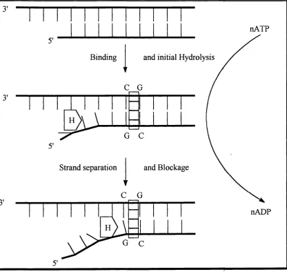
nATP
Binding and initial Hydrolysis
Strand separation and Blockage
nADP
fig 13 The helicase binds to the ssDNA. ATP is a requirement o f the process as the enzyme requires energy to overcome the stability o f the DNA helix; and to separate
Chapter 1 42
2.2.3 In vivo Reduction48
It is known that a variety o f mild reducing agents can cause the cleavage o f anthracyclines via a radical pathway (fig 14). In vivo, molecular oxygen can promote a similar pathway which produces radicals within the cell. This can pqtentially cause DNA scission as in the enediyne class of antibiotics."^^
OH OH
e" from
COCH3 m ild reducing
agent
OH OH
OH OR OH OR
sem iquinone
H +
OH OH OH OH
COCH:
OH
OH Or
hydroquinone quinone methide
OH
RSH
OH
7-deoxyaklavinone OH
OH OH
COCH
OH
SH
OCH OH
[O] M olecular O xygen
OH
OH SH
Chapter 1 43
The pathway goes via a semiquinone and a hydroquinone st^te at which time it is thought that the aglycone cleaves leaving a quinone methide. This quinone methide is susceptible to attack from variety o f nucleophiles whiclt exist in the cell including -SH, -NH2 and -OH groups. This quinone methide can, therefore, feasibly
be a reactive intermediate which attaches itself to the DNA and then the resultant adduct acts as an inhibitor o f cellular processes. Indeed, some worl^ers propose that metabolic activation is a prerequisite for cross linking o f anthracyclines to DNA by means o f a radical pathway although no structure was given for thç adduct, just the results o f dénaturation studies.
Initial studies using a daunomycin 9 and an oxidising agent as a treatment for leukaemia in vitro have proved to be successful, and this suggests that the mechanism as a proposed mode o f action is a valid one. With, a derivative of nogalamycin 13 called menogaril (nogalamycin aglycone withouf sugars at C l - replaced by OMe) it has been shown that, with in vivo reduction it is possible to get addition to N2 of guanine.^ ^ However, it must be noted, that for in vivo models to succeed, a method o f delivery which protects the agents from molecular oxygen must be devised. This will most probably take the form o f liposomes which are hydrophobic and may also aid delivery into the cell.
Whatever the mode o f action, there is a pronounced effect when these compounds are used in the fight against cancer. The drug aclacinopiycin 14, one of the less well studied anthracyclines, has been marketed for nearly twenty years as aclacin/aclarubicin. It has efficacy as a single agent, or combined with other drugs and can be used in a wide variety of illnesses including acute lymphocytic leukaemia and solid gastric, lung, breast and ovarian tumours. It is clear that there are very important effects in vivo associated with the anthracyclines, anc^ the majority o f evidence points to an effect as a result o f binding to dsDNA.
2.3 The binding of anthracyclines to DNA 2.3.1 Kinetics and thermodynamics
_____________________________________________________________________________ Chapter 1 44
binding kinetics as well. DuVemay noted an apparent two fold, increase in the binding constant per sugar residue added, with ATgpp being in the ran^e 1 - 6 x 10^ M*
1 52
This increase in binding constant with the number o f sugar residues has been mooted as grounds for increased sequence selectivity. However, sequence selectivity cannot be just a factor o f the final DNA-drug complex but rather q combination of kinetic and thermodynamic factors for both the drug in its bound conformation and the local DNA structure. For a drug to show sequence selectivity if must show fast association kinetics and slow dissociation kinetics as a result of favourable interactions in the binding site.
For intercalation to occur, in the case o f nogalamyciq 13, molecular modelling has shown that DNA breathing has to occur to allow the bqse pairs to open 10Â for the aglycone to thread through the DNA helix.^"^ This process is necessarily sequence dependent. However the binding o f the sugars in the mino^ groove o f DNA will be dependent on the local groove geometry, and may play a moi^e important role than the intercalation in the overall thermodynamics of the binding process.
CH
4 '
OH
OCH
OH" CH:
CH5
OH OH
CH:
Nogalamycin 13
______________________________________________________ Chapter 1 45
These separate pieces of work seem to have a consensus opinion in that the binding to DNA is a multistep process although the precise number o f steps remains unclear. Chaires postulates that that a three step process occurs ip the binding of daunomycin 9 to DNA (fig 15).^^
D + S
Cl
C
D = DNA
S = Substrate
C = Complex
fig 15 Mechanism for the kinetics o f binding (1)
Kinetics and thermodynamics point to the first step being thp formation of a loosely bound outside complex. This is seen to be endothermie and driven by the entropically favourable condensation of water from the phosphate backbone (AH = +2 . 1 kcal mol'% E ^ i = +1 2 . 8 kcal mof^) which is consistent with qther data for this
type of p r o c e s s . T h i s step as expected is found to be bimoleoular with a rate constant o f 2 . 8 x 1 0^ M '^s'\
The equilibrium constant for the second step (8.3) is equivalent to that found in the intercalation of proflavine l / ^ This most probably occurs^ via the thermal breathing o f the DNA having a large enough amplitude to accommodate the aglycone between the base pairs.
The third step is then postulated to be some form o f organisation within the complex with the rate seen to be similar to those found from studying the mode of binding of actinomycin.^^
Chapter 1 46
D + S
Cl
C
fig 16 Mechanism for the kinetics o f binding (2)
For the proposed mechanism of Chaires/^ the formation o f C l and C2 should
be dependent on the ionic strength of the solvent whilst the organisation o f the complex should be relatively indifferent to ionic strength. However this was not found to be the case, suggesting a different method of binding showp below (fig 17).
D + S
C
fig 17 Mechanism for the kinetics o f binding (3)
In this proposed mechanism both C2 and Cg are intercalate^ forms from the
outside complex C i and some form o f interplay can exist between the two. This could correspond to binding at two different sites and imply sequence specificity if the thermodynamics o f binding in mode C2 outweigh those for C3 ^nd both of these
are not just simply a different conformation of a complex bound to the same binding site.
The exact mechanism for the general binding of anthracyclines to DNA is still somewhat unclear. Nevertheless, since an overall negative enthalpy o f binding for daunomycin 9 to DNA was observed there must be a total negative enthalpy for the formation o f states C2 and C3 .
C h a p t e r 1 4 7
2.3.2 Structural aspects o f binding
2.3.2.1 Daunomycin 9
The major source o f information into how this class o f antibiotics interacts
with dsDNA comes from X-ray diffraction data and nmr data.^’’^"'Much attention has
been focused on daunomycin 9, as this was the first m em ber to have a crystal
structure published with it in its bound state. Daunomycin 9 is the simplest m em ber
o f the class, only possessing one sugar residue. It was co-cr>'stallisçd with the self-
complementary DNA hexamer d(CGTACG) and the X-ray diffraction pattern
determined to 1.2 The structure shows the 6 bp double helix o f DNA in its
B-fonn with two daunomycin 9 molecules intercalated, one at each end o f the
sequence in the d(CpG) step. The aglycone chrom ophore is orientated such that it is
perpendicular to the long axis o f the DNA, with the D ring protruding into the m ajor
groove o f the helix and the non-aromatic A ring positioned in tlje minor groove
(lig 18).
____________________________________________________________________________________C h a p t e r 1 4 8
1 he m ajor stabilising effect between drug and DNA arises as a result o f
interactions via direct hydrogen bonds to points above and below the site o f
intercalation.
fhere are two direct hydrogen bonds from 0 9 to N3 and N2 o f a
neighbouring guanine base (G2) (fig 19), which act as anchoring points within the
complex. In the m inor groove two bridging water molecules between the drug and
DNA stabilise the complex. In the major groove, a hydrated sodium cation is co
ordinated to N7 o f the terminal guanine (G1 ) and the 0 4 and 0 5 o f 9 with a distorted
octahedral geometry.
tig 19 The binding o f daunomycin 9 to DNA (2)
T he am ino sugar was found to lie in the minor groove without hydrogen
bonding to the DNA, with its functionalities pointing out into the aqueous medium.
However, this sugar was found to lie at distances o f between 1.99 Â and 3.49 Â
from the D N A helix in 21 instances (out o f a total o f 43 close contacts for the overall
complex). Since close van der Waals contacts are generally considered to be at
Chapter 1 49
playing a vital role in stabilising the complex. The role o f this sug^r in binding was highlighted by two further studies. It was found that the charged atnino group plays an important role in binding, in that its removal reduces the binding affinity by a factor of about 10^ (AT = 1 x 10^), but, the introduction o f an iodo substituent restores the binding constant, AT, to a value o f 1 x 10*.” Comparative binding studies were also used to evaluate the electrostatic contributions to DNA bindipg. In going from daunomycin 9 to hydroxyrubicin the charged amino group is replaced by a hydroxyl functionality. However, the observed decrease in free energy was larger than could be explained purely by the loss of a positive charge. Rather, it was ascribed to the unfavourable interaction between the negative potential around a hydroxyl group and the negative potential of the minor groove floor. Thus, although there is no direct contact between the sugar and DNA, the interactions present do play a major role in determining the binding.^^
It has been shown experimentally that the sugar is important for in vivo
effects. A daunomycinone aglycone itself has no biological activity^^ and it is thought that this is as a result of the amino group being able to interfere with the interaction o f topoisomerase and/or helicase with the DNA-daunomycin 9 complex.
Chapter 1 50
2.3.Z.2 Aclacinomycin 1468
OCH.
OH
OH OH
Me
NMe-Me'
HO
Me'
Aclacinomycin A 1 14
_____________________________________________________________________________ Chapter 1 51
complex, several interesting features show themselves in the structure derived from nmr data/*^
A stable 2:1 aclacinomycin 14:d(CGTACG) complex was found to exist in solution, similar to the work on daunomycin 9. The anisotropic shift of the protons in the anthracycline, as a result of the base pairs ring current, indicated that the aglycone was fully buried within the ClpG2 and C5pG6 steps, with H ll o f the aglycone at closest proximity. The structural refinement gave 10 possible binding structures all o f which showed a buckle at the intercalation.
The aglycone itself was found to be shifted 1.2 Â towards the minor groove. When compared to daunomycin 9 this shift in aglycone positioning alters the geometry o f the sugar side chain in the minor groove. The first sugar, rhodosamine, lies over the G2:C11 base pair, with the N- methyl groups proxipial to A 10. The remaining sugars were seen to show several nOe cross peaks indicating a distance of 4 Â-5 Â to the minor groove. More importantly, there was seen to be a 20° kink in the DNA backbone at the T3pA4 step due to a crowding between the 2'-deoxyfucose residue and the A4 deoxyribose. This forces a widening of the minor groove, and causes the A4N6 to form two hydrogen bonds to T904 (interstrand) and T304 (intrastrand) simultaneously. This effect may be, however, a copipounded effect from two species being intercalated in a small hexamer, and a§ a result o f the changed aglycone orientation as compared to daunomycin 9. This is a consequence of the CIO methoxycarbonyl group. Computer modelling showed that the addition o f a methoxycarbonyl group to the data for daunomycin 9 would lead to clash between it and the Cl:G 12 base pair. The complex for aclacinomycin 14 has this functionality 2.71 Â from N2 of G12. Work on MAR 70, which contains the daunomycin 9 aglycone and the first two sugars of aclacinomycin 14, showed no kink further confirming that it is the aglycone alignment which is causing the kipk.^^
It is interesting to note that there is no noticeable change in the anthracycline itself upon binding, which would tend to indicate that the ability to bind via
Chapter 1 52
Nmr experiments allow the dynamics o f binding to be probed. It was found that even in the 1:1 drug-DNA complex intercalation occurred at the CpG step. Since this is a more realistic guide to binding than sites seen by X-diffraction work, it has been suggested that this is evidence for a preferred binding site. However, the number of sequences surveyed was very limited. Any suggestion o f sequence selectivity is very tentative and further work is required to confirm or refute this.
It is also worth noting an earlier nmr paper on aclacinomycin 14 binding. This paper, although not as thorough as later papers in giving a model o f the binding, concentrated on the resonances o f the backbone o f the DNA. By studying the binding of aclacinomycin 14 to the oligonucleotide d(CCTAGG), it was possible to infer a binding site o f TpA. Although very limited, it does seem %o indicate that a TpA (or ApT) binding site is preferable to a CpC (GpG) and is preliminary evidence for sequence selectivity in this drug. However, no GpC steps arç present in this particular oligonucleotide to correlate with the work of Wang.
23,2.3 Nogalamvcin 13
The other widely studied anthracycline is nogalamycin 13. This is somewhat different from those already described, as it represents an increase in structural complexity. The aglycone chromophore is substituted at both ends by bulky sugar residues. At one end it has a nogalose sugar residue attached as in other anthracyclines at C7 whilst at the other end it has a positively charged bicyclic aminoglucose ring attached to the C l hydroxyl group and to
_____________________________________________________________________________________ C h a p t e r 1 53
26"). However, this may be as a result o f there being more helical constraints when a
species binds at the middle o f the sequence than at the ends. This is further
contlnned by the obseiwation that the c>1osine sugar ring is not seen to be puckered
in the nmr solution study.
In the X-ray diffraction study it was found that the DNA was in a distorted
B-form configuration, with the nogalose sugar residing in the m inor groove and the
amino glucose being positioned in the major groove. Once again it was seen that
there is very little difference between the free and bound drug, and the orientation o f
the two sugars on the same side o f the aglycone mirrors the turn o f the minor groove
and major groove around the intercalation site. Both sugars were found to be
pointing towards the AT section o f the DNA giving the m olecule a dumbbell-like
shape (tig 20).
______________________________________________________________________ Chapter 1 54
In the major groove there is an obvious hydrogen bond from the amino glucose hydroxyl to N7 of G2 (2.8 Â). This is the main stabilising influence at that end o f the molecule. At the extremity of the aglycone, the A ripg was found to protrude into the minor groove and is in a distorted conformation such that the hydroxyl at C9 is pseudo-equatorial, pointing away from the DNA and does not interact. The carbonyl group o f the methyl ester functionality at CIO, on the other hand, adopts an equatorial position, perpendicular to the aglycone and points directly at the DNA. As a result, it is seen that there are two hydrogen bonds from the amino group of G12 (or G6) at a distance o f 3.0 Â to this carbonyl group, The complex is
further stabilised by a hydrogen bond to the glycosidic ether linkagç from the amino group of G2 (G8) at a distance of 3.4 Â. It is noteworthy that if th^ compound was
intercalated between an AT sequence, adenine has no amino group in the minor groove and hence no minor groove hydrogen bond would be possible. This may explain any binding selectivity seen.
The nogalose sugar fills the minor groove, almost forming a continuous hydrophobic domain and is held by van der Waals forces. However, unlike many other minor groove binders, e.g. netropsin 3, which bind at AT riçh sequences and cause a slight narrowing, the nogalose sugar causes an expansipn o f the minor groove with the phosphorus-phosphorus distance being seen to increase from 11.5- 13 Â up to 15 Â (netropsin reduces it to 1 0 A).
In comparison to daunomycin 9, the aglycone, like in aclaçinomycin 14, is pulled towards the minor groove by a distance o f 2.0 A, and again the unwinding is seen to be very low at 11° (ethidium 2 unwinds DNA by 26°).
In the nmr study similar degrees o f unwinding were seen. It is useful to note that no direct interactions were found to the positively charged A^A-dimethyl amino group which is in the major groove. However, the positive charge was found to be close to the N 7/06 o f GIO at a distance of 4 A which could suggest water mediated hydrogen bonds. In comparison to daunomycin 8 this positive charge is in a very
similar position to where a sodium cation was found in the crystal structure.