DIAGNOSTIC DILEMMAS AND CLINICAL REASONING
A 2-Year-Old With 4 Weeks of Daily
Fever
John B. Darby, MDa, Lucette Liddell, BAb, Marietta DeGuzman, MDc, Kenneth L. McClain, MDd, Jared Rubenstein, MDa,
Lindsay Chase, MDa, Lucila Marquez, MD, MPHa,e
abstract
A 2-year-old female presents for evaluation of 4 weeks of daily fevers. When the fevers began, she had mild upper respiratory tract symptoms, which quickly resolved. The fevers persisted, however, with a maximum of 40°C. The child’s review of symptoms was significant for a 1-kg weight loss over the past month. Ten months before presentation, she had moved from Saudi Arabia with her family. One week before the onset of symptoms, she had visited a petting zoo. During episodes of fever, the patient was ill-appearing and had an elevated heart rate and respiratory rate. On examination, she was found to be thin, febrile, tachycardic, and with scattered lymphadenopathy. Results of laboratory tests were remarkable for an elevated white blood cell count of 16 100 cells per uL with a neutrophilic predominance. Erythrocyte sedimentation rate (ESR) and C-reactive protein (CRP) were elevated at 99 mm/h and 27 mg/dL, respectively. A chest radiograph indicated a small amount offluid in the interlobarfissures. Our expert panel examines her case, offers a definition of fever of unknown origin, and makes diagnosticconsiderations.
CASE HISTORY WITH SUBSPECIALTY INPUT
John Darby, MD, Moderator, Pediatric Hospital Medicine:
A 2-year-old girl was transferred for evaluation and management of fevers. She was well until 4 weeks before admission, when she developed symptoms of an upper respiratory tract infection (URTI) and intermittent fevers up to 40°C. Although the URTI symptoms resolved over 3 days, the patient continued to have daily fever spikes for the next 4 weeks. When febrile, she was ill-appearing, tachypneic, and tachycardic, but between fever spikes, she was a normal 2-year-old playing with her siblings. She had been evaluated in outpatient settings several times over the past month, with no definitive diagnosis, and had received ceftriaxone and clindamycin for unclear reasons, with
no improvement in symptoms. Her parents initially attributed the fevers to a viral illness she acquired from day care.
Review of systems was remarkable for weight loss of 1 kg (according to parental report) and poor appetite. Her medical history was unremarkable, and her immunizations were up to date through 1 year.
The child was born in Toronto but lived in Saudi Arabia until 10 months before the onset of symptoms, when the family moved to Toronto. She then moved to Texas with her parents and younger siblings 1 month before presentation. The child attends day care and had visited a petting zoo ∼1 week before the symptoms began. Although she had a negative reaction to purified protein derivative (PPD) after her time in Saudi Arabia, her parents have never been tested for tuberculosis (TB).
aSection of Hospital Medicine,cSection of Immunology, Allergy & Rheumatology, dSection of Pediatric Hematology and Oncology, Texas Children’s Cancer and Hematology Centers, and eSection of Infectious Disease, and bDepartment of Pediatrics, Baylor College of Medicine/ Texas Children’s Hospital, Houston, Texas
Dr Darby contributed to the design and execution of the case conference, drafted and edited the original manuscript, and made revisions to the manuscript; and Ms Liddell and Drs DeGuzman, McClain, Rubenstein, Chase, and Marquez contributed to the design and execution of the case conference, and reviewed and made revisions to the manuscript. All authors approved thefinal manuscript.
www.pediatrics.org/cgi/doi/10.1542/peds.2014-3692
DOI:10.1542/peds.2014-3692 Accepted for publication Mar 4, 2015
Address correspondence to John B. Darby, MD, Texas Children’s Hospital, 6621 Fannin St, Suite A210, Houston, TX 77030. E-mail: jbdarby@bcm.edu
PEDIATRICS (ISSN Numbers: Print, 0031-4005; Online, 1098-4275).
Copyright © 2015 by the American Academy of Pediatrics
FINANCIAL DISCLOSURE:The authors have indicated they have nofinancial relationships relevant to this article to disclose.
FUNDING:No external funding.
On presentation, the patient was thin and ill-appearing with a temperature of 38.5°C. Her pulse was 137 beats/min, her blood pressure was 100/55 mm Hg, and she had a respiratory rate of
64 beats/min. Her weight was 10.2 kg (fourth percentile for age) and height was 84 cm (15th percentile for age). She appeared ill but was in no acute distress. Cardiovascular examination was notable for tachycardia with no murmurs. Her lungs were clear, her liver was palpable 1 cm below the right costal margin, and results of the musculoskeletal examination were unremarkable. She had scattered lymphadenopathy in the anterior cervical, supraclavicular, and inguinal regions.
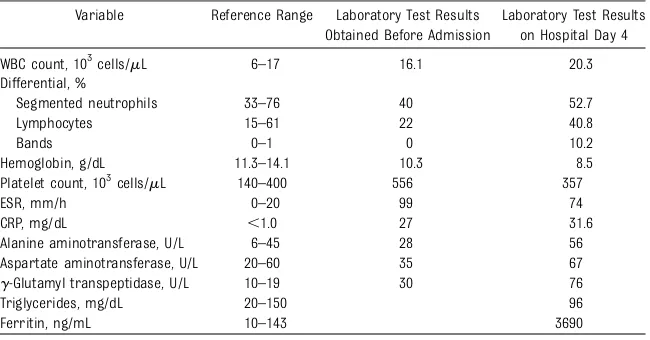
Results of the patient’s laboratory evaluation (completed at an outside facility) are shown in Table 1. They are notable for an elevated white blood cell count, platelet count, ESR, and CRP and mildly decreased hemoglobin level. Results of urine, blood, and stool cultures were negative. A chest radiograph indicated a small amount offluid in the interlobarfissures. A computed tomography (CT) scan indicated maxillary sinusitis; an abdominal ultrasound was normal.
With our patient in mind, I would like to hear a definition of fever of unknown origin (FUO).
Jan Drutz, MD, Academic Medicine:
I do not know if FUO has clearly been defined, particularly in children. From my 40 years of experience, I would say that a fever of.2 weeks in excess of 38.3°C, in which the patient has an otherwise normal history and physical examination, could be considered FUO. In addition, whatever evaluation was done (complete blood cell count, blood culture, urinalysis) is also normal. Recently, our colleague, Dr Palazzi, wrote that FUO was defined as fever
.38.3°C that lasted.8 days for which no diagnosis is apparent after initial evaluation.1
Dr Darby:
Does our patient meet the definition, in your mind?
Dr Drutz:
Yes, she certainly does.
Dr Darby:
How do you frame a differential for FUO in a pediatric patient?
Dr Drutz:
I would frame the differential depending on the age of the child. With a child younger than this patient, I would be concerned more about infectious diseases. For a 2-year-old, however, there are other possibilities. It could be any of the
collagen vascular diseases, an inflammatory condition, infectious disease, or malignancy.
Dr Darby:
Does the nature of the fever matter? Should we consider height, duration, and frequency when thinking about FUO cases?
Martin Lorin, MD, Academic Medicine:
In the era before immunizations againstHaemophilus influenzae type b andPneumococcus, patients with fever.40°C had a statistically significant greater chance of serious bacterial infection than those with a fever,40°C. Most of these data were in young infants, however.2
Nowadays, occasionally the fever pattern is helpful. This child clearly does not have 1 of the periodic fever disorders in which the fever chronicity is usually much longer. The fever may range from 2 to 3 days in familial Mediterranean fever up to 15 days in tumor necrosis factora receptor-1–associated periodic syndrome, and the periodicity/recurrence of fevers occurs over months to years. The fact that she has a daily febrile spike suggests possible bacterial infection but is absolutely classic for juvenile idiopathic arthritis (JIA) as well.
Dr Darby:
Is a connective tissue disorder therefore a real possibility?
Dr Lorin:
Statistically, infection isfirst, followed by rheumatologic disorders and malignancy. The longer the fever persists without explanation, the higher the possibility of the latter categories.
Dr Darby:
How do we decide to admit a patient with FUO?
TABLE 1 Laboratory Data Before Admission and on Hospital Day 4
Variable Reference Range Laboratory Test Results Obtained Before Admission
Laboratory Test Results on Hospital Day 4
WBC count, 103cells/mL 6–17 16.1 20.3
Differential, %
Segmented neutrophils 33–76 40 52.7
Lymphocytes 15–61 22 40.8
Bands 0–1 0 10.2
Hemoglobin, g/dL 11.3–14.1 10.3 8.5
Platelet count, 103cells/mL 140–400 556 357
ESR, mm/h 0–20 99 74
CRP, mg/dL ,1.0 27 31.6
Alanine aminotransferase, U/L 6–45 28 56 Aspartate aminotransferase, U/L 20–60 35 67 g-Glutamyl transpeptidase, U/L 10–19 30 76
Triglycerides, mg/dL 20–150 96
Ferritin, ng/mL 10–143 3690
Juan Juarez, MD, Pediatric Emergency Medicine:
The decision depends on whether the child’s condition is stable. For a child who is in stable condition, or who comes to the emergency department solely for a second opinion before release, this evaluation would likely be conducted as an outpatient. For an infant of a few weeks or months, the decision is less obvious as rapid clinical deterioration may occur.
There is also a consideration of resource availability. We often will send the child to outpatient care as long as we can guarantee proper follow-up with the primary care provider or referral to infectious disease or rheumatology, for example.
Lindsay Chase, MD, Pediatric Hospital Medicine:
I agree with Dr Juarez that the availability of reliable outpatient care is paramount in our decision to admit or discharge this patient. It would be important to contact the primary medical physician and ascertain key information about the child’s growth and existing evaluation and establish if it is reasonable to advance the evaluation as an outpatient. Aside from resource availability, however, there are some redflags that need to be considered. Even if“just”
according to parental report, a weight loss of 1 kg in a 2-year-old is concerning. We need to act more quickly for a 2-year-old losing weight than for a child with a fever only. In addition, the high elevation of CRP and ESR makes me concerned for a malignant or rheumatologic process. For these reasons, I would admit the patient.
Dr Darby:
Would you have included any other studies in the initial evaluation?
Dr Chase:
I would have sent serologic samples for cytomegalovirus and Epstein-Barr virus evaluation because they may
affect—or be affected by—future treatments. In addition, my infectious disease, rheumatology, and oncology colleagues often like to have baseline titers. There may also be infection-triggered systemic inflammatory conditions. I probably would not have initially included the assay for rheumatoid factor (without a specific recommendation for it from
a pediatric rheumatologist) because it is not sensitive for assessing
rheumatologic processes in children.
Dr Darby:
A CT scan performed before admission revealed maxillary sinusitis. Can we say that the diagnosis is bacterial sinusitis and send the patient home with
amoxicillin or amoxicillin/clavulanic acid?
Derek Zhorne, MD, Pediatric Hospital Medicine:
The American Academy of Pediatrics recently endorsed an updated clinical practice guideline for the diagnosis and management of acute bacterial sinusitis that made the diagnostic criteria more stringent.3According to these guidelines, we can make a presumed diagnosis of acute bacterial sinusitis in a patient with viral URTI symptoms with persistent illness, defined as having a daytime cough or nasal discharge, or both, for
.10 days. This patient had several days of congestion, rhinorrhea, and fever, and the nasal symptoms resolved after 3 days. This clinical picture seems more consistent with a viral URTI, despite the CT findings.
Dr Darby:
What is our next step? Do we involve consultants?
Dr Chase:
Considering the child’s age and that the 3 most probable etiologies are infection, rheumatologic disorder, and hematologic malignancy, I would start by consulting infectious disease, as infection often needs to be ruled out
to safely pursue treatments for malignant and rheumatologic conditions.
Dr Darby:
Dr Palazzi, how do you approach FUO?
Debra Palazzi, MD, Pediatric Infectious Diseases:
Because these cases evolve, it is important to continually repeat the history and physical examination. I would start with common viral serology tests and add laboratory tests according to any relevant items of history obtained. For this patient, it is important to know whether there was animal exposure at the petting zoo. Considering the length of time that has passed, and that most children with such infections present with gastrointestinal symptoms, the petting zoo is unlikely to be the culprit. There are, however, rare reports of Q fever associated with petting zoo exposures.
Dr Darby:
For the sake of discussion, what other pathogens would you consider?
Dr Palazzi:
The most common reports for petting zoo infections have cited enteric pathogens causing diarrhea or, less likely, constipation.
Aside from the petting zoo, I would also consider infections carried by kittens, as cat scratch disease is often the culprit in FUO cases. The clinical picture of hepatosplenic cat scratch fever is the ill-appearing patient with high-spiking fevers interrupted by periods of wellness, which is consistent with the patient’s presentation. Although her ultrasound was negative, the hypoechoic hepatosplenic lesions associated withBartonellainfection do not always appear on imaging.
than 10 months after her move to the United States, but a few may present months to years after exposure. Visceral leishmaniasis is an infection that comes to mind, and the diagnosis is sometimes made only after
observing organisms in tissue samples, such as from the bone marrow. These patients generally have substantial hepatosplenomegaly, however, which was not evident in this patient.
Dr Darby:
Would you consider TB as a possibility?
Dr Palazzi:
It is possible the patient could have active TB, but there is no focal evidence to pursue a TB diagnosis. She has lymphadenopathy, but it is diffuse rather than mediastinal or a single node. In addition, there were no infiltrates on the chest radiograph, and she has not progressed to more severe illness over these 4 weeks. The patient eventually had a negative
“T-SPOT.TB”test in addition to a negative PPD. T-SPOT.TBis an enzyme-linked immunospot used to assess for T cells in the peripheral blood that respond to TB antigen by producing interferon-g.
Dr Darby:
Results of the patient’s blood cultures were negative for aerobic and anaerobic pathogens. Also negative were the evaluations forToxoplasma, Brucella,Mycobacterium,Histoplasma, Bartonella, diphtheria, HIV, Epstein-Barr virus, cytomegalovirus, malaria, andCryptococcus. As mentioned, the TB evaluation (including a PPD and T-SPOT.TB) was negative. In addition, QuantiFERON-TB Gold (QFT), which is a specific interferon-grelease assay (IGRA), was sent and was reported as indeterminate.
What is IGRA, and what is meant by an indeterminate result?
Lucila Marquez, MD, MPH, Pediatric Infectious Disease:
The IGRA is essentially a PPD test in vitro. A sample of the patient’s
blood is taken, and the white blood cells are tested for their reactivity to TB antigen in the laboratory. Specifically, QFT provides the quantity of interferon-gproduced by T cells in reaction to those antigens. An indeterminate result equates to PPD anergy. The assays have both positive and negative controls. If the positive control does not react, we do not know if it is because the patient has never been exposed to TB antigens or because the patient does not have the ability to react because of immune suppression. Therefore, from an indeterminate result, we cannot exclude anything. The QFT is different from the T-SPOT.TBin that the number of cells producing interferon-gis counted in the T-SPOT. TBtest (in contrast to a measurement of the amount of interferon-g produced in the QFT test).
Dr Darby:
Are IGRAs accurate for a 2-year-old patient?
Dr Marquez:
The guidelines of the Centers for Disease Control and Prevention state that children aged ,5 years should be tested by using a PPD.4There are data for use of IGRAs in younger children, however. Anecdotally, we have seen neonates with congenital TB react on these assays.
Dr Darby:
Is the BCG immunization still part of preventive care in Saudi Arabia? How do you interpret those results if the patient was vaccinated with BCG?
Dr Palazzi:
IGRAs do not react to BCG, unlike a PPD. The IGRA has different antigens not included in the BCG immunization.
Dr Darby:
With results of urine and stool cultures negative, and the
aforementioned studies, we have at this point a negative infectious
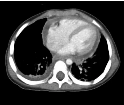
evaluation. There was evolution of her inpatient serum laboratory test results, as shown in Table 1. In addition, an antinuclear antibody profile was negative. A chest CT demonstrated a moderate pericardial effusion, thickening of the
pericardium, and bilateral small pleural effusions (Fig 1).
Although there was no joint pain or rash, and the antinuclear antibody profile was negative, the patient continued to spike daily fevers to 39.4°C during her hospital stay. Should we be concerned for a rheumatologic process?
Marietta DeGuzman, MD, Pediatric Rheumatology:
Yes, this possibility is concerning. We need to look at the patient during the height of the fever because some cutaneous changes in rheumatologic diseases will only be seen when the patient is febrile. When febrile, this child had a stiff, immoveable neck. These changes regressed when the fever subsided.
The patient’s anemia with increasing ferritin and elevated liver enzyme levels make rheumatologic disorder a strong possibility, although there was no arthritis at the time of the examination. We need to be concerned about vasculitides in this age group, particularly polyarteritis nodosa and Kawasaki disease. It is important to look at the coronary arteries. Early-onset sarcoidosis may present with unexplained prolonged fever. The negative antinuclear antibody profile, lack of renal disease, and the child’s age render the possibility of systemic lupus
erythematosus unlikely. We must also ensure that this is not a systemic reactive inflammatory process secondary to infection, such as Adenovirus,Parvovirus, or Streptococcus.
Dr Darby:
effusion with mild thickening of the pericardium. The child had elevated ferritin levels. Should we be concerned for the possibility of hemophagocytic lymphohistiocytosis (HLH)?
Kenneth McClain, MD, Pediatric Hematology-Oncology:
HLH is a syndrome of pathologic inflammation caused by a breakdown in the control of cytokine release. The clinical presentation encompasses fever and multisystem dysfunction. HLH often enters the differential of the FUO as infectious etiologies and others are ruled out.5Elevated levels of ferritin, D-dimer, and transaminase support a diagnosis. Aside from a drop in the hemoglobin, no cytopenias have yet occurred; often with autoimmune disease, however, the patient begins with a thrombocytosis, and the platelet count does not drop as quickly.
HLH is a syndrome diagnosed by using a synthesis of clinical and laboratoryfindings. The diagnosis is easiest if the patient has a molecular mutation in 1 of the 7 implicated genes found in familial cases. If molecular diagnosis is not confirmed, the patient should fulfill 5 of the
following 8 criteria: fever;
splenomegaly; cytopenias (affecting at least 2 of 3 cell lines);
hypertriglyceridemia; evidence of hemophagocytosis in bone marrow, spleen, or lymph nodes; low or absent natural killer cell activity; ferritin level .500mg/L; and soluble CD25/interleukin (IL)-2 receptor level .2400 U/mL.
In a child with mild
hepatosplenomegaly and ferritin levels .3000 mg/L, we should be suspicious for HLH and request further evaluation.6I would order a serum IL-2 receptor assay, which is an indication of T-cell
hyperactivation. An indication of hemophagocytosis also makes the diagnosis of HLH more compelling, although the diagnosis can be made without it.
Patients with HLH typically present with FUO and are ultimately diagnosed with the hemophagocytic syndrome when enough criteria are met. Their cytopenias are initially not dramatic, but the laboratory tests must be revisited frequently because HLH is a syndrome that evolves in its severity. Critically, the level of ferritin continues to rise in HLH. This rise is in contrast to shock, liver disease,
chronic transfusion, autoimmune disease, and bacterial infection, in which ferritin levels are high but stable. The combination of D-dimer,
g-glutamyl transpeptidase, and ferritin is helpful for looking at the diagnosis of HLH and response to therapy. We have found,
retrospectively, in our patients whose ferritin levels drop rapidly within 2 weeks of starting therapy that the mortality rate is below the average of 55%. By contrast, a decline,50% is associated with horrible mortality.7
HLH is difficult to diagnose and treat, and it requires constant vigilance and attention to levels of ferritin, D-dimer,g-glutamyl transpeptidase, and serum IL-2 receptors, as well as progressive cytopenias and organomegaly.8If a patient meets only 2 or 3 of the HLH criteria but develops renal or pulmonary failure, hypotension, and central nervous systemfindings, the consideration of HLH may be strengthened.
In this case, the patient had only a mild elevation in her ferritin level, and it did not rise. Her liver enzyme levels were also mildly elevated, and there was no significant thrombocytopenia or coagulopathy. At the onset of the hospitalization, the diagnosis was not consistent with HLH.
Dr Darby:
Dr DeGuzman, the patient remained febrile, and we still did not have a diagnosis. Can you tell us the diagnosis you made and why?
Dr DeGuzman:
The patient’s clinical profile of prolonged fever with organomegaly, serositis noted on chest CT as pericardial and pleural effusions, anemia of prolonged inflammatory process, and laboratory indicators of systemic inflammation, as well as a most reasonable diagnostic exclusion of infection, malignancy, and the primary vasculitides, define the most likely diagnosis of systemic-onset FIGURE 1
juvenile idiopathic arthritis (SoJIA). During her hospital stay, the child developed the characteristic rheumatoid rash. Although she has had no peripheral arthritis, our patient developed stiffness of her cervical spine in association with the febrile episodes. It is not unusual that persistent arthritis, a diagnostic criterion for JIA, develops weeks or months after the onset of the systemic features.
During the latter part of the patient’s hospital stay, her fever became characteristically that of a quotidian type, with 1 to 2 spikes per day, occurring in the early morning. In between the febrile episodes, she was well-appearing, a characteristic feature that differentiates SoJIA from infectious and malignant disorders and other systemic rheumatologic disorders.
With the diagnosis of SoJIA, this patient’s treatment initially included corticosteroids
(intravenous methylprednisolone) and anakinra (an IL-1 receptor antagonist). This treatment resulted in improvement in both the
clinical and laboratory features of systemic inflammation.
In summary, SoJIA is a diagnosis made clinically with the most reasonable exclusion of the infectious and malignant processes.
Dr Darby:
This case is a very interesting example of SoJIA. Can you tell us more about this disease?
Dr DeGuzman:
JIA is the most common chronic rheumatic disease of childhood. It is a heterogeneous disorder
encompassing different forms, and its etiology is still unclear. Diagnosis is defined before age 16 years, with occurrence of persistent arthritis of
.6 weeks and for which other causes for arthritis are not identified. Subsequent classification is based on the number of joints involved during
thefirst 6 months of the disease and on the extra-articular features.9
SoJIA has markedly distinct clinical and laboratory features from the other subsets of JIA, suggesting a different pathogenesis.10Fever of prolonged nature is the hallmark systemic feature, the nature of which often cannot initially be differentiated from infectious or malignant causes. The characteristic quotidian pattern, seen in fewer patients, may not be apparent until the latter phase of the illness. Other manifestations include a salmon-colored, evanescent rash (rheumatoid rash) that comes and goes with the fever, serositis, lymphadenopathy, and
hepatosplenomegaly. Muscle and joint pain are common with the febrile episodes. The presence of at least 1 active synovitis is a mandatory supportivefinding for the diagnosis. It is, however, common for the arthritis to not be clinically appreciated until weeks after the onset of the systemic features. Increased markers of systemic inflammation (CRP and ESR) and the absence of autoantibodies (eg, rheumatoid factor, cyclic citrullinated peptides) are important laboratory
findings in SoJIA. Hyperferritinemia, anemia, leukocytosis, and
thrombocytosis are also common laboratory features.
Another distinct feature of SoJIA is its association with macrophage
activation syndrome, or a secondary HLH, which may occur on
presentation or later in the clinical course. It is a dangerous syndrome and is associated with increased mortality in patients with SoJIA. Hyperferritinemia is a common feature of SoJIA, and in association with cytopenia, transaminase elevation, hypofibrinogenemia, declining ESR, and elevated
triglycerides, it defines a diagnosis of macrophage activation
syndrome.11,12
New insights into the pathogenesis of SoJIA have illuminated the role of
uncontrolled activation of phagocytes with hypersecretion of IL-1 and IL-6 and have changed the paradigm of this illness as an autoinflammatory disorder.13Better understanding of the pathogenesis likewise has provided targeted treatments (anti–IL-1 and anti–IL-6 agents) that are more effective and safer than earlier medications.14,15
SoJIA has a variable outcome, and a monocyclic course with minimal or absent articular complications has been noted in∼50% of
reported cases, with the rest having a polyphasic or polyarticular course.
Dr Darby:
How is our patient doing?
Dr DeGuzman:
She responded well to anakinra, the IL-1 receptor antagonist, and had resolution of fevers and joint pain and improvement in her inflammatory markers. The corticosteroids were stopped, and she achieved disease remission after several months of IL-1 blockade therapy.
ACKNOWLEDGMENTS
We thank Jan Drutz, MD, Martin Lorin, MD, Juan Juarez, MD, Derrick Zhorne, MD, and Debra Palazzi, MD.
REFERENCES
1. Palazzi DL. Fever of unknown origin in children: Evaluation. Houston, TX: UpToDate, 2013. Available at: http:// www.uptodate.com/contents/fever-of-unknown-origin-in-children-evaluation. Accessed July 30, 2013
2. Bonadio WA, McElroy K, Jacoby PL, Smith D. Relationship of fever magnitude to rate of serious bacterial infections in infants aged 4-8 weeks.Clin Pediatr (Phila). 1991;30(8):478–480
3. Wald ER, Applegate KE, Bordley C, et al; American Academy of Pediatrics. Clinical practice guideline for the diagnosis and management of acute bacterial sinusitis in children aged 1 to 18 years.
www.pediatrics.org/cgi/content/full/132/ 1/e262
4. Department of Health and Human Services, Centers for Disease Control and Prevention (CDC). Updated guidelines for using interferon gamma release assays to detectMycobacterium tuberculosisinfection - United States, 2010.MMWR Morb Mortal WklyRep. 2010; 59(RR_5). Available at: www.cdc. gov/mmwr/PDF/rr/rr5905.pdf. Accessed March 18, 2015
5. Palazzi DL, McClain KL, Kaplan SL. Hemophagocytic syndrome in children: an important diagnostic consideration in fever of unknown origin.Clin Infect Dis. 2003;36(3):306–312
6. Allen CE, Yu X, Kozinetz CA, McClain KL. Highly elevated ferritin levels and the diagnosis of hemophagocytic lymphohistiocytosis.Pediatr Blood Cancer. 2008;50(6):1227–1235
7. Lin TF, Ferlic-Stark LL, Allen CE, Kozinetz CA, McClain KL. Rate of decline of ferritin in patients with hemophagocytic lymphohistiocytosis as a prognostic variable for mortality.Pediatr Blood Cancer. 2011;56(1):154–155
8. Jordan MB, Allen CE, Weitzman S, Filipovich AH, McClain KL. How I treat hemophagocytic lymphohistiocytosis.
Blood. 2011;118(15):4041–4052
9. Petty RE, Southwood TR, Manners P, et al; International League of Associations for Rheumatology. International league associations for rheumatology classification of juvenile idiopathic arthritis: section revision.J Rheumatol. 2004;31(2):390–392
10. Mellins ED, Macaubas C, Grom AA. Pathogenesis of systemic juvenile idiopathic arthritis: some answers, more questions.Nat Rev Rheumatol. 2011;7(7): 416–426
11. Davì S, Consolaro A, Guseinova D, et al; MAS Study Group. An international consensus survey of diagnostic criteria for macrophage activation syndrome in systemic juvenile idiopathic arthritis.
J Rheumatol. 2011;38(4):764–768 12. Minoia F, Davì S, Horne A, et al; Pediatric
Rheumatology International Trials Organization; Childhood Arthritis and Rheumatology Research Alliance; Pediatric Rheumatology Collaborative Study Group; Histiocyte Society. Clinical
features, treatment, and outcome of macrophage activation syndrome complicating systemic juvenile idiopathic arthritis: a multinational, multicenter study of 362 patients.
Arthritis Rheum (Munch). 2014;66(11): 3160–3169
13. Pascual V, Allantaz F, Arce E, Punaro M, Banchereau J. Role of interleukin-1 (IL-1) in the pathogenesis of systemic onset juvenile idiopathic arthritis and clinical response to IL-1 blockade.J Exp Med. 2005;201(9):1479–1486
14. Nigrovic PA, Mannion M, Prince FHM, et al. Anakinra asfirst-line disease-modifying therapy in systemic juvenile idiopathic arthritis: report of forty-six patients from an international multicenter series.Arthritis Rheum. 2011;63(2):545–555
15. De Benedetti F, Brunner H, Ruperto N, Calvo N, Cuttica I, Malattia R, et al. Tocilizumab in patients with systemic juvenile idiopathic arthritis: efficacy data from the placebo-controlled 12-week part of the phase 3 TENDER trial.
DOI: 10.1542/peds.2014-3692 originally published online April 27, 2015;
2015;135;902
Pediatrics
Rubenstein, Lindsay Chase and Lucila Marquez
John B. Darby, Lucette Liddell, Marietta DeGuzman, Kenneth L. McClain, Jared
A 2-Year-Old With 4 Weeks of Daily Fever
Services
Updated Information &
http://pediatrics.aappublications.org/content/135/5/902 including high resolution figures, can be found at:
References
http://pediatrics.aappublications.org/content/135/5/902#BIBL This article cites 12 articles, 5 of which you can access for free at:
Subspecialty Collections
oskeletal_disorders_sub
http://www.aappublications.org/cgi/collection/rheumatology:muscul
Rheumatology/Musculoskeletal Disorders
b
http://www.aappublications.org/cgi/collection/infectious_diseases_su
Infectious Disease
following collection(s):
This article, along with others on similar topics, appears in the
Permissions & Licensing
http://www.aappublications.org/site/misc/Permissions.xhtml in its entirety can be found online at:
Information about reproducing this article in parts (figures, tables) or
Reprints
DOI: 10.1542/peds.2014-3692 originally published online April 27, 2015;
2015;135;902
Pediatrics
Rubenstein, Lindsay Chase and Lucila Marquez
John B. Darby, Lucette Liddell, Marietta DeGuzman, Kenneth L. McClain, Jared
A 2-Year-Old With 4 Weeks of Daily Fever
http://pediatrics.aappublications.org/content/135/5/902
located on the World Wide Web at:
The online version of this article, along with updated information and services, is
by the American Academy of Pediatrics. All rights reserved. Print ISSN: 1073-0397.