OF IONISING RADIATIONS
Lawal Abdul Kadiri
A Thesis Submitted for the Degree of PhD
at the
University of St Andrews
199
1
Full metadata for this item is available in
St Andrews Research Repository
at:
http://research-repository.st-andrews.ac.uk/
Please use this identifier to cite or link to this item:
http://hdl.handle.net/10023/13336
BIOLOGICAL EFFECTIVENESS
OF IONISING RADIATIONS.
A thesis presented for the degree of Doctor of Philosophy .
University of St. Andrews.
St.Andrews, Fife, KY16 9SS,
United Kingdom.
May, 1990
BY
Lawal Abdul KADIRI
All rights reserved
INFORMATION TO ALL USERS
The quality of this reproduction is dependent upon the quality of the copy submitted.
In the unlikely event that the author did not send a com plete manuscript and there are missing pages, these will be noted. Also, if material had to be removed,
a note will indicate the deletion.
uest
ProQuest 10170779
Published by ProQuest LLO (2017). Copyright of the Dissertation is held by the Author.
All rights reserved.
This work is protected against unauthorized copying under Title 17, United States C ode Microform Edition © ProQuest LLO.
ProQuest LLO.
789 East Eisenhower Parkway P.Q. Box 1346
Declaration ii
Certificate iii
List of publications iv
Acknowledgements v
Dedication vi
Abstract vii
Table of text context viii
List of Tables xii
List of Figures xiii
DECLARATION.
I Lawal Abdul KADIRI hereby declare that this thesis has been composed by myself, that it is a record of my own work, and that it has not been accepted in partial or complete fulfilment of any other degree or professional qualification.
This research was earned out in the Department of Physics and Astronomy in the University of St. Andrews under the supervision of Dr. D. E. Watt.
Lawal Kadiri. Date.
I was admitted to the Faculty of Science of the university of St. Andrews under Ordinance General No 12 on 3rd Febuary, 1987 and as a candidate for the degree of Ph.D on 1st October, 1987.
Lawal Kadiri. Date...
--CERTIFICATION
I hereby certify that the candidate Lawal Abdul KADIRI, has fulfilled the conditions of the Resolutions and Regulations of the University of
St. Andrews appropriate to the Degree of Doctor of Philosophy.
David E. Watt Date...î.T/.î:/.?.S.
Research Supervisor
D.E.Watt, C.Z.Chen, L.A.Kadiri and A.Younis. Towards a unified system for the | expression of Biological damage by ionising radiation. IN: Health effects of low dose of
ionising radiation-Recent advances and their implications. BNES, London, 1988, pages 37-41.
D.E. Watt and L.A. Kadiri. Physical quantification of the biological effectiveness of ionising radiations. Int. J. Quantum Chemistry. In press.
L.A.Kadiri and D.E.Watt. Common mechanism in the induction of various biological effects of ionising radiations. Presented at the ARR meeting. St.Andrews, 3-5 January,
1990. Abstract published in Int, J. Radiat. Biology, 57 (1990) 1259.
i
L.A.Kadiri, D.E. Watt and I.A.M . Al-Affan. Biophysical calculations of the initial yield
"Yettore tabbitino ha Jaumirawo". I wish to express my gratitude to the following.
My supervisor. Dr. D. E. Watt, for the valuable guidance, help, advice, encouragement and stimulating discussions throughout the course of this work. His accessibility, friendliness, willingness to assist in both academic and nonacademic matters are exemplary manners of a supervisor and a gentleman.
The academic staff and students of Physics and other departments of the University of St.Andrews for a variety of assistance. Messes R. McGraw, Myles White, Tom McQueen, J. Clark, M. Robertson and their collaborators at the Mechanical and Electronic workshops who have offered valuable technical assistance. E. Bell and L. Mordi of the Library and many other supporting staff of the Physics Department for excellent services.
Elias, Chen, Younis, Yusuf, Aba-Umar, Ibrahim and many fellow colleagues at the Radiation Research Laboratory for their understanding, discussions, and encouragement which provided an atmosphere conducive for research. Aminu, Abdullahi, Garba, JK, Yahya, ThankGod, Ralphel, Wale and many other friends who have made my stay in St. Andrews easier.
Ahmadu Bello University, Zaria, in collaboration with the Federal Government of Nigeria, for the fellowship that enabled this work to be done.
Dr. S. B. Elegba, Dr. J. Adetunji, Prof. J. Kiefer, Prof. D.E. Ajakaiye, Mallam H. Mijinyawa and many other teachers, supervisors and Mends at Zaria and elsewhere for providing continuing moral and academic guidance.
Jika, Sa*ad, Habu, Mahmud and many other friends for offering me cushions of support at difficult times.
VI
JJ2J3WAT10N.
^
Thts work, is to dedicated to Mappa,
N an d u , H am m a an d J'taUum ,
[
r,I
ABSTRACT
It was shown that the conventional radiation dosimetric system which is based on RBE and LET is incapable of determining the likely consequences of ionising radiation exposure. Analyses of data on the induction of the chromosome aberrations, mutations and transformation in mammalian cells by radiations of different types and energies has indicated that (a) the induction of double strand breaks (dsb) in the DNA is their common critical lesion, (b) Fast ions and neutrons radiations are by order of magnitude more damaging than photons and electrons of equal mean free path, (c) Damage is through intra track action of the charged particles.
A new system of radiation dosimetry, which does not require a radiation quality parameter, was proposed. It was based on the observation that for each of the biological endpoints considered an Absolute Biological Effectiveness (ABE) for damage by the charged particles can be defined as the product of the charged particle fluence and the saturation effect cross section, scaled with the efficiency (e) of damage by radiation of mean free path (X). eis given by l-exp-( Xo/X), where Xq, about l.Snm, is the mean
inter-strand distance of the DNA.
The physical requirements for its instrumentation, basically the emulation and quantification of the induction of dsb in the DNA, were defined. The feasibility for its realisation using detectors based on gas ionisation, superconductivity, secondary electron emission, and semiconductivity was assessed. Ultrathin films of rectified, organic semiconductors appeared to have the best potential, but such materials are not yet available in the physically characterised form as may be required for detector construction; investigations were made with available films of plastic scintillators.
TABLE OF TEXT CONTEXT
CHAPTER ONE:
INTRODUCTION AND SCOPE 1
1.1 THE NEED FOR AND THE PROBLEMS OF RADIATION DOSIMETRY 1
1.2 THE ROLE OF RADIOBIOLOGICAL, MODELS 2
1.3 RADIATION QUALITY: THE LET 4
1.4 THE CONVENTIONAL DOSIMETRIC SYSTEM: ITS HANDICAPS 6
1.5 THE NEED FOR ALTERNATIVE RADIATION DOSIMETRIC SYSTEM 10
1.6 SCOPE OF THE THESIS 12
CHAPTER TWO:
FUNDAMENTAL PHYSICAL MECHANISMS OF RADIATION ACTION 14
2.0 PREFATORY REMARKS 14
2.1 DIRECT AND INDIRECT ACTION 16
Z2 REVIEW: RADIOSENSITIVE SUES AND LESIONS FOR CELL DEATH 19
2.2.1 RADIOSENSITIVE SITES AND THE CELL NUCLEUS 19
2.2.2 DNA AS THE TARGET 20
2.2.3 DNA STRAND BREAKS AS THE CRITICAL LESIONS 21
2.2.4 THE POSSIBILITY OF OTHER TARGETS FOR CELL DEATH 22
2.3 REVIEW OF PREVIOUS WORK OF W ATT AND COLLABORATORS 19
2.4 CALCULATION OF PHYSICAL DATA 26
2.4.1 MEDIUM 26
2.4.2 FAST IONS 26
2.4.3 PHOTONS AND ELECTRONS 27
2.4.4 NEUTRONS 28
2.5 CHROMOSOME ABERRATIONS 29
2.5.1 SIGNIFICANCE AND DATA TREATMENT 29
2.5.2 RESULTS 37
2.6 CELLULAR MUTATIONS: 39
2.6.1 TREATMENT OF DATA 39
2.6.2 RESULTS 44
2.7 CELLULAR TRANSFORMATION 49
2.7.1 TREATMENT OF DATA 29
2.8 DNA STRAND BREAKS: 52
2.8.1 TREATMENT OF DATA 52
2.8.2 RESULTS 55
2.9 GENERAL DISCUSSIONS 57
2.9.1 INTRA-TRACK AND INTER-TRACK ACTION 57
2.9.2 INAPPROPRIATENESS OF ABSORBED DOSE, RBE AND LET 58
2.9.3 THE ROLE OF ELECTRONS AND DELTA-RAYS 60
CHAPTER THREE:
APPROACHES TO INSTRUMENTATION IN UNIFIED DOSIMETRY 63
3.1 REQUIREMENTS OF A DETECTOR FOR UNIFIED DOSIMETRY 63
3.1.1 PHYSICAL REQUIREMENTS 64
3.1.2 MODE OF OPERATION AND INTERPRETATION OF RESPONSE 66
3.2 GAS BASED DETECTORS 69
3.2.1 THE BASIC PRINCIPLES OF THE ROSSI COUNTER 69
3.2.2 THE ROSSI COUNTER AND NANODOSIMETRY 69
3.2.3 THE VARIANCE AND VARIANCE-COVARIANCE TECHNIQUES 70
3.2.4 DETECTORS BASED ON ION RECOMBINATION 71
3.2.5 THE MULTISTEP AVALANCHE COUNTERS 72
3.2.6 THE GAS-SCINTILLATOR PROPORTIONAL COUNTER 73
3.3 DETECTORS BASED ON. SECONDARY ELECTRON EMISSION 73
3.4 DETECTORS BASED ON SEMICONDUCnVITY 75
3.5 DETECTORS BASED ON SUPERCONDUCTIVITY 79
3.5.1 PRINCIPLE 79
3.5.2 TUNNEL JUNCTIONS AS RADIATION DETECTORS 79
3.5.3 JOSEPHSON JUNCTIONS AS FAST SWITCHES 81
3.5.4 REMARKS ON SUPERCONDUCTIVE DETECTORS 82
3.6 OTHER SOLID STATE DETECTORS 84
3.7 REMARKS ON THE VARIOUS DETECTION SYSTEMS 85
CHAPTER FOUR:
PLASTIC SCINTILLATORS FOR ABSOLUTE DOSIMETRY 4.1 BASIC CONSIDERATIONS
4.2 REVIEW OF EXPERIMENTAL DATA
4.2.1 RESPONSE OF THICK ORGANIC SCINTILLATORS 4.2.2 RESPONSE OF THIN PLASTIC SCINTILLATORS
4.3 SPECIFIC LUMINESCENCE AND LINEAR PRIMARY IONISATION 93
4.4.1 INPUT DATA AND CALCULATION 94
4.4.2 RESULTS 95
4.5 DISCUSSIONS 97
4.5.1 DIFFERENCE IN DAMAQE PROCESSES 97
4.5.2 THE ROLE OF DELTA RAYS 99
4.5.3 Z*2/p^ AND LINEAR PRIMARY IONISATION 102
4.5.4 CHARACTERISTIC DISTANCES ESI PLASTIC SCINTILLATORS 103
4.5.5 SCINTILLATION AND UNIFIED DOSIMETRY 103
CHAPTER FIVE:
EXPERIMENTAL WORK 105
5.0 OBJECTIVES 105
5.1 TH IN FILM FABRICATION 105
5.1.1 OVERVIEW AND SELECTION OF FABRICATION TECHNIQUE 105
5.1.2 THE MAKING OF THIN FILMS 107
5.1.3 RESULTS AND COMMENTS ON FILM FABRICATION 108
5.2 THICKNESS AND UNIFORMITY MEASUREMENTS 110
5.2.0 OVERVIEW AND SmjECnON OF PERTINENT METHODS 111
5.2.1 OPTICAL MEASUREMENTS OF FILMS THICKNESS 111
5.2.2 WEIGHING 112
5.2.3 ALPHA PARTICLE TRANSMISSION METHOD 113
5.2.4 DETERMINATION OF FILM UNIFORMITY 116
5.3 THE PHOTOMULTIPLIER AND ITS PARAPHERNALIA 117
5.3.1 THE PHOTOMULTIPLIER 118
5.3.2 THE VOLTAGE DIVIDER NETWORK 120
5.4 THE DETECTOR ASSEMBLY 122
5.4.2 THE ALUMINIUM MIRROR 122
5.4.3 THE LUCITE LIGHT GUIDE 124
5.5 OPTIMISATION OF EXPERIMENTAL CONDITIONS 125
5.5.1 OPERATING CONDITIONS OF THE PHOTOMULTIPLIER 126
5.5.2 MODERATION OF THE ALPHA PARTICLE ENERGY 127
5.5.3 DETERMINATION OF COINCIDENT SCINTILLATION SPECTRA 127
5.5.4 DETERMINATION OF OTHER SCINTILLATION SPECTRA 128
5.6.1 DARK NOISE AND COINCIDENCE COUNTING 130
5.6.2 ENERGY LOSS AND FILM THICKNESS 132
5.6.3 THE RESPONSE AND THICKNESS OF THIN FILM DETECTOR 134
5.6.4 RESOLUTION AND THICKNESS OF THIN FILM DETECTOR 137
5.6.5 EFFICIENCY AND THICKNESS OF THIN FILM DETECTOR 140
5.6.6 INCIDENT ENERGY AND RESPONSE CHARACTERISTICS 143
5.6.7 TOD AND CONVENTIONAL SCINTILLATOR DETECTOR 146
5.7 THIN FILM DETECTOR AND UNIFIED DOSIMETRY 149
5.8 ENHANCEMENT OF THE LUMINESCENT RESPONSE 152
CHAPTER SIX:
CONCLUSIONS AND SUGGESTIONS FOR FUTURE WORK 154
6.1 INFERENCES MADE IN CHAPTER TWO 154
6.2 INFERENCES MADE IN CHAPTER THREE 155
6.3 INFERENCES MADE IN CHAPTER FOUR 156
6.4 INFERENCES MADE IN CHAPTER FIVE 157
6.5 SUGGESTIONS FOR FUTURE WORK 157
REFERENCES 160
H
LIST OF TABLES
2.1 The seven most common elements of a Veference'man 14
2.2 The yield of the primary radiolsis products 17
2.3 Doses required in the nucleus, cytoplasm and membrane for inactivation of
CHO cells at 50% survival level 19
2.4 Values of physical parameters taken for liquid water as the main
component of cellular mass 26
2.5 Radiation types and sources of the data collated for figures 2.8 to 2.13 30
4.1 The Chemical composition of NE102A 94
I
FIGURE
LISTL OF FIGURES
CAPTION 1.1 1.2 2.1 2.2 2.3 2.4
The survival fraction expressed as a function of dose for V79 Chinese Hamster cells irradiated with radiations of different type and energy The relationship between RBE and LET for cellular inactivation by radiations of different quality
The 'normalised* effect cross section for cellular inactivation plotted against the radiations linear primary ionisation
The linear coefficient, a (per gray ) for dicentric aberrations induction plotted against the track-averaged LET.
The linear coefficient, a (per gray ) for dicentric aberrations induction plotted against the dose-averaged LET,
The linear coefficient, a (per gray ) for dicentric aberrations induction plotted against the linear primary ionisation.
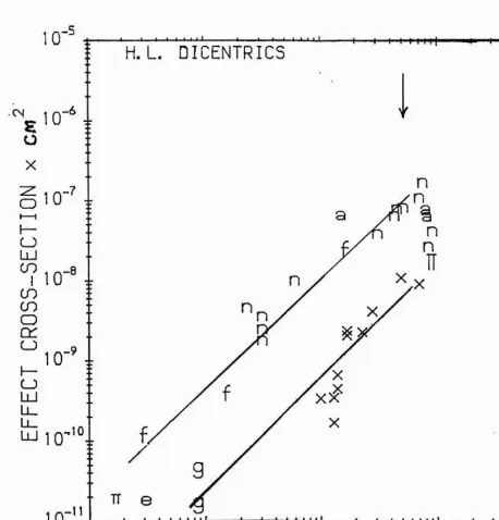
2.5 The effect cross section for dicentric induction plotted against the dose-average LET.
2.6 The effect cross section for dicentric induction plotted against the track-average LET.
2.7 The effect cross section for dicentric induction plotted against the linear primary ionisation.
2.8 The effect cross section for 6-TG*^ mutation induction in mammalian cells plotted against the track-averaged LET.
2.9 The effect cross section for the induction of mutations in yeast cells plotted against the track-averaged LET.
2.10 The effect cross section for induction of 6-TG*" in mammalian cells plotted against the linear primary ionisation.
2.11 The effect cross section for mutation induction in yeast cells plotted against the linear primary ionisation.
2.12 The effect cross section for induction of 6-TG** in mammalian cells plotted against the maximum energy of the 5-rays generated by the ions.
I
2.13 The effect cross section for mutation induction in yeast cells plotted against
the maximum energy of the 8-rays generated by the ions. 48 2.14 The effect cross section for the induction of transformation in C3H10T1 /2
cells plotted against the linear primary ionisation of the charged particles. 50 2.15 The effect cross section for the induction of strand breaks in mammalian
cells plotted against the linear primary ionisation. 53 2.16 The effect cross section for the induction of strand breaks in a wild and
inactivation in repair deficient yeast cells and the linear primary ionisation. 54 3.1 Current-voltage characteristics of a Josephson tunnel junction 80 4.1 Specific fluorescence versus calculated specific energy loss in NE102 87 4.2 Scintillation efficiency versus energy in NE102A 89 4.3 Light response of TFD to transiting ions and the specific energy loss 90 4.4 The specific fluorescence and the specific energy loss for various ions in Nal 92 4.5 The conversion efficiency and the specific energy loss for various heavy
ions in Nal 92
4.6 Specific luminescence and linear primary ionisation for ions stopped in NE102 95 | 4.7 Light response of TFD to transiting ions and the linear primary ionisation 96
4.8 Specific fluorescence and linear primary ionisation of fast ions stopped in Nal 97
4.9 The specific luminescence and the delta-ray maximum energy for ions stopped |
in NE102A. 99
J 4.10 Light response and energy of the delta-rays for heavy ions transversing the
TFDofNE102A. 100
4.11 Light response and energy of the delta-rays for light ions transversing the TFD
ofNE102A 100
4.12 The specific luminescence of ions stopped in NaI(Tl) and the ion's delta-ray I
energy of the ions. 101
^ 4.13 The relationship between specific luminescence and the yield of delta-rays
inN alO l) 102
5.1 The dip coating method of making thin films 108
5.2 Film thickness and withdrawal speed of a microscope slide out of the
scintillator solution. 109
5.3 The set up of the Michelson interferometer for measuring optical thickness. 112 5.4 The vacuum chamber for film's uniformity and thickness measurements 114 5.5 The electronic set up for the measurement of films thickness and uniformity 114
5.6 Calibration and linearity test of the MCA 115
bialkali photocathode and the reflectivity of evaporated aluminium 118 5.8 The voltage divider network for the photomultiplier. 121 5.9 The irradiation chamber for thin film detector 123
5.10 The Aluminium mirror. 124
5.11 The Perspex block film support 125
5.12 The electronic set up for scintillation counting 127 5.13 The set up for the irradiation of thick films mounted directly on
the photomultiplier. 129
5.14 The response of 5um thick NE102A film in the absence of coincidence
and the noise spectrum. 130
5.15 The spectra of 5um NE102A film obtained with and without the
use of coincidence. 131
5.16 Film's thickness and energy loss in the film. 132 5.17 Energy loss and energy loss straggling in air and in thin film. 133 5.18 Thin film detector light output and thickness. 134 5.19 The coincident spectra of NE104 films of different thickness. 136 5.20 Pulse-height resolution of TFD and film thickness. 137 5.21 The TFD response resolution and film thickness. 138
5.22 Detection efficiency and film thickness. 141
5.23 Relative scintillation efficiency and energy of the radiations. 143 5.24 The energy loss in films and the energy of the radiations. 144 5.25 The relative dL/dE and the energy of the radiations 144 5.26 The pulse-height resolution and the energy of the alpha particles. 145 5.27 Light response of a film in the TFD set up compared with that laid directly
on the photomultiplier. 146
5.28 The light response of TED set up compared with that directly laid
on the photomultiplier. 147
5.29 The light output of a 50um and a lOOum thick films, in the TFD set up
as the incident energy is varied. 148
5.30 The light output of a 50um and a lOOum thick films, laid directly
on the Photomultiplier, as the incident energy is varied. 149
LIST OF ABBREVIATIONS
ABE Absolute biological effectiveness ADC Analouge digital converter AC (or DC) Alternating (or Direct) current DNA Deoxyribonucleic acid
dsb double strand break
CHO Chinese hamster ovary (cells).
csda continous slowing down approximation cps counts per second
FWHM Full width at half maximum HPRT hypoxanthinetransferabusyl HVL Half value layer
ICRP International commission on radiological protection
ICRU International commission on radiological measurements and units LET Linear energy transfer
MCA Multichannel analyser MS AC Mutistep avalanche counter
MWPC Mutiwire proportional counter \
PM Photomultiplier
PPAC Pairallel plate avalanche counter RBE Relative Biological Effectiveness RQI Radiation Quality Index
QE Quantum efficiency SBD Surface Barrier Detector sc sacchramomyces cerevisiae SCA Single Channel Analyser SEE secondary electron emission SNR signal-to-noise ratio
ssb single strand break
STJ Superconductive tunnel junction TE Tissue equivalent
TFD Thin film detector (plastic scintillator) 6-TG^ 6-thioguanine ressistance
CHAPTER ONE
INTRODUCTION AND SCOPE.
1.1 THE NEED FOR AND THR PROBLEMS OF RADIATTON DOSIMETRY
The basic objective of radiation dosimetry is to predict the most probable effects of radiation perturbations on an object of interest. The attainment of this goal is complicated by the various physical and chemical effects induced in the interaction of ionising radiations
with matter. The nature and the extent of the effects under a given irradiation condition t depend on the characteristics of that radiation (energy, momentum, etc) and of the object
irradiated (its constitution, size, etc). In principle, any phenomenon which results from the action of radiation may be relevant for radiation detection. The phenomena which may be practically employed for radiation dosimetiy are limited, mainly due to the requirement that, for the detector's response to be a useful indicator of radiation action on an object of interest, it should emulate it.
This restriction is particularly severe when living matter, say man, is exposed, for the initial physical and chemical effects can interact with the complex processes of life and may
lead to complicated biological responses being manifested over a wide time scale (Boag, # 1975). Genetic effects such as leukemia are manifested a decade or more after exposure.
Biological detectors such as the retrospective scoring of dicentric aberrations (Lloyd and
Purrot, 1981) can be used, but are of limited value due to poor reproducibility and I durability. With physical detectors, on the other hand, problems arise not in the detection i and quantification of the radiation exposure, but in the interpretation of the observed
changes in terms of the probability of inducing significant genetic and somatic effects.
Medically they are used in therapy, tomography, diagnosis of bone fracture, determination of bone mineral content and volumes of body fluids, etc.
In order to sustain this expansion it is necessary to minimise the associated health risks and to optimise the efficiency of the radiations. Both objectives can be realised through a comprehensive understanding of the mechanisms of radiation interaction with matter, and with living tissue in particular. The knowledge will then be used in the design of
(a) relevant and reliable radiation detectors,
(b) a system for the interpretation of the detector response in terms of the likelihood of inducing significant health hazards to man, and
(c) a set of mles and regulations for radiation protection.
1.2 THE ROLE OF RADIOBIOLOGICAL MODELS.
The knowledge hitherto available on the deleterious action of ionising radiation with living matter is limited and inconclusive. There are two sources of data: Epidemiological survey and experimentation with rodents and cultured cells. The data available from uranium mine workers, accidental over-exposures and from atomic bomb survivors of Hiroshima and Nagasaki is difficult to assess (UNSCEAR, 1977). Due to the length of the observation periods, complications with other factors such as carcinogens create uncertainties. Experimental investigations with rodents could be informative, but large number of animals and high doses have to employed, and their relevance to man is in doubt. Except for occasional, accidental exposures and in fractionation high dose therapy (Elkind, 1984), the situation of usual interest is that of low doses (less than O.lGy) of prolonged exposure, as exist in the vicinities of nuclear power reactors, radioactive waste containments. X-ray sources, accelerators etc. The biological effects of radiations at such doses is not well known. Extrapolation of their likely consequences from data obtained with high doses (more than IGy) is difficult and may not be based on sound grounds (Upton, 1977). The improvisational approach is to develop biophysical models of radiation action based on the statistically more reliable high dose data. The models are then used as a guide in the extrapolation of the possible effects of low doses (e.g., Kellerer and Rossi, 1972).
systems are built and that all manifestable effects may be initiated at cellular level provides justification for their use. Furthermore, in these systems, the conditions of exposure and in
some cases the cells themselves can be manipulated and modified in a predetermined # manner. The biological end point most widely employed is cellular inactivation or
inhibition of reproductive capacity (e.g., Cox et al, 1977). The other effects sometimes investigated are the induction mutations (Kiefer et ai, 1982), strand breakage (Kampf and
Eichhom, 1983), chromosomal aberrations (Edwards et al, 1985) and transformations |
(M iller et al, 1989). 1
The general methodology is to determine the yield of a specified endpoint as a function of the absorbed dose, D, defined loosely as the energy absorbed per unit mass, for radiations
of different types and/or energies. Often the exposure conditions may be varied. The 4 analysis of the relationship between the radiation dose and the manifested biological
response is the basis of most biophysical models of radiation action and dosimetry. Two basic types of dose response are observed. In one the increment in effect is linearly proportional to a dose (D) increment.
Y = + a .D 1.1.1
Where Y is the yield of specified effect, oq, the spontaneous frequencies of the specified ^
end point, is often negligible. In the other the response is a non linear function of the dose and is a polynomial of the type
Y = a^ + oD + pD^ 1.1.2
The actual values of the empirical parameters a, «o, and p depend upon the system and the
I
end point, the dose rates and dose range used.The shape of the dose-response curves is often associated with the mechanism of radiation 4 injury (for example: Chadwick and Leenhouts, 1981). Dose response data describable by
expressions of the type (1.1.1) and (1.1.2) are associated with the induction of damage by the passage of a single and multiple charged particles across the radiation susceptible site, respectively (Kellerer and Rossi, 1972; Neary, 1965). But more complicated dose response curves have been observed (Elkind, 1984). There are also alternative interpretations based on the involvement of cellular repair processes (Alper, 1979; Goodhead et al, 1980).
threshold (IC R U -40,1986). Of particular relevance to the theme of the present study is the need for models to identify the critical radiosensitive sites and their sizes. This is significant in the design of radiation dosimeters, for the interaction of radiation with matter is a random process. Consequently, the ability of characterising the interaction with mean quantities such as the absorbed dose depends on the volume of the absorber. For volumes with mean diameter of micron or less energy deposition by both low and high LET radiations can not be described with mean values (Rossi, 1968).
1.3 RADIATION QUALITY: THE LET.
Figure 1.1 illustrates a typical set of dose response curves obtained by irradiating mammalian cells with radiations of different type.
10^ T3
10^ 250kVp X-rays
-1
I
<0 eI 10-'
10
10" r3
atpha-particias
- O 1.90 Mevprotoixs e 1.15 MeV V - A 0.76 MeV
I_____I_____1 I
0 1 2 3 4 5 6 7 8
dose /Gy
FIGURE 1.1: The survival fraction of V79 cells irradiated with radiations of different type and energy plotted as a function of dose. Adapted from Folkard et al (1989).
[image:24.613.45.499.315.646.2]a given dose. Photon and electron radiations are generally less efficient than fast charged particles and neutrons on a unit dose basis. The inherent damage capability of different radiations in inducing a specified endpoint, in a given system, under given conditions of exposure is referred to as the radiation quality. An ideal quality parameter should be capable of predicting the biological response to different radiations and provide a functional relationship between the responses of physical or biological detectors to a given radiation. But such a parameter is yet to be unambiguously identified. Various parameters such as lineal energy (ICRU-36, 1983), z*2/p2 (Butts and Katz, 1967) and linear primary ionisation (Watt et al, 1985) have been proposed. The present system of radiation dosimetry, used for legislation purposes, is based on the parameter linear energy transfer (LET).
In its original form (ICRU-16,1970), the LET is defined as the energy transferred per unit length of the primary particle track. In this form, it is termed as the total LET, Loo. It is quantitatively equal to, but conceptually different from, the collisional stopping power (dE/dX). The dE/dX refers to the loss of energy by the incoming charged particle whereas the LET characterises energy transfer to the absorber. In close collisions, energetic electrons (6-rays) having ranges that exceed the dimension of the absorber may be ejected. Thus not all the energy lost by the incoming particle is absorbed "locally" in the medium of interest, and the two parameters are not equal.
The difference depends on the radiation type and the size of the absorber. For photon and electron irradiation a maximum of half the kinetic energy of the primary particle may be transferred to the secondary electron. The maximum energy transferable to the 6-ray (Tô,max) ill the case of heavy charged particles of mass M and energy E is
T = (m + hi)^fT
6,max (4m.M)
Where m is the mass of the electron. The 6-rays can in turn ionise and generate other electrons or escape from the system. In consideration of this, the restricted LET (L^) is introduced (ICRU-16,1970),
transfers less than some specified value A. The lim it, A, usually lOOeV; is the energy of
secondary electrons considered to have ranges exceeding the dimensions of the pertinent site. The 6-rays having energies greater than the cut off are considered as separate tracks (ICRU-16,1970) and their LET is appropriately evaluated. Although an energy cut off is normally employed a radial distance cut off may also be imposed (ICRU-16,1970).
Practical radiation fields, except in the case of photon or electron irradiation, are usually comprised of a mixture of charged particles of different type, energy and, therefore, LET. Nuclear reactor workers are for instance exposed to gamma rays and neutrons having energies ranging from a few hundreds of keV to a few MeV. Thus the LET is not single valued. Two LET averages are defined (RBE Committee, 1963); the track averaged or frequency averaged LET (Lt):
|Lt(L)dL'
L r = 1.1.3
Jt(L)dL 0
and the dose averaged LET (Lp):
jL V )d L
~ "TÔ5 1.1.4
jLt(L)dL 0
where t(L) is the fraction of track length with LET between L and L+dL. Restricted dose averaged LET and restricted frequency averaged LET can be analogously defined (ICRU-16, 1970). One problem with the use of these quantities is that whereas the total LET distributions are measurable the restricted LET distributions have to be calculated analytically for a given situation (ICRU-16,1970; Howard-FIanders, 1958). This, therefore, creates complications in the implementation and limits the usefulness of the present radiation dosimetric system.
1.4 THE CO m ^NnO NAL DOSIMETRIC SYSTEM: ITS HANDICAPS.
ICRU-4^, 1986) radiations are primary characterised in terms of the absorbed dose, and the linear energy transfer (LET). The latter is used for the specification of radiation quality. The parameter conventionally used for expressing the biological effectiveness of ionising radiations is the relative biological effectiveness (RBE).
The RBE of a radiation for a given type and severity of effect, is the ratio of the absorbed dose of some reference radiation which produces that effect, to the absorbed dose of the test radiation that produces an identical effect under similar conditions of irradiation. The reference radiation can be any stated radiation but is usually 250kV X-rays or ^Co gamma rays. Thus, under the present practices, correlation between the biological effectiveness of radiation and the physical quantities measurable and/or derivable from physical radiation detectors, is obtained by taking the RBE as a function of the total LET or one of the LET averages. The RBE generally varies with the type and severity of biological effect, the cell type and its physiological condition. It also varies with the ionising density of the radiation and its rate of delivery.
g
(Ml) d eut crons and a-particles^^ (A ) He ions
( # ) Protons
L.i„i,LlX
10
#
LET(keV/pml
FIGURE 1.2: The relationship between RBE and LET for the induction of cellular inactivation by radiations of different quality and type. Adapted from Belli et al (1989).
LET. It was observed (Barendsen, 1964) that in the case of loss of reproductive capacity of asynchronous human cells, the RBE initially increases with LET until a maximum value (greater than one), at about lOOkeV/\m to 200keV/pm, is attained, after which it decreases or increases less rapidly. Similar trend was observed for other biological endpoints. But a unique functional relationship between the two parameters was not obtained. Recent investigations (see Figure 1.2 ) have shown that at the same LET protons are more damaging than a-particles. Other results (Kraft, 1987) have shown that the value of the LET at which the efficiency of cellular inactivation saturates depends on the ion type and the RBE maxima shifted to higher LET values for heavier ions. These suggest, and it is shown in Chapter two, that the parameters LET and RBE are unsuitabile for a clear characterisation of biological damage.
It is expected that Lj^ would be a suitable quality parameter if RBE is a linear function of LET, and the L^, if RBE is independent of LET or decreases with the inverse of the frequency mean LET. Bewley (1968) has shown that neither the total LET nor either the Lp or Lt uniquely determines the biological effectiveness of fast neutrons. A wide
relationship was also reported between the induction of chromosome aberration in lymphocytes and LET by Edwards et al (1985).
The limitations of LET are both conceptual and practical (Kellerer, 1987; Kellerer and Chemlevsky, 1975; ICRU-16,1970). It is a mean value pertaining to a single particle of specified charge and velocity which degrades its energy in accordance with the continuous slowing down approximation (csda). It describes neither random fluctuations in the interaction of a given particle nor fluctuations among identical particles. For the more usual heterogeneous radiation fields, the mean values and restrictions are not conclusive since the weighting factors of the different components are usually unknown and the selection of the energy cut off lim it for delta rays has to be done with due consideration of the size of sensitive targets. The smaller the size of the site considered, the greater the variation in the absorbed dose and LET. The implications of these in radiotherapy and practical instrumentation for radiation protection are as follows.
or subcellular dimensions of micrometers or nanometers is unimportant, due to the diffused pattern of ionisation of these radiations. Oxygen effects are however important in the
interactions of these low LET radiations (Raju, 1980). In radiotherapy treatment with high # LET radiations oxygen effects are reduced and better depth-dose distribution can be
obtained, but fluctuations of energy deposition at subcellular level are of practical & significance, particularly towards the end of the particle's ranges. Isodose contours having | resolution of the order of nanometers are therefore needed. In this respect LET and | conventional ionisation chambers are inadequate.
The existing system legislation for radiation protection is based on the concept of -4 dose-equivalent (H), with which limits for the exposure to radiation under given
circumstances are set By definition
H = N.Qp.D 1.1.5
where N is equal to one, except in the case of exposure to the eyes; D is the absorbed dose of a given radiation type (in grays) and Qp is the quality factor. The quality factor is a fixed value but selected in such a way as to represent a conservative judgment of the maximum
RBE. The quality factors currently recommended by the ICRP (1977) are specified as a | function of the total LET as follows;
Q^= 0.8 + 1.6.L_ 1.1.6
There is a subjective judgment in the the selection of the in built safety factors (Rossi, 1977). The practical implementation of this guideline is also beset by the problem of
determining the effective LET and of assigning a suitable quality factor. In the case of a 4 single radiation field of low LET radiation, say y- or X-rays, a linear averaged LET
deduced from the absorbed dose provides an adequate description of the radiation field for the practical assessment of radiation exposures.
parameter H in correlating the effectiveness of any radiation field in a unified way, and is due to the inappropriateness of the concepts of dose and LET as a basis of the radiation dosimetric system.
There are also inherent problems in the application of the concepts of absorbed dose in internal dosimetry as in nuclear medicine where auger emitting electron capture radionuclides are incorporated into the body. There are also problems in the dosimetry of alpha particles ingested through radioactive aerosols. Furthermore, the quality factors assigned to neutrons having energy in the range of MeV do not explicitly take into account the influence of neutron energy on biological effectiveness (ICRU-26,1977). The present system does not cater for the dosimetry at interferences, eg bone-tissue, and cannot describe the recently observed phenomenon of reverse dose effect (Sykes and Watt, 1989).
1.5. THE NEED FOR AN ALTERNATIVE RADIATION DOSIMETRIC SYSTEM.
The problems cited above suggest the need for a new system of radiation dosimetery with 4 appropriate detectors, based on the recently improved understanding of radiobiological 4 action (see chapter two). The new system should overcome the problems inherent in the
existing system and enable the direct assessment of absolute biological effectiveness (ABE) I of ionising radiation without preknowledge of the radiation type or intensity. The proposed
system would be a system of unified dosimetry (termed unidosimetrv. in this workj).
There are at present various radiation dosimeters which are based on various physical | principles and with which quantities of interest are measured by modifying the response
of some radiation detection system in a suitable manner. Thus absorbed dose in tissue is
determined from ionisation chambers and proportional counters made tissue equivalent.
I
The dose equivalent to neutron exposure can be directly obtained by modifying theresponse with neutron moderators.
It is now well established that the biological effects of ionising radiations are determined by its interaction within the cellular and subcellular components of living matter (see Chapter two). The diameters of a typical mammalian cell and of the DNA, the carrier of genetic information, are respectively of the order of micrometer and nanometer. In volumes of such order of magnitude the stochastic in the energy deposition processes are significant. Macrodosimeters and the concepts of dose and LET which are mean values are not suitable.
Realisation of this has lead to the introduction of microdosimetry and microdosimeters for % use in radiobiology, heavy particle therapy, radiation protection in mixed radiation fields,
microelectronics, etc.
Two parameters introduced by Rossi (1968) as microdosimetric counterparts to the absorbed dose and LET are the specific energy (z), and the lineal energy (y). The specific energy is defined as the ratio of the energy imparted (e) to the matter in a given volume by a single energy deposition event divided by the mass (m) of the matter in that volume:
= e / m 1.3.1
The lineal energy is defined as the energy imparted to the matter in a specified volume by a if
single energy deposition event divided by the mean chord length (1) through that volume:
y = e / 1 1.3.2
To enable the measurement of these quantities and their distributions in volumes of less
4
than 10pm mean diameter, Rossi and collaborators (Rossi, 1968) introduced gas basedmicrodosimeters. Basically, these are tissue equivalent, spherical proportional counters which simulate the radiation's interaction, with tissues of 0.3-10pm diameter, by appropriate reduction of the gas pressure (see section 3.2). The micron is the order of size of the critical cellular structures deduced from the The Dual Theory of Radiation Action' in its 'site' version (Kellerer and Rossi, 1972).
'f
the specified volume and its distribution.
Recently Watt and co-investigators (Cannell and Watt, 1985; Chen and Watt, 1986; Watt et Ë al 1985, Watt, 1989) showed that the dominant mechanism of radiation action as
manifested by loss of cellular clonegenic capacity is determined by the probability that the mean interionisation distance of the charged particle tracks matches the DNA strand separation. It was deduced that the DNA double strand breaks are the critical lesions. This contention was extended and confirmed for chromosome aberration, mutation and transformation in the present work (Chapter Two). These data provide an overwhelming justification for radiation nanodosimeters but with a much more subtle requirement, that is, it should simulate the simultanoes damage to both DNA strands by the ionising radiation.
Because of this difference and that in the proposed system of dosimetry, the quality f parameter is irrelevant. The terms unidosimetry (shortened from unified dosimetry) and | unidosimeter are preferably used in this work.
1.6. SCOPE OF THE THESIS.
In the formulation of the unified dosimetric system three related aspects have to be considered. First, a clear justification for the new system has to be presented. Second, appropriate physical and biological parameters (radiation quality) with which the biological
effectiveness of radiation can be unambiguously predicted have to identified. Third, an 4
appropriate physical system for its instrumentation, and for interpretation of the instrument’s response, have to be presented. The objective of this work is to consider these requirements.
In chapter three the requirements for appropriate instrumentation based on the deductions of chapter two are defined. It is inferred that the appropriate detector need not measure the total energy deposited in the sensitive volume, but to count the frequencies of coincident interactions in two nanometer size volumes that simulate the DNA duplex. The threshold of energy deposits that need to be detected is that corresponding to the energy involved in the induction of a DNA single-strand break (about 30eV).
Also made in chapter three is an appraisal of the potentials of various radiation detection systems in satisfying the outlined criteria. It is deduced that gas based detectors may not satisfy the geometrical requirements of the detector. Radiation detection based on secondary electron emission, thermoluminescence, radiopholuminescence were judged to lack the required sensitivity. On the other hand detectors based on superconductivity do have the desired configuration, have the advantage of high sensitivity but the role of the substrate on which they are supported is not clear. Films of semiconductors made using modem techniques of fabricating submicron films may be useful, if they can be facilitated with rectifying contacts. The organic semiconductors appear to have the best potential. For reasons of availability, only thin film scintillator detectors were practically assessed.
The objective of chapter four is to determine whether the response of a scintillator to the presence of ionising radiation bears a resemblance to the response of a cell (a biological detector) to the same radiation. In other words, the degree to which the physical detector can simulate the interaction of radiations with cellular targets as shown in the second chapter is sought. It is deduced that the two differ considerably. This indicates a fundamental weakness in the use of detectors based on light response for unidosimetry.
Reported in the fifth chapter is an experimental assessment of organic scintillators in the form of thin film plastic detectors. The method of fabricating the high quality films, the assessment of its quality and the ability to mount the films into a detector assembly are discussed. The response of thin films of various thickness to the passage of natural alpha particles of different energies was determined. The data were used to determine the minimum thicknesses that can give a detectable signal and the detection efficiency of the system. Methods of modifying the system in such a way as to obtain a nanodosimeter were sought.
CHAPTER TWO
FUNDAMENTAL PHYSICAL MECHANISM
OF RADIATION ACTION
2.0 PREFATORY REMARKS.
Radiations of interest in dosimetry are y-rays, hard and soft X-rays, electrons, protons,
neutrons, natural a-particles and accelerated nuclei. Their minimum energies may be considered as lOeV (Inokuti, 1983 ); accelerated ions may have kinetic energies up to the
order of their rest mass energy. The interaction of these radiations with matter is 4 predominantly with the electrons of the target medium. Electrons, natural a-particles and
accelerated nuclei are directly ionising. Photons act through the agency of secondary electrons produced in compton and photoelectric interactions and the subsequently
generated electrons. Neutrons induce damage mainly via the interaction of the recoil 4 protons. The carbon, hydrogen and oxygen recoils may play a smaller role (ICRU-26,
1977).
Table 2.1 The seven most common elements of a ’reference’ man, according to ICRP-23 (1974).
EkmsoL Atomic.numbgr, .^.wsighi.of.ihg.total body
Oxygen 8 16
Carbon 6 23
Hydrogen 2 10
Nitrogen 7 2.6
Calcium 20 1.4
Phosphorous 16 1.1
[image:34.612.77.493.238.596.2]atomic number (Z) constituents the contribution to the total energy loss through Bremsstrahlung and Cerenkov radiation generation is small and and therefore neglected. Rutherford scattering which may play a fundamental role in determining the scattering of charged particles, makes a trivial contribution to the total energy loss. The density or polarisation effect (ICRU-37, 1984) can also ignored. At the other end of the energy range, it can be inferred that direct transfer of energy to translational, rotational or vibrational modes of the target electrons is also insignificant (Inokuti, 1983). The energy involved in the attachment of an electron is too small for ionisation of biological matter. In superexcitations energies in excess of the mean ionisation potential of water (about 12.6eV) are involved, but they are never more than 30eV (the mean energy expected to ionise a water molecule, the main constituent of the cell). In water, superexcited states may form a third of the nonionising primary events (Platzman, 1967), but are most probably irrelevant in terms of inducing biological effects.
Excitations events, involving energies of one to ten electronvolts, are generally considered less effective than ionisations in the induction of significant biological effects. Experimental investigations (Jagger, 1967) with ultraviolet (UV) radiation, which has insufficient energy for ionising biological molecules show that they are an order of magnitude less effective for inactivation of viruses than ionising radiations. Thus, it is probable that only ionisation may play a significant role in biological damage (Rossi,
1970).
However, a given manifested effect, such as the exhibition of exponential dose-response curves (Elkind, 1984) may be prompted by different basic mechanisms, each having a separate kind of dependence on some physical factors of the radiation. So the deductions are valid only if supported by separately obtained evidence.
The objective of the present chapter is to discern the fundamental physical properties of the radiation which determine the biological action, and to identify the characteristics which need to be considered in the design of detectors for a unified system of radiation dosimetry. The three aspects of the problem are the identification of the dominant mechanisms, the radiosensitive sites and a suitable quality parameter. Previous attempts at fulfilling some of these objectives (section 2.3) and current understanding of the role of water molecules, the main constituent of cellular mass, and its radiolysis products are first briefly reviewed.
2.1. DIRECT AND INDIRECT ACTION.
The direct interaction of radiation with water molecules results in excitation, superexcitation and ionisation. Their respective G-values (the yield per lOOeV of absorbed energy) are 0.54,0.92 and 0.48 (Platzman, 1967). Excitation and ionisation lead mainly to H and OH radicals and the hydrated electron ( e^q). Some of the radicals recombine to form H2 and H2O. These radiolytic products diffuse about and react with the DNA, proteins and other biological molecules by extracting the side groups such as H, CH3, OH, COOH, NH2, SH and H2PO4 forming solute (DNA) radicals. Solute radicals may also be formed
through dissociation and addition reaction. Stable products of the reactions may be formed through demerisation and addition, disproportionate reactions, hydrolysis, oxygen addition and hydrogen transfer. Thus, various products, many of which may have little significance in the induction of manifested cellular effects may be produced (For reviews: Sonntag,
1987 and Hutchinson, 1985).
TABLE 2.2: The yield of the primary radiolytic products. Adapted from 41 Sonntag (1987).
Product G-value.
OH 5.08
H+ 0.88
Caq 5.00
H3O+ 5.00
The OH radical is produced at all times; the production of the reducing radicals is dominated by the hydrated electron. The H3O+ usually dissociates into H+ and H2O. In aerated solutions H2O2 is produced but its concentration is so low that it may have little
significance (Prise et al, 1989). In oxygenated conditions, H atoms also add to double bonds or extract an H atom from a C-H bond. The hydrated electron may form a negative ion in conjugated systems. The e^q is known for reacting quickly with bases, but not with sugars (Sonntag, 1987), while H atoms react more slowly (Hutchinson, 1985).
The hydrated electron does not extract H atoms from C-H bonds and it does not produce strand breaks (Sonntag, 1987). However, Alper (1979) suggested that e^q may play an important role in damaging the cell membranes, unless oxygen (a scavenger) is present. Since the yield of the H" is much smaller than of e^q, it may therefore be expected that it is only the OH radical that may be significant in damaging the DNA.
The OH radical attacks mainly the bases, but it can also induce single strand breaks, whose role may depend on the type of cell. In viruses with single stranded DNA a single break may lead to cell death, the radicals formed in the medium may therefore cause inactivation. Johansen and Howard-Flanders (1965) showed that in the killing of bacteria the OH radicals play an important role. Mammalian cells have much lower surface-to-volume ratio than viruses and have double stranded DNA. For the radicals to induce inactivation they
should be capable of inducing a double strand break (dsb) either by one radical action or | two radical acting together. It is probable that in aqueous cell suspensions of more
organised cellular systems that it is only the endogenous radicals (those formed from the water molecules that are bound to the DNA) that are pertinent. The exogenous radicals
(those from the bulk water radicals) may have little significance on cellular inactivation | (Jacobs et al, 1983), There are studies which in broad terms appear to concur with this.
Roots and Okada (1975) reported experimental and analytical investigations on the
radiolysis of water with hard X-rays in the presence and absence of radical scavengers. % Their results show that the interactions, which occur within a few nanoseconds after
irradiation, of the free radicals and molecular products, the OH radical in particular, largely (75%) determine the loss of the reproductive capacity of the cell in the case of the low LET radiations. In subsequent work Roots et al (1985) showed that the OH free radicals causing the damage are produced within 4 and 9nm of the target. However, there are studies that cast doubts on the mechanism of the radical action.
Chatteijee and Magee (1985) reported results of Monte Carlo simulation of the production of strand breaks in dilute aqueous solution of SV40 DNA exposed to high energy electrons. The major radicals were followed from their point of production to the various reaction sites on the DNA molecule. They scored a double-strand break as the occurrence of two breaks on opposite strands separated by less than 10 or 12 base pairs. Their results indicate that there is a negligible probability for the production of a double-strand break through the action of two OH radicals from the same track or through OH radicals from two different tracks, and concluded that double-strand break induction must depend on
some other mechanisms. Ward (1985) argued that a fast radical can induce a break in the
i
first strand, but will be subsequently slowed and interference by scavengers may hinder it from interacting with the next strand. He inferred that the production of a double-strand
break by a single radical action is not plausible except within the interaction volumes of | high radiation energy deposition.
Okada (1970) had made an extemsive review of experimental investigations that indicates that the critical radiation target is the nuclear DNA. The following is a brief summary of the more recent data.
2.2.1 RADIOSENSmVE SITES AND THE CELL NUCLEUS
Cole and collaborators (Cole et al, 1980) used radiation beams of different penetration depth to irradiate cultured mammalian cells, yeast cells and dry and wet bacteria. They inferred that the cell membrane and cytoplasm are less likely to be targets, and belittled the role of water radicals, for the cellular loss of reproductive capacity.
Table 2.3 : Doses required in the nucleus, cytoplasm and membrane for inactivation of Chinese hamster ovary cells at at 50 per cent survival level. Adapted from Sonntag (1987):
Radiation Mode of Doses to Doses to Doses to 5QHTÇS,- irradiation thsnMugS the cvtoplasm membrane
X-rays External 3.3 3.3 3.3
3H-TdR Internal 3.8 0.27 0.01
1251-concavalin Internal 4.1 24.7 516.7
Evidence indicative of the significance of the nucleus have also been obtained from the so called "suicide" experiments. Waiters et al (1977) compared the level of damage made to cells when their DNA, their outer membrane-bound protein and their cytoplasm are labelled. Table 2.3, which is based on their results, suggests that, compared to the cytoplasm and the nucleus, the outer membrane is not a sensitive target, and that the nucleus is more sensitive than the cytoplasm. Yusui and Hofer (1986) extended this work and showed that the nuclear DNA is more sensitive than mitochondrial DNA. Using lOSiudR-labelled cells, a non random distribution of radiosensitive sites within the nucleus was also shown by Yusui et al (1985) and Burki (1976).
inactivation of cells with fast ions of different quality. By comparing the estimated cross sections for inactivation with independently obtained areas of various cellular organelles,
information on the nature of the radiosensitive component is obtained (e.g. Barendsen, f 1964). However, the inferences made from this approach are often inconclusive, for the
derived cross-sections depends on the ion type, particularly when the ions are saturated (Kraft, 1987; Todd, 1967). It will be shown in subsequent sections that by correlating the
effect cross-section to the mean free path of the radiations such ambiguities will be | removed.
2.2.2 DNA AS THE TARGET.
The role of the DNA as the critical target for reproductive death is better established in 4 viruses than in mammalian cells. Viruses have essentially three components: the coat (the f capsid and tail), some proteins and the nucleic acids (which may be DNA or RNA).
Depending on the viruses, the nucleic acids could be single or double stranded. To 4 proliferate, viruses inject their DNA into host cell within which they multiply. The % inactivation of viruses may therefore arise from damage to proteins that form the injection | system, crosslinking between the nucleic acids with the proteins or one nucleic acid with 4
another, and damage to the nucleic acid itself.
By irradiating the double-stranded viruses. Bacteriophage T l, prior to their injection into their bacterial host, Bohne et al (1970) reported that only one in twenty of the inactivated phages fail to attach to their host This indicates that in the case of this system the damage
to the coat and the proteins are of minor importance, compared to that of the nucleic acids, I However, Ward (1980) reported that in the case ofrpolivirus injection into heLa cells
about a third of the exposed viruses failed to infect their HeLa cell hosts, suggesting that damage to the proteins, though smaller, is not negligible.
The basis for deducing that the DNA is also the important target in mammalian and other | cells is provided by studies (for review: Okada, 1970) that correlate the cellular mass with ‘ ■ radiosensitivity (Dq). For example, Kaplan and Moses (1964) showed that the logarithms
volume.
2.2.3. DNA STRAND BREAKS AS THE CRITICAL LESIONS FOR CELL DEATH The alterations that ionising radiations can induce in the DNA includes single-strand breaks (ssb), double-strand breaks (dsb), base changes, base losses, denatured zones, intermolecular cross links, intramolecular cross links such as DNA-proteins, etc. The occurrence of these lesions is not mutually exclusive. Many, for example ssb with dsb, and base losses with denatured zones, occur concomitantly. It is therefore impractical to isolate the lesions for quantitative analysis. The ssb and dsb are probably the most important and better quantified lesions (Kiefer, 1985).
Frankenberg et al (1981) showed that in yeasts, killing is related to unrepaired dsb. In the case of mammalian cells most of the evidence on the role of dsb as the critical lesions for loss of reproductive capacity is largely circumstantial. Ritter et al (1977) showed that the level of cell killing correlated with the yield of nonrejoined breaks. Radford (1986) reported a series of investigations which strongly suggested that DNA dsb were the critical lesion in mouse cells exposed under a number of different environmental conditions; in all the level of cell killing is directly proportional to the observed yield of DNA dsb. He also showed that DNA single strand breaks, base damage, and DNA-proteins cross links have little or no effect on the level of cell killing.
2.2.4 THE POSSIBILITY OF OTHER TARGETS FOR ΠLL DEATH
The role of the sulphryl groups was also inferred by Okada (1970) to be as modifiers of radiosensitivity but not as important targets for loss of reproductive capability. The DNA is, therefore, the most significant target for loss of reproductive capability. Other evidences, obtained from different considerations on the role of the DNA as the target for loss of reproductive capacity in mammalian cells is given in section 2.3. The extension and confirmation of the role of the DNA in the induction of other cellular endpoints, carried out by the present author is given sections 2.5 to 2.8.
2.3 REVIEW OF PREVIOUS WORK OF WATT AND COLLABORATORS.
The main impetus to the reanalysis of published biophysical data by Watt and Coworkers is an earlier theoretical treatment of the hit and target theory (Watt, 1975) which indicated that energy based parameters may not be the most appropriate for the description of radiation effects in biological systems. This contention was later proved experimentally through the inactivation of enzymes in the dry state and in vacuum (Watt et al, 1984 and 1981). The results showed that for fast charged particles, the whole enzyme molecule constituted the target; inactivation followed the single hit in the single target model, where the hit is a single ionisation and the target is the entire biomolecule; the LET appeared as a reasonable indicator of radiation action; the 6-rays could efficiently induce damage. However for slow heavy particles in an energy region where the LET is approximately constant, but where the type of physical interaction changed from mainly ionising to predominantly elastic events, the effect cross-section could differ by an order of magnitude. This demonstrated that the degree of radiation damage was determined predominantly by the frequency of the physical interaction and not necessarily by the amount of energy transfer.