8 MeV Electron Induced Changes in Structural
and Thermal Properties of Lexan Polycarbonate
K. Hareesh, Ganesh Sanjeev
*Microtron Centre, Department of Studies in Physics, Mangalore University, Mangalagangotri, India. Email: *[email protected]
Received September 20th, 2011; revised October 29th, 2011; accepted November 8th, 2011.
ABSTRACT
Lexan polycarbonate films were irradiated by 8 MeV electron beam at different fluences and characterized using X-ray Diffractogram (XRD), UV-Visible spectroscopy, Fourier Transform Infrared (FTIR) spectroscopy, Differential Scan- ning Calorimetry (DSC) and Thermogravimetric Analysis (TGA). The structural parameters such as degree of crystal- linity and crystallite size were found to decrease after irradiation due to chain-scission. The UV-Visible spectroscopic study show the formation of chromophore groups upon irradiation and was reduced at larger wavelength. FTIR Study shows the carbonate linkage was found to be the radiation-sensitive linkage and benzene ring does not undergo any changes after irradiation. The DSC studies showed a decrease in glass transition temperature and heat of fusion after irradiation due to chain-scission which indicates polymer was moving towards more disordered state. Thermal decom- position temperature of Lexan polycarbonate increases upon irradiation.
Keywords: Lexan Polycarbonate, Irradiation, Structural, Thermal Properties
1. Introduction
In the recent few years, studies related to irradiation ef-fects on polymers have emerged as an important area of basic as well as applied research [1]. The irradiation of polymers can modify their structure and properties due to chain-scission and cross-linking. Therefore, some of the irradiation-based polymer processing methods have emerged such as radiation assisted diffusion, growth of polymer nano-composites, polymer blends, polymer grafting etc. [2,3]. Systematic investigations on effects of a wide range of radiations on various polymers are thus important for providing useful information for radiation processing of polymers. The radiation induced changes in the properties are very specific to type of polymer, radiation and its environment [4]. Amongst the range of low to high energy radiations, electron beam are of spe- cial interest. The polymers, which are difficult to process by chemical methods, can be easily modified using elec- tron irradiation [5,6]. This leads to significant changes in structural [7-11] and thermal [12] properties due to ra- diochemical alterations such as unsaturation, evolution of gases [13], formation of carbon clusters, change in free volume [6], creation of defects and amorphization [6,14] etc. As a result, electron beam irradiation of polymers has shown great potential for the fields such as microe-
lectronics, biomedical, device technology, nano-materials and materials science [15-17].
Lexan polycarbonate (Lexan) is the most promising polymer utilized in all fields of science and technology, especially in optical, medical [18] and electronic applica- tions due to its low cost, easy processability and low weight. Literature survey indicates that the effects of radiation on Lexan are being extensively studied. Expo- sure to gamma [13,18,19] and RF plasma [20] shows significant optical, chemical and surface modifications. However, to modify the polymer, energy loss of ion beam is very important and specific to its nature, energy and environment. The effects of such parameters of elec- tron beam on structural and thermal properties of Lexan have not been paid much attention. Therefore, in this paper, the effects of 8 MeV electron on structural and thermal properties of Lexan have been studied in detail.
2. Experimental
measured using current integrator calibrated against ap- propriate radiation dosimeters. XRD diffractogram were recorded in the 2θ range (4˚ - 60˚) with a scanning speed and step size of 1˚/min and 0.01˚ respectively using a Advance X-ray diffractometer (Bruker AXS D8) with CuKα radiation of wavelength λ = 1.5406 Å. UV-Visible
spectra of the pristine and electron irradiated Lexan sam- ples were analysed using, UV-Visible spectrophotometer (UV-1800, Shimadzu, JAPAN) in the wavelength range 190 - 1100 nm with reference to air. FTIR spectra of all samples were recorded using spectrophotometer (NIC- OLET 5700, FTIR, USA) in the wave number range 400 - 4000 cm–1 having a resolution of 4 cm–1. The ther-
mograms i.e. plot of heat flow as a function of tempera- ture for pristine and electron irradiated Lexan samples weighing 1.3 - 2.3 mg were obtained using DSC (Q20 TA Instruments, USA) in the temperature range of 27˚C - 200˚C at a constant predetermined heating rate of 10˚C /min under nitrogen atmosphere. The most reliable tech- nique for obtaining thermal degradation parameters for polymer is the Thermogravimetric analysis. The appara- tus consists of a microbalance within a furnace, allowing the weight of the sample to be continuously monitored while the temperature is controlled. So, the weight (%) of the sample is recorded as a function of temperature. In this work, the thermograms of the decomposing poly- mers were recorded using SDT (Q600 TA Instruments, USA) heated at a rate of 10˚C/min under Nitrogen at- mosphere.
3. Results and Discussions
[image:2.595.321.523.202.370.2]The schematic representation of the mechanism of elec- tron irradiation induced reaction of Lexan is as shown in
Figure 1.
3.1. X-Ray Diffraction Study
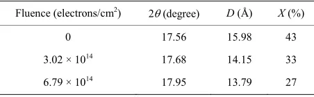
XRD technique has been utilized to detect changes in crystalline and amorphous regions along with the degree of crystallinity. It is used to measure crystallite size, per- centage of crystallinity, microstrain and dislocation den- sity of materials through interaction of X-ray beams with samples. The XRD spectra of pristine and electron irra- diated Lexan films is as shown in Figure 2. The diffract-
tion pattern clearly indicates that the Lexan is semicrys- talline in nature with main peak at 17.56˚. For irradiated samples also identical peaks were obtained but they slightly shift towards higher angel indicating decrease in lattice spacing. As compared to pristine sample, the dif- fraction pattern of irradiated films show the broadening of main peak indicating the decrease in crystallite size [22,23]. The crystallite size (D) was calculated using following equation [24],
cos
K D
b
(1)
where K is shape factor and is 0.9, is wavelength of X-ray beam and is 1.5406 Å, b is full width at half maximum, is diffraction angle or Bragg angle. The calculated values of crystallite size are listed in Table 1.
[image:2.595.310.537.408.588.2]The percentage of crystallinity (X) can be calculated using equation [25],
Figure 1. Schematic representation of chemical reaction induced in Lexan after electron irradiation.
Figure 2. XRD pattern for pristine and electron irradiated Lexan.
Table 1. Crystallite size (D) and percentage of crystallinity (X) of pristine and electron irradiated Lexan.
Fluence (electrons/cm2) 2(degree) D (Å) X (%)
0 17.56 15.98 43
3.02 × 10¹⁴ 17.68 14.15 33
[image:2.595.310.536.654.724.2]100
c
c a
A X
A A
(2)
where Ac and Aa are the area of crystalline and amor-
phous halos respectively. The percentage of crystallinity for all the samples is listed in Table 1. It has been ob-
served from Table 1 that both crystallite size and per-
centage of crystallinity were decreases after irradiation due to breakage of polymeric bonds and emission of some volatile gases, which may form the disordered state in the polymeric film. The decrease in crystallinity can be further confirmed from DSC studies.
3.2. UV-Visible Spectroscopic Study
The optical absorption spectra of pristine and electron irradiated Lexan samples are shown in Figure 3. After
electron irradiation, the polymer surface showed a visible colour change from transparent to yellow and the inten- sity of the colour increases with increase in electron flu- ence. This is also confirmed with UV-Visible absorbance spectra where the absorption edge shifts progressively towards longer wavelength. This may be attributed to the formation of chromophore groups (conjugated double bonds) as a consequence of the beam induced bond breaking and reconstruction. If there are enough chro- mophore groups in the conjugation within the sample, absorption edge is expected to move towards the visible region [25,26]. But in the present study, the absorption edge was in the UV region indicating less formation of chromophore groups.
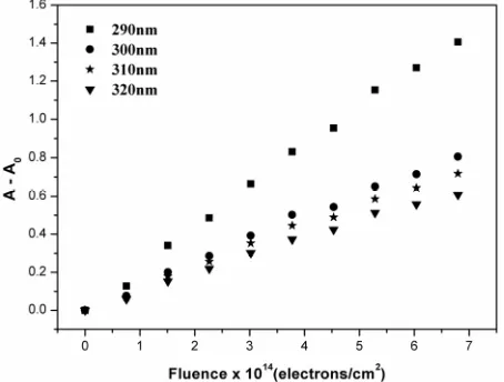
The absorbance difference (A-A0) versus electron
fluence was plotted in Figure 4 at wavelengths 290 nm,
300 nm, 310 nm and 320 nm. The linear dependence of absorbance with the electron fluence was observed. The solid lines in Figure 4 fit the experimental data using the
relation [25],
Figure 3. Absorbance spectra for pristine and electron irradiated Lexan.
Figure 4. Plot of absorbance difference (A-A0) versus electron
fluence for Lexan samples.
0
A k n A (3) where Aλ() is the absorbance at a wavelength λ and at a
fluence ; kλ is absorption coefficient of the chromopho-
res produced (cm–2); n is the number of chromophores
which absorb at a particular λ created per incident pri- mary ion and per unit area, and σ is the cross-sectional area of a cylinder within which n chromophores are cre- ated. Thus nσ represent the efficiency of the chromo- phore groups formation induced by ion beam and there fore, the slope kλnσ of the plot (A-A0) versus electron
fluence () represents the efficiency multiplied by kλ. The
value of kλnσ for electron bombardment for λ = 290, 300,
310 and 320 nm are found to be 2.10 × 10⁻¹⁵ cm2, 1.18 ×
10–15 cm2, 1.05 × 10⁻¹⁵ cm2 and 0.91 × 10⁻¹⁵ cm2 respec- tively. It is observed that the value kλnσ decreases with
increase in wavelength. The (A - A0) value at wavelength
290 nm is significantly more when compared to 300nm. This indicates that the absorption profile of the chromo- phore groups where different absorption at different wavelengths are due to difference in the absorption coef- ficients of the chromophore groups, for example, the absorption coefficient (k) at 290 nm is maximum when
compared to the absorption coefficient (k) of other
wavelengths.
3.3. FT-IR Spectral Analysis
The Vibration modes of chemical bonds are character- ized by the absorption bands [27]. Figure 5 shows the
FT-IR spectra for the pristine and electron irradiated Lexan films at different fluences. There is almost no change in the spectra of samples irradiated at the fluence 3.02 1014 electrons/cm2. On irradiation to the fluence of
6.79 1014 electrons/cm2, the intensity of the peak cor-
[image:3.595.311.538.88.260.2] [image:3.595.60.283.521.694.2]Figure 5. FT-IR spectra of pristine and electron irradiated Lexan.
from this, the intensity of the peak band corresponding to C=O stretch at 1700 cm–1 decreases slightly on irradia-
tion. This may be due to chain-scission at the carbonate site (as shown in Figure 1) with the probable elimination
of carbon dioxide or carbon monoxide. The corrobora- tion of chain-scission can be deduced from the decrease in the intensity of absorption bands around 1160 cm–1
attributed to carbonate stretch. No change in the C-H stress has been observed which indicates that the electron irradiation have not influenced the benzene ring.
3.4. Differential Scanning Calorimetry Study
One of the important parameters of polymer namely glass transition temperature can be determined using DSC data. Glass transition temperature is a temperature where the polymer goes from rigid glassy state to rub- bery state as chain becomes more flexible. At the glass transition temperature, the weak secondary bonds that stick the polymer chains are broken and macromolecule starts to move. DSC thermograms of pristine and electron irradiated Lexan samples were measured and are shown in Figure 6. The endothermic transformation of the unir-
radiated Lexan film occurs in a temperature range 145˚C to 154˚C with a glass transition temperature (Tg) of
150˚C and the corresponding heat of fusion was found to be 4.43 J/g. At the electron fluence 3.02 1014 elec-
trons/cm2, the DSC curve show a shift of T
g to 146˚C and
for 6.79 1014 electrons/cm2 T
g was observed at 143˚C,
i.e. the glass transition temperature decreases with in- crease in electron fluence. After irradiation, the shift in the Tg to lower temperature reveals that the electron irra-
diation leads to chain-scission and subsequently reduce- tion in molecular weight. As a result, the polymeric sys- tem was changing towards more disordered state [28] which is also confirmed from XRD studies. It was found
3.02 ×10¹⁴ electrons/cm2 6.79 ×10¹⁴ electrons/cm2
[image:4.595.309.538.86.253.2]Temperature (˚C)
Figure 6. DSC pattern of pristine and electron irradiated Lexan.
that the heat of fusion for unirradiated Lexan was found to be 4.43 J/g. After irradiation, the heat of fusion de- creases with increase in electron fluence, i.e. for 3.02
1014 electrons/cm2 it was found to be 4.34 J/g and for 6.79 1014 electrons/cm2 it was found to be 4.15 J/g. This decrease in heat of fusion may be due to bond breaking during electron irradiation.
3.5. Thermogravimetric Analysis Study
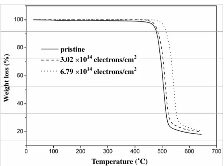
The thermal degradation is a very important process which helps to know the influence of the polymer struc- ture on the thermal stability and the temperature at which the polymer can be used. Sinha et al. [12]have reported that the thermal stability of the polycarbonate detector decreases at high dose of gamma, and the weight loss starts at around 420˚C and continues up to 700˚C. Figure 7 shows the thermograms of the pristine and the electron
irradiated Lexan samples showing the weight (%) as a function of temperature. From Figure 7 it was found that
the pristine sample thermogram shows a small change in weight loss at 250˚C and sample is thermally stable up to 440˚C. This weight loss was due to water vaporization and was not significant. After 440˚C, the film experi- enced a great weight loss up to 530˚C because of thermal decomposition, and about 76% of the sample decom- posed in to volatiles. After 530˚C, there is a small de- crease in weight loss due to emission of volatile gases. The thermal decomposition temperature for pristine Lexan was found to be 495˚C. Lexan sample irradiated at fluence 3.02 1014 electrons/cm2 was thermally stable up
3.02 ×10¹⁴ electrons/cm2
6.79 ×10¹⁴ electrons/cm2
[image:5.595.60.285.81.248.2]Temperature (˚C)
Figure 7. TGA thermogram of pristine and electron irradiated Lexan.
502˚C. The Lexan sample irradiated by electron of flu- ence 6.79 1014 electrons/cm2 was stable up to 480˚C
and then sudden decrease in the weight loss was ob- served up to 570˚C. After 570˚C, the weight loss was less and the corresponding thermal decomposition tempera- ture was found to be 516˚C. From this study it is clear that, the thermal stability of Lexan increases after elec- tron irradiation.
4. Conclusions
The conclusion drawn from the studies of 8 MeV elec-tron irradiation effects on the structural and thermal properties of Lexan are as follows. The XRD results show decrease in crystallite size and percentage of crys-tallinity upon irradiation indicating that the polymeric system is moving towards more disordered state which is also supported by DSC studies. Using UV-Visible stud-ies, the chromophore group formation were obtained by linear fit and the obtained values for the formation of chromophore groups is decreased at longer wavelength. The carbonate linkage is found to be the radiation-sensitive linkage and benzene ring does not undergo any changes after irradiation as studied from FTIR studies. TGA re-sults reveal that thermal stability of Lexan increases after irradiation.
5. Acknowledgements
The authors are thankful to Board of Research in Nuclear Science, Department of Atomic Energy, Govt. of India for financial support through a project No. 2001/34/21 -BRNS/453. Authors are thankful to research scholar and staff of Microtron Centre for their help and support dur- ing the course of this work. The authors are also thankful to staff of USIC, Karnatak University and staff of STIC, Cochin University for extending the experimental facil- ity.
REFERENCES
[1] R. L. Clough, “High-Energy Radiation and Polymers: A Review of Commercial Processes and Emerging Applica- tions,” Nuclear Instruments and Methods in Physics Re- search B, Vol. 184, No. 1-8, 2001, pp. 8-33.
doi:10.1016/S0168-583X(01)00966-1
[2] M. R. Cleland, L. A. Parks and S. Cheng, “Applications for Radiation Processing of Materials,” Nuclear Instru- ments and Methods in Physics Research B, Vol. 208, 2005, pp. 66-73.doi:10.1016/S0168-583X(03)00655-4 [3] Andrzej G. Chmielewski, M. Haji-Saeid and S. Ahmed,
“Progress in Radiation Processing of Polymers,” Nuclear Instruments and Methods in Physics Research B, Vol. 236, No. 1-4, 2005, pp. 44-55.
doi:10.1016/j.nimb.2005.03.247
[4] A. Chapiro, “General Consideration of the Radiation Chemistry of Polymers,” Nuclear Instruments and Meth- ods in Physics Research B, Vol. 105, No. 1-4, 2005, pp. 5-7.doi:10.1016/0168-583X(95)00861-6
[5] Sangappa, T. Demappa, Mahadevaiah, S. Ganesha, S. Divakara, M. Pattabi and R. Somashekar, “Physical and Thermal Properties of 8 MeV Electron Beam Irradiated HPMC Polymer Films,” Nuclear Instruments and Meth-ods in Physics Research B, Vol. 266, No. 18, 2008, pp. 3975-3980. doi:10.1016/j.nimb.2008.06.021
[6] A. Harisha, V. Ravindrachary, R. F. Bhajantri, Ismayil, G. Sanjeev, B. Poojary, D. Dutta and P. K. Pujari, “Electron Irradiation Induced Microstructural Modifications in BaCl2 Doped PVA: A Positron Annihilation Study,”
Polymer Degradation and Stability, Vol. 93, No. 8, 2008, pp. 1554-1563.
doi:10.1016/j.polymdegradstab.2008.05.003
[7] A. Rivaton and J. Arnold, “Structural Modifications of Polymers under the Impact of Fast Neutrons,” Polymer Degradation and Stability, Vol. 93, No. 10, 2008, pp. 1864-1868.
doi:10.1016/j.polymdegradstab.2008.07.015
[8] S. Shah, A. Qureshi, N. L. Singh, P. K. Kulriya, K. P. Singh and D. K. Avasthi, “Structural and Chemical Modification of Polymer Composite by Proton Irradia- tion,” Surface & Coatings Technology, Vol. 203, No. 17-18, 2009, pp. 2595-2599.
doi:10.1016/j.surfcoat.2009.02.052
[9] R. C. Ramola, S. Chandra, A. Negi, J. M. S. Rana, S. Annapoorni, R. G. Sonkawade, P. K. Kulriya and A. Srivastava, “Study of Optical Band Gap, Carbonaceous Clusters and Structuring in CR-39 and PET Polymers Ir-radiated by 100 MeV O7+ Ions,” Physics B, Vol. 404, No.
1, 2009, pp. 26-30.doi:10.1016/j.physb.2008.09.033 [10] G. M. Wallner, W. Platzer and R. W. Lang, “Structure-
Property Correlations of Polymeric Films for Transparent Insulation Wall Applications. Part 2: Infrared Optical Pro- perties,” Solar Energy, Vol. 79, No. 6, 2005, pp. 593-602. doi:10.1016/j.solener.2005.05.007
Vol. 65, No. 1-4, 1992, pp. 413-422. doi:10.1016/0168-583X(92)95077-5
[12] D. Sinha and K. K. Dwivedi, “Radiation-Induced Modi- fication on Thermal Properties of Different Nuclear Track Detectors,” Radiation Measurements, Vol. 36, No. 1-6, 2003, pp. 713-718. doi:10.1016/S1350-4487(03)00232-4 [13] R. Navarro-Gonzalez, P. Coll and R. Aliev, “Pyrolysis of
-Irradiated Bisphenol-A Polycarbonate,” Polymer Bulle-tin, Vol. 48, No. 1, 2002, pp. 43-51.
doi:10.1007/s00289-002-0004-4
[14] A. Qureshi, N. L. Singh, A. K. Rakshit, F. Singh and D. K. Avasthi, “Swift Heavy Ion Induced Modification in Polyimide Films,” Surface & Coatings Technology, Vol. 201, No. 19-20, 2007, pp. 8308-8311.
doi:10.1016/j.surfcoat.2006.10.055
[15] E. Balanzt, N. Betz and S. Bouffard, “Swift Heavy Ion Modification of Polymers,” Nuclear Instruments and Methods in Physics Research B, Vol. 105, No. 1-4, 1995, pp. 46-54.doi:10.1016/0168-583X(95)00521-8
[16] J. Jagielski, A. Turos, D. Bielinski, A. M. Abdul-Kader and A. Piatkowska, “Ion-Beam Modified Polymers for Biomedical Applications,” Nuclear Instruments and Methods in Physics Research B, Vol. 261, No. 1-2, 2007, pp. 690-693.doi:10.1016/j.nimb.2007.03.021
[17] D. Fink, P. S. Alegaonkar, A. V. Petrov, M. Wilhelm, P. Szimkowiak, M. Behar, D. Sinha, W. R. Fahrner, K. Hoppe and L. T. Chadderton, “High Energy Ion Beam Ir- radiation of Polymers for Electronic Applications,” Nu- clear Instruments and Methods in Physics Research B, Vol. 236, No. 1-4, 2005, pp. 11-20.
doi:10.1016/j.nimb.2005.03.243
[18] J. Y. J. Chung, “Stabilization of Gamma-Irradiated Poly-carbonate,” Medical Plastics and Biomaterials Magazine Technical paper series, 1997.
[19] S. Singh and S. Prasher, “The Optical, Chemical and Spectral Response of Gamma-Irradiated Lexan Polymeric Track Recorder,” Radiation Measurements, Vol. 40, No. 1, 2005, pp. 50-54.
doi:10.1016/j.radmeas.2004.11.005
[20] B. Jaleh, P. Parvin, N. Sheikh, M. Hajivaliei and E. Hasani, “Surface Modification of Lexan Treated by RF Plasma,” Surface & Coatings Technology, Vol. 203, No.
17-18, 2009, pp. 2759-2762. doi:10.1016/j.surfcoat.2009.02.133
[21] Ganesh, K. C. Prashanth, Y. N. Nagesha, A. P. Gnana Prakash, D. Umakanth, M. Pattabi, K. Siddappa, S. Sal-kalachen and A. Roy, “Modification of Power Diode Characteristics Using Bremsstrahlung Radiation from Microtron,” Radiation Physics and Chemistry, Vol. 55, No. 4, 1999, pp. 461-464.
doi:10.1016/S0969-806X(99)00268-6
[22] K. D. Pae, S. K. Bhateja and J. R. Gilbert, “Increase in Crystallinity of Poly (Vinylidene Fluoride) by Electron Beam Radiation,” Journal of Polymer Science Part B:
Polymer Physics, Vol. 25, No. 4, 1987, pp. 717-722. doi:10.1002/polb.1987.090250402
[23] Y. Rosenberg, A. Siegmann, M. Narkis and S. Shkolnik, “Low Dose -Irradiation of Some Fluoropolymers: Effect of Polymer Chemical Structure,” Journal of Applied Polymer Science, Vol. 45, No. 5, 1992, pp. 783-795. doi:10.1002/app.1992.070450504
[24] B. Jasinska, A. L. Dawidowicz and S. Pikus, “Application of Positron Annihilation Lifetime Spectroscopy in Studies of Crystallization Processes,” Physical Chemistry Che- mical Physics, Vol. 5, 2003, pp. 3289-3293.
doi:10.1039/b304588a
[25] L. Relleve, N. Nagasawa, L. Q. Luan, T. Yagi, C. Aranilla, L. Abad, T. Kume, F. Yoshii and A. dela Rosa, “Degradation of Carrageenan by Radiation,” Polymer Degradation and Stability, Vol. 87, No. 3, 2005, pp. 403-410.doi:10.1016/j.polymdegradstab.2004.09.003 [26] D. Sinha, T. Swu, S. P. Tripathy, R. Mishra, K. K.
Dwivedi and D. Fink, “Spectroscopic and Thermal Stud- ies of Gamma Irradiated Polypropylene Polymer,” Radia- tion Effects and Defects in Solids, Vol. 158, No. 7, 2003, pp. 531-537.doi:10.1080/1042015031000074101 [27] G. Aruldhas, “Molecular Structure and Spectroscopy,”
Prentice-Hall of India, New Delhi, 2004.
[28] R. Mishra, S. P. Tripathy, D. Fink and K. K. Dwivedi, “Activation Energy of Thermal Decomposition of Proton Irradiated Polymers,” Radiation Measurements, Vol. 40, No. 2-6, 2005, pp. 754-757.