WHAT DOES THE BONE SAY ABOUT IMMEDIATE IMPLANT!! ANALYZING THROUGH
*,1
Dr. Neeraj Chandra,
2Dr. Shivangi Chandra
1
Department of Periodontology and Implantology, Institute of Dental Sciences, Bareilly
2
Department of Pedodontics and Preventive Dentistry,
3
Department of orthodontics and Dentofacial Orthopedics,
4
Department of Public Health Dentistry,
ARTICLE INFO ABSTRACT
Aim: Immediately placed implant is lucrative and offers several advantages. This study was done to find out any hard tissue changes around the implants in bucco
Materials and Methods:
implant placement. Implant system used was Alpha
scan of the jaw, OPG and IOPA were done to assess the quality and quantity of the bone, proximity with anatomical structures and dimention of the tooth to be replaced and facio
3 mm apical to the crest and 6 mm apical to the crest was calculated. Second stage surgery was carried out after 4 to 6 months depending upon the quality of the bone.
Results: (Kolmogorov
before implant placement and 5.35±0.78 after 6 months, 3 mm apical to the crest was 9.08±1.25 and after 6 months it was 8.87±1.21 and 6mm apical to the crest was 10.81±1.61 and after 6 months 10.55±1.64mm.
present study showed statistically significant bone loss in bucco Conclusion:
bound to occur after implant placement but the facio
Copyright©2016, Yash Raj. This is an open access article distributed under the Creative distribution, and reproduction in any medium, provided the original work is properly cited.
INTRODUCTION
The use of dental implants for treatment of single tooth replacement cases was first introduced in 1986.
2007) Since then osseointegrated implants have become a standard of care in daily practicein oral rehabilitation, and implants are now a realistic and well documented treatment alternativefor partially and fully edentulous patients. Implants for single tooth replacement can conserve sound tooth structure by reducing the need to prepare adjacent teeth as abutments.
(Marlin E. Gher et al., 1994)The classical way to evaluate the
success rates of dental implant are the lack of mobility, infection, discomfort, absence of pain and continuous
*Corresponding author: Dr. Neeraj Chandra,
Department of Periodontology and Implantology, Institute of Dental Sciences, Bareilly – 243006, Uttar Pradesh, India.
ISSN: 0975-833X
Article History: Received 14th March, 2016
Received in revised form 26th April, 2016
Accepted 15th May, 2016
Published online 30th June,2016
Key words:
Computed tomography, Denta Scan,
Immediate Implant, Facio-palatal/lingual.
Citation: Yash Raj, 2016. “What does the bone say about immediate implant!! Analyzing through dent 8, (06), 33579-33586.
RESEARCH ARTICLE
WHAT DOES THE BONE SAY ABOUT IMMEDIATE IMPLANT!! ANALYZING THROUGH
DENTA SCAN
Dr. Shivangi Chandra,
3Dr. Yash Raj Bahadur,
4and
3Dr. Gaurav Chaudhary
Department of Periodontology and Implantology, Institute of Dental Sciences, Bareilly
Uttar Pradesh, India
Department of Pedodontics and Preventive Dentistry, Seema Dental College and Hospital,
Uttarakhand, India
Department of orthodontics and Dentofacial Orthopedics, Seema Dental College and Hospital,
249203, Uttarakhand, India
Department of Public Health Dentistry, Dr. Hedgewar Dental College and Hospital, Hingoli,
ABSTRACT
Immediately placed implant is lucrative and offers several advantages. This study was done to find out any hard tissue changes around the implants in bucco-lingual direction before and after 6 months of implant placement. Materials and Methods: This study was done on 18 patients who has undergone extraction and immediate implant placement. Implant system used was Alpha-Bio®. Pre- operative test includes computed tomography (CT)
scan of the jaw, OPG and IOPA were done to assess the quality and quantity of the bone, proximity with anatomical structures and dimention of the tooth to be replaced and facio-palatal/lingual width of bone
3 mm apical to the crest and 6 mm apical to the crest was calculated. Second stage surgery was carried out after 4 to 6 months depending upon the quality of the bone.
Results: Statistical analysis was done on SPSS ver 15 software. The resul
(Kolmogorov-smirnov & shapiro-wilk). The mean value bucco-lingual width, at the crest was 5.62±0.87 mm before implant placement and 5.35±0.78 after 6 months, 3 mm apical to the crest was 9.08±1.25 and after 6 months
was 8.87±1.21 and 6mm apical to the crest was 10.81±1.61 and after 6 months 10.55±1.64mm. present study showed statistically significant bone loss in bucco -lingual/palatal direction.
Conclusion: We can say that immediate implant placement is a safe and predictable option, and bone remodelling bound to occur after implant placement but the facio-palatal/lingual changes are clinically not significant.
is an open access article distributed under the Creative Commons Attribution License, which distribution, and reproduction in any medium, provided the original work is properly cited.
The use of dental implants for treatment of single tooth
replacement cases was first introduced in 1986. (Todd et al.,
Since then osseointegrated implants have become a standard of care in daily practicein oral rehabilitation, and implants are now a realistic and well documented treatment alternativefor partially and fully edentulous patients. Implants oth replacement can conserve sound tooth structure by reducing the need to prepare adjacent teeth as abutments. The classical way to evaluate the success rates of dental implant are the lack of mobility,
absence of pain and continuous
Department of Periodontology and Implantology, Institute of Dental 243006, Uttar Pradesh, India.
periapical radiolucency. These features accounted for the popularity of the immediate implants.
proven that when implants placed immediately after tooth extraction has proven to be a predictable treatment protocol with a high success rate. Immediate implant placement has several advantages, such as reduction of the number of surgical treatments, reduction of the time between tooth extraction, and the placement of the definitive prosthesis.
Immediate placement of the dental implant, at the time of extraction, can direct the positioning of the implant and reduce encroachment on anatomic structures such as the maxillary sinus and the inferior alveolar canal.
1994) It is seen that for osseointegration of implant, bone substitutes, bone grafting, me
these have been used to achieve bone formation in the defect formed due to gap junction. (David
materials like xenogenic grafts, autogenous bone and various
Available online at http://www.journalcra.com
International Journal of Current Research
Vol. 8, Issue, 06, pp.33579-33586, June, 2016
INTERNATIONAL
What does the bone say about immediate implant!! Analyzing through denta scan”, International Journal of Current Research
z
WHAT DOES THE BONE SAY ABOUT IMMEDIATE IMPLANT!! ANALYZING THROUGH
4
Dr. Shivani Shokeen
Department of Periodontology and Implantology, Institute of Dental Sciences, Bareilly – 243006,
ge and Hospital, Rishikesh – 249203,
Seema Dental College and Hospital, Rishikesh –
Hingoli, Maharashtra, India
Immediately placed implant is lucrative and offers several advantages. This study was done to find out any lingual direction before and after 6 months of implant placement. This study was done on 18 patients who has undergone extraction and immediate operative test includes computed tomography (CT) scan of the jaw, OPG and IOPA were done to assess the quality and quantity of the bone, proximity with palatal/lingual width of bone at the crest, 3 mm apical to the crest and 6 mm apical to the crest was calculated. Second stage surgery was carried out after 4
Statistical analysis was done on SPSS ver 15 software. The results were tested using Normality tests lingual width, at the crest was 5.62±0.87 mm before implant placement and 5.35±0.78 after 6 months, 3 mm apical to the crest was 9.08±1.25 and after 6 months was 8.87±1.21 and 6mm apical to the crest was 10.81±1.61 and after 6 months 10.55±1.64mm. Results from this
lingual/palatal direction.
t is a safe and predictable option, and bone remodelling palatal/lingual changes are clinically not significant.
ribution License, which permits unrestricted use,
These features accounted for the
popularity of the immediate implants. (Joly et al., 2003)It is
proven that when implants placed immediately after tooth extraction has proven to be a predictable treatment protocol Immediate implant placement has es, such as reduction of the number of surgical treatments, reduction of the time between tooth extraction, and
the placement of the definitive prosthesis. (Covani et al., 2007)
Immediate placement of the dental implant, at the time of ct the positioning of the implant and reduce encroachment on anatomic structures such as the maxillary
sinus and the inferior alveolar canal. (Marlin E. Gher et al.,
It is seen that for osseointegration of implant, bone substitutes, bone grafting, membranes or a combination of these have been used to achieve bone formation in the defect
David et al., 2012)Varies graft
materials like xenogenic grafts, autogenous bone and various
INTERNATIONAL JOURNAL OF CURRENT RESEARCH
other allografts have been used in conjunction with immediate implants. However, animal studies have shown that osseointegration of immediate implants can be achieved
without bone augmentation procedures. (Schropp et al., 2003)
Bone augmentation is a term which generally used to describe variety of procedures that are used to describe variety of procedures that are used to “build” bone so as implants can be placed.These procedures typically involve grafting (adding) bone or bonelike materials to the jaw, and waiting for the grafted material to fuse with the existing bone over several months. Now to achieve this safe, predictable and cost effective mechanism of rehabilitation, Branemark developed a list of clinical recommendation regarding treatment protocols (Adell
et al., 1990; Adell et al., 1985). According to one of the recommendations, there should be waiting time of atleast 12 months was necessary following tooth extraction before an endosseous dental implant could be placed. The rationale for this reasoning was to allow resolution of any hard or soft tissue pathology in a proposed recipient site. The goal of modern dentistry is to restore the tooth to normal contour, function, comfort, esthetics and health.The use of dental implants to replace missing teeth is becoming a preferred alternative for restorative dentists without involving adjacent teeth. Patients have gained awareness of the new options that they increasingly request modification or replacement of existing dental restorations (eg: dentures, fixed partialdentures, and
removable partial dentures). (Botticelli et al., 2003; Akimoto
et al., 1999) Considering all the above factors the present study has been taken up with the following aims & objectives: To evaluate the hard tissue profile around the immediate Implant using denta scan software.
MATERIALS AND METHODS
The study population consisted of 18 patients; 8 females & 10 males, ranging in age from 20 to 55 years. The patients were scheduled for extraction & immediate replacement with an
Implant. The Implant system was used Alpha-Bio®
(manufactured in Israel). In all the cases root form implants were selected.The patients included in this study on the basis of the following criteria: absence of any local or systemic factors that would inhibit or jeopardize the healing process needed for osseointegration. The age were between 18 to 50 years of age, patients who were co-operative and those patients who were having good oral hygiene method. In this study implants placed in both upper and lower jaw were included, keeping in mind implants were to be placed in a single rooted teeth. The patients were excluded from the study who were medically compromised, having Para functional habits like bruxism, patients who were on medications that might interfere with the peri-implant healing process. Patients who were pregnant and lactating mothers were also excluded. Patients suffering from psychiatric disorders were also excluded from the study. Before implant placement study cast. OPG and denta- scan evaluation was done.
Pre-operative evaluation of implant site
Before starting the cases we have evaluated the soft and hard tissue. Gingiva was examined to see the consistency, texture and thickness. The occlusion, periodontal integrity of the dentition, an alignment and the interocclusal space was
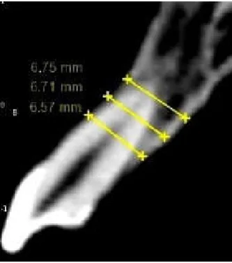
assessed. In all the cases pre-operative computer tomographic (CT) scan of the jaw, IOPA and OPG, were taken to assess the quantity and the quality of the bone at the implant placement site, proximity of the implant site to vital anatomical structure, dimension of the tooth to be replaced and the bucco-lingual width of bone at the crest, 3mm apical to crest and 6 mm apical to the crest was measured. (Figure 1,2,3,4,5,6)
Surgical Procedure
All the patients were planned to be operated under local anaesthesia. The tooth scheduled for immediate implant was carefully removed by either periotome or piezotome. (Figure2) Implant placement was performed only when there is labial cortical plate is present. The Osteotomy was initiated with 2 mm pilot drill. The osteotomy procedure was extended atleast 3-4 mm beyond the apex of the socket so that the implant will be in contact with lingual or palatal wall. By using the sequential large drill sizes, the osteotomy site was enlarged according to the width of implant to be used, keeping one thing in mind that the width of last drill should be 0.5 mm short of the width of the implant. With the use of the rachet the implant is tightened in a clockwise direction. The implants were placed at the crest level. (Figure 7,8,9,10,11,12,13) Patient is prescribed with oral antibiotics, anti-inflammatory analgesics as and when required postoperatively. Chlorhexidine 0.2% mouth wash was given for 2 weeks postoperatively. The patient was evaluated on a monthly basis. Second stage surgery was carried out after 4 to 6 months depending upon the quality of the bone. This procedure was also carried out under LA with number 15 blade, a circular incision was given over the implant site. After exposing the implant site, cover screw was removed and healing cap was placed. The soft tissue was then suture back. Before starting a second stage surgery the denta scan evaluation was done to calculate the bucco-lingual width
of the bone at the crest (which is 0.5 mm apical to crest). (Figure 3)
Prosthetic phase
After performing second stage surgery, after 15 days the healing cap was removed and a two- piece internal hex abutment was placed in the implant. Impression was taken with a elastomer impression materials using the open tray technique and a PFM crown was given.
RESULTS
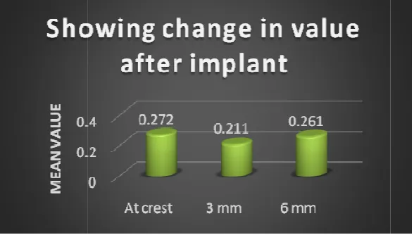
After six month no complications requiring surgical intervention or antibiotic therapy has been observed during the healing period. At the second stage surgery, all of the implants were clinically stable and asymptomatic. The results were tested using Normality tests (Kolmogorov-smirnov & shapiro-wilk). Dentascan examination failed to show any kind of peri-implant bone loss. If we talk about the bucco-lingual width, the mean value at the crest was 5.62±0.87 mm before implant placement and 5.35±0.78 after 6 months, 3 mm apical to the crest the mean value was 9.08±1.25 and after 6 months it was 8.87±1.21 and 6mm apical to the crest the mean value before was 10.81±1.61 and after 6 months it was 10.55±1.64mm. The mean change was 0.27±0.13mm at crest, 0.21±0.12mm at 3mm apical to the crest and the 0.26±0.13mm 6mm apical to the crest. Results from this present study also indicate that
there is statistically significant bone loss present in bucco -lingual/palatal direction but the results are not much of clinical significant.
The vertical bone loss have also occurred in the healing period and the average bone loss was 0.45 mm which is not statistically or clinically significant. (Figure 14,15)
[image:3.595.160.437.126.271.2]Dentascan image of immediate implant placement
[image:3.595.214.381.416.605.2]Figure 1. 3-D image before placement
Figure 2. Panaromic view before placement
Figure 3. Saggital section before placement
Figure 4. 3-D Image after 6 months of placement
[image:3.595.164.436.632.761.2]Figure 5. Panaromic view after 6 months of placement
[image:4.595.230.374.203.377.2]Figure 6. Saggital section after 6 months of placement
Figure 8. Photograph showing the reflection of flap on buccal side
33582 Dr. Neeraj Chandra et al. What does the bone say about immediate implant!! analyzing through denta scan
Figure 5. Panaromic view after 6 months of placement
[image:4.595.184.415.409.557.2]Figure 6. Saggital section after 6 months of placement
[image:4.595.184.412.590.769.2]Figure 7. Pre-operative photograph
Figure 8. Photograph showing the reflection of flap on buccal side
Figure 9. Photograph showing the reflection of
Figure 10. Photograph showing atraumatic extraction with the help of piezotome
Figure 11. Photograph showing the extracted tooth
[image:5.595.149.441.532.766.2]33583 International Journal of Current Research,
Figure 9. Photograph showing the reflection of flap on palatal side
Figure 10. Photograph showing atraumatic extraction with the help of piezotome
Figure 11. Photograph showing the extracted tooth
International Journal of Current Research, Vol. 08, Issue, 06, pp.33579-33586, June, 2016
Figure 12. Photograph showing placement of implant with cover
[image:6.595.166.427.287.460.2]Level Before implant At crest 5.622 ± 0.874 3 mm 9.083 ± 1.254 6 mm 10.811 ± 1.618
Figure 14. This table shows the Bucco
Figure 15. The mean change was 0.27±0.13mm at crest, 0.21±0.12mm at 3mm apical to the crest and the 0.26±0.13mm 6mm
[image:6.595.153.447.593.760.2]33584 Dr. Neeraj Chandra et al. What does the bone say about immediate implant!! analyzing through denta scan
Figure 12. Photograph showing placement of implant with cover
Figure 13. Post-operative photograph
After 6 months Change ‘t’ value 5.350 ± 0.788 0.272 ± 0.136 8.470 8.872 ± 1.218 0.211 ± 0.128 7.007 10.550 ± 1.640 0.261 ± 0.133 8.301
This table shows the Bucco-Lingual/Palatal changes before and 6 months after implant Placement. The results were statistically significant
The mean change was 0.27±0.13mm at crest, 0.21±0.12mm at 3mm apical to the crest and the 0.26±0.13mm 6mm apical to the crest
al. What does the bone say about immediate implant!! analyzing through denta scan
P value <0.001** <0.001** <0.001**
Lingual/Palatal changes before and 6 months after implant Placement.
The mean change was 0.27±0.13mm at crest, 0.21±0.12mm at 3mm apical to the crest and the 0.26±0.13mm 6mm
DISCUSSION
Extensive work by the Swedish orthopedic surgeon Per-Ingvar Branemark led to the discovery that commercially pure Titanium, when placed in the bone, get fixed in plane because of close bond that has developed between the two, this phenomenon was later described as osseointegration. (Adell
et al., 1990; Adell et al., 1985) The implant-supported restoration has proven to be an efficacious means of replacing a missing tooth. Implant-supported restorations in the esthetic zone are considered successful only when an inconspicuous result is obtained. In order to be considered successful, an implant-supported restoration must achieve a harmonious balance between functional, aesthetic & biological imperatives. (Becker and Becker, 1996; Becker and Becker, 1990) Optimal implant restorations depend not only on prosthetic and technical parameters but also on biologic and surgical considerations. It is seen that success of implant treatment relies on maintenance and presence of bone adjacent to implants. The crestal bone area is usually a significant indicator of implant health. Crestal bone loss during healing indicates the
need for preventive therapy. (Botticelli et al., 2003; Botticelli
et al., 2004) Albrektsson et al. have included interproximal crestal bone loss as one criterion for implant success. According to their criteria, bone loss of less than 0.2 mm annually following the implant’s first year of function is
essential for long-term success. (Carlsson et al., 1988) The loss
of crestal bone could be attributed to the fact that whenever bone is stripped of its periosteum, its nutrition is affected, which could result in some amount of resorption of the crestal
bone. (Chen et al., 2005) This loss of crestal bone during the
first year after placement of the implant could also be attributed to the process of wound healing at the bone-implant interface. The dentascan was used in this study to calculate the parameters. Dentascan is a software program, which provide computed tomographic imaging of mandible and maxilla in three planes i.e. axial, panaromic and oblique sagittal. Dentascan provides accuracy, clarity and identical scale which permits the uniformity of measurements. It also provides cross-referencing of anatomical structures. The facio-palatal/lingual width can be measured with the help of sagittal view and it also provides the clear visualization of internal structures, such as
the incisive and inferior alveolar canals. (Bhatia et al., 2012;
Siddhartha et al., 2013)As per my knowledge so far this is a
first study of its kind in which hard tissue parameters were calculated using denta-scan software.
It has been noted that there is a marked reduction of bucco-lingual width statistically. The implants experienced more extensive buccal bone remodeling as compared to lingual site.This remodeling could be due to either because of Regional Axillarated phenomena (RAP) or it could be because we have reflected flap in all the cases. It could also be possible that the result of simultaneous new bone apposition to fill the peri-implant defect and buccal and lingual bone resorption. Such kind of remodeling leads to reduction of the width of alveolar bone and can occurred around all the implants studied.
(Bra°nemark et al., 1977)We have measured the bucco-lingual
width before implant placement, the measurements were made at the crest, 3mm apical to the crest and 6 mm apical to the crest.The facio-palatal/lingual measurements were again taken
after 6 months of placement of the implant. One study done by
Covani et al. in 2007 to analyze bone healing and vertical bone
remodeling for implants placed immediately after tooth removal without guided bone regeneration. They found that peri – implant bone defects had healed completely 6 months after implant placement. The pattern of bone healing around the neck of implants showed an absence of peri-implant defects. The vertical distance between the implant shoulder and bone crest ranged from 0 to 2mm. they concluded that bone remodeling of implants placed in fresh extraction sockets showed a healing pattern with new bone apposition around the implants neck and horizontal and vertical bone reabsorption. The vertical reabsorption, which had been observed at buccal sites, was not associated with any negative esthetic
implications. (Covani et al., 2007)
Immediate implant placement reduces the number of surgical procedure, the implant can be placed in fresh extraction socket, in same location as the extracted tooth, which minimizes the need for angled abutments and also facilitates the positioning of final restoration. Immediate placement of implants provides us with better esthetics, by preserving the bony receptors, to prevent atrophy of the alveolar ridge, which leads to prevention of recession of the mucosal and gingival tissue. So, we can say it stimulates preservation of gingival aesthetics.
Conclusion
It is concluded that the implants should be placed atleast 2-3 mm apical to the crest in immediate implant placement cases. Immediate implant placement has been studied extensively over many years. Evidence available till date indicates that it is a recognized and successful procedure that might benefit patients. In conclusion, within the limits of this study we can say that immediate implant placement is a safe and predictable option, and bone remodelling bound to occur after implant placement but the bucco-lingual changes are clinically not significant. However, one should do careful planning and case selection to ensure implant success and final esthetic outcomes.
REFERENCES
Adell R, Eriksson B, Lekholm U, Branemark PI, Jemt T. 1990. Long-term follow-up study of osseointegrated implants in
the treatment of totally edentulous jaws. Int J Oral
MaxillofacImplants, 5: 347–359.
Adell R, Lekhholm U, Branemark PI. 1985. A 15-year study of osseointegrated implants in the treatment of the edentulous
jaw. Int J Oral Surg., 10: 387–418.
Akimoto K, Becker W, Donath K, Becker BE, Sanchez R. 1999. Formation of bone around titanium implants placed into zero wall defects: pilot project using reinforced
e-PTFE membrane and autogenous bone grafts. Clin Implant
Dent Relat Res., 1: 98–104.
Becker W, Becker BE, Handelsman M. 1990. Guided tissue regeneration for implants placed into extraction sockets and for implant dehiscences: surgical techniques and case
report. Int J Periodontics Restorative Dent, 10: 376–391.
Becker W, Becker BE. 1996. Flap designs for minimization of recession adjacent to maxillary anterior implant sites: A
clinical study. Int J Oral Maxillofac Implants, 11: 46–54.
Botticelli D, Berglundh T, Buser D, Lindhe J. 2003. Appositional bone formation in marginal defects at
implants. Clin Oral Implants Res., 14: 1–9.
Botticelli D, Berglundh T, Buser D, Lindhe J. 2003. The jumping distance revisited: an experimental study in the
dog. Clin Oral Implants Res., 14: 35–42.
Botticelli D, Berglundh T, Lindhe J. 2004. Resolution of bone defects of varying dimension and configuration in the marginal portion of the peri-implant bone. An experimental
study in the dog. J Clin Periodontol., 31: 309–317.
Bra°nemark PI, Hansson BO, Adell R, Breine U, Lindstro¨m J, Halle´n O, Ohman A. 1977. Osseointegrated implants in the treatment of the edentulous jaw. Experience from a
10-year period. Scand J Plast Reconstr Surg Suppl., 16(1): 1
132.
Branemark PI, Svensson B, van Steenberghe D. 1995. Ten-year survival rates of fixed prostheses on four or six
implants admodum Branemark in full edentulism. Clin
Oral Implants Res., 6: 227–231.
Carlsson L, Rostlund T, Albrektsson B, Albrektsson T. 1988. Implant fixation improved by close fit. Cylindrical
implant– bone interface studied in rabbits. Acta Orthop
Scand, 59(8): 272–275.
Chen ST, Darby IB, Adams GG, Reynolds EC. 2005. A prospective clinical study of bone augmentation
techniques at immediate implants. Clin Oral Implants Res.,
16: 176–184.
Covani et al. 2007. Connective Tissue Graft Used as a
Biologic Barrier to Cover an Immediate Implant. J
Periodontol., 78: 810-815
David PO, Demarchi CL, Maestre-Ferrín L, Peñarrocha DM. 2012. Comparision of Immediate and Delayed Implants in
maxillary molar region: Int J Oral Maxillofac Implants,
May-Jun;27(3):604-10
HP Bhatia, S Goel, B Srivastava. 2012. Denta Scan: J Oral
Health Comm Dent, 6(1)25-27
Joly et al. 2003. Clinical and radiographic evaluation of soft
tissue changes around implants : a pilot study. J
Periodontol., 74 : 1097-1103
Marlin E. Gher et al. 1994. Bone Grafting and Guided Bone
Regeneration for Immediate Dental Implants in Humans. J
Periodontol.
Schropp et al. 2003. Bone Healing Following Immediate
Versus Delayed Placement of Titanium Implants into
Extraction Sockets: A Prospective Clinical Study. J Oral
Maxillofac Implants, 18:189–199.
Siddhartha C, Amiya A, Nishi S, Ankita S. 2013. Dentascan: A
Diagnostic Boon: J Dent Sci Res., 4(1):13 – 17
Todd M. Mayer et al. 2007. A Single Tooth Implants: A
Viable Alternative For Single Tooth Replacement. J
Periodontol., 73:687-693