Diffusional Anisotropy of the Human Brain Assessed with
Diffusion-Weighted MR: Relation with Normal Brain Development and Aging
Yoshiyuki Nomura, Hajime Sakuma, Kan Takeda, Tomoyasu Tagami, Yasuyuki Okuda, and Tsuyoshi Nakagawa
PORPOSE: To analyze diffusional anisotropy in frontal and occipital white matter of human brain quantitatively as a function of age by using diffusion-weighted MR imaging. METHODS: Ten neonates (<1 month), 13 infants (1-10 months), 9 children (1-11 years), and 16 adults (20-79 years) were examined. After taking axial spin-echo images of the brain, diffusion-sensitive gradients were added parallel or perpendicular to the orientation of nerve fibers. The apparent diffusion coefficient parallel to the nerve fibers (0) and that perpendicular to the fibers (90) were computed.
The anisotropic ratio (90/0) was calculated as a function of age. RESOL TS: Anisotropic ratios of frontal white matter were significantly larger in neonates as compared with infants, children, or adults. The ratios showed rapid decrease until 6 months and thereafter were identical in all subjects. In the occipital lobe, the ratios were also greater in neonates, but the differences from other age groups were not so prominent as in the frontal lobe. Comparing anisotropic ratios between frontal and occipital lobes, a significant difference was observed only in neonates.
CONCLOSIONS: Diffusion-weighted images demonstrated that the myelination process starts earlier in the occipital lobe than in the frontal lobe. The changes of diffusional anisotropy in white matter are completed within 6 months after birth. Diffusion-weighted imaging provides earlier detection of brain myelination compared with the conventional T1-and T2-weighted images.
Index terms: Brain, growth and development; Brain, magnetic resonance; Magnetic resonance,
diffusion-weighted scanning; Myelin
AJNR Am J Neuroradio/15:231-238, Feb 1994
lntravoxel incoherent
motion imaging
enables
quantitative assessment of
water
diffusion
in the
tissue
by measuring
the
apparent diffusion
coef-ficient,
which
reflects
the mobility of
water
mol-ecules in
the
tissue
(1-3).
Moseley
et al
(4)
inves-tigated anisotropic water diffusion
in white matter
of the cat brain with a 2.0-T experimental
mag-netic resonance
(MR)
magnet.
They
demon-strated
that
signal attenuation depends on
the
relationship
between
the
orientation of
nerve
fi-bers
and
the
direction of
the
diffusion-sensitive
gradients. Greater signal attenuation
(faster
dif-fusion) was
observed
when
their
direction was
Received September 22, 1992; accepted pending revision December 4; final revision received February 25, 1993.
From the Department of Radiology, Mie University School of Medicine, Mie, Japan.
Address reprint requests to Kan Takeda, MD, Department of Radi ol-ogy, Mie University School of Medicine, 2-174 Edobashi, Tsu, Mie 514, Japan.
AJNR 15:231-238, Feb 1994 0195-6108/94/1502-0231
© American Society of Neuroradiology
23
1
parallel, compared
with that
obtained
with
per-pendicular
alignment. Chenevert
et
al
(5)
esti-mated
an apparent diffusion
coefficient
of
the
human adult brain with
a
1
OX
1
0-mm
column
location technique
and
demonstrated the
diffu-sional
anisotropy in the white matter
quantita-tively. In
our
previous paper (6), we reported that
the diffusional anisotropy in white matter of neo
-natal brain was quite weak compared with that
of
infants
or adults.
These data suggested that
the
diffusional anisotropy
is related to the
devel-opment of
myelination. In this
study
we tried to
evaluate diffusional anisotropy
in white matter of
the
brain
in a large number of subjects without
neurologic abnormalities from newborns to an
elderly adults. The purpose of this paper is to
investigate the relation between the changes of
diffusional anisotropy in the white matter as a
function of normal brain development and aging
.
Materials and Methods
The work was done between October 1989 and Novem
cardiac trigger, phase-encoding steps of 128, 2 excitations). Then diffusion-sensitive gradient pulses (duration of 44 msec, gradient separation of 10.8 msec, amplitude of 0.9 G/cm) were added on either side of a 180° pulse in the sequence. The direction of diffusion-sensitive gradient pulses was changed between X (readout) and Y
(phase-encoding) axes depending on the direction of diffusion to
be detected. Diffusional anisotropy of frontal and occipital white matter was visually evaluated by comparing a refer-ence spin-echo image and two diffusion-weighted images having orthogonal diffusion-sensitive gradients. In addition, at least four regions of interest were taken in frontal and occipital white matter, and the apparent diffusion
coeffi-cient in each region of interest was calculated (Fig 1). We
calculated apparent diffusion coefficient (90), in which the diffusion-sensitive gradients were perpendicular to the nerve fibers, and apparent diffusion coefficient (0), in which the gradients were parallel, using the following equation:
apparent diffusion coefficient = -ln(S 1 /SO)/b,
where S 1 is signal intensity in the presence of diffusion-sensitive gradients, and SO is signal intensity without dif-fusion-sensitive gradients ( 1 ). The gradient factor b was
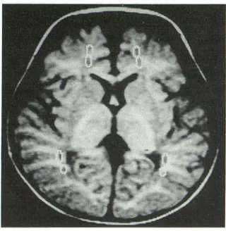
Fig. 1. Regions of interest are demonstrated on a
diffusion-weighted image (white circles). At least two regions of interest
were chosen in the frontal and occipital lobes in order to calculate
the apparent diffusion coefficient.
groups was assessed by the new multiple range test (Dun-can method) (7). For the comparison between the frontal and occipital lobe in each age group, Student paired
t
test was used. A value of P < .05 was considered significant.Results
In all adults, diffusional anisotropy of frontal
and occipital white matter was clearly seen. When
the diffusion-sensitive gradients were
perpendic-ular to the nerve fibers, the diffusion-induced
reduction in signal intensity was small (Fig 2A).
In contrast, when the gradients were parallel to
the nerve fibers, a large signal reduction was
observed, reflecting a rapid diffusion along the
nerve fibers {Fig 28). Diffusional anisotropy in
children and infants more than 6 months of age
was also as obvious in the adults {Figs 3A and
38)
.
In
neonates, however, diffusion anisotropy
was quite obscure in deep and cortical white
matter compared with the adults or infants,
al-though it was slightly more obvious in the
occip-ital lobe than in the frontal lobe {Figs 4 and 48).
The anisotropic ratios (apparent diffusion
coef-ficient
90/0)
in
frontal and occipital white matter
are plotted against the ages in
all subjects (Fig 5)
.
The ratios in the frontal lobe are about
0.8
at the
neonatal period and then show rapid decrease
until approximately 6 months of age
.
After 6
months the
ratios
do not differ significantly
throughout
infants,
children, and adults. The
ra-tios
in the
occipital lobe, by contrast, are higher
in neonates and young infants under 6 months of
age than other subjects, but
their
difference is
not so striking compared with those in
the frontal
lobe
.
By comparison between
frontal
and
occipi-tal
lobes,
the ratios
are
higher in
the frontal lobe
than the occipital lobe just at the neonatal period.
After
1
month they became similar
.
The mean
anisotropic ratios
in frontal
and
occipital lobes in
individual age groups are shown
[image:2.612.94.254.529.693.2]AJNR: 15, February 1994 DIFFUSIONAL ANISOTROPY
233
A
B
c
Fig. 2. Diffusion-weighted MR images in normal adult brain of a 28-year-old subject.
A, Diffusion-sensitive gradients are applied in a left to right direction.
B, The gradients are applied in an anterior to posterior direction. Large signal
attenuation is demonstrated in occipital white matter, frontal white matter, and corpus
callosum when the gradients were applied parallel to the direction of nerve fibers
(arrows).
C, Tl-weighted image (spin-echo, 600/20 [repetition time/echo time]).
D, T2-weighted image (two cardiac cycles, echo time 120 msec).
D
ratio is 0
.
79
±
0.05 in neonates, being
signifi-cantly higher than that in infants, children, or
adults
(P
<
.01, respectively)
.
In occipital lobe
the ratio is still higher in neonates than infants
(P
<
.05) or adults
(P <
.01), but the difference was
less marked compared with the frontal lobe.
Com-paring the ratios between occipital and frontal
lobes in each age group, a significant difference
was observed
only
in neonates
(P
<
.01)
.
Absolute values of apparent diffusion
coeffi-cients (0) and (90) in frontal and occipital white
matter are plotted against the ages in all subjects
(Figs 6A and 68)
.
In the frontal lobe, apparent
diffusion coefficient (0) does not differ
signifi-cantly from neonates to elderly adults
.
By
con-trast, apparent diffusion coefficient (90) in
neo-nates is higher than older subjects and shows
rapid decrease until 6 months and then keeps
fairly constant
.
In the occipital lobe, on the other
hand, neither apparent diffusion coefficient (0)
nor (90) differ significantly throughout all
sub-jects.
Discussion
Since Moseley et al (4) obtained
diffusion-weighted MR images in the cat brain using a 2-T
CSI system, diffusion-weighted imaging has an
increasing interest because of its potential to
show diffusional anisotropy along the nerve fibers
in the white matter. They speculated that the
diffusional anisotropy reflects the degree of mye
-lination in white matter. Recently, anisotropically
restricted diffusion of water was demonstrated in
the brains of neonates and infants by Bydder's
group
(8-1
0)
.
They reported that restricted
A
used had a low magnetic field (0. 15 T), and their
gradient system was not self-shielded. In this
study, we obtained diffusion-weighted images
and determined the apparent diffusion coefficient
in the subjects ranging in age from neonates to
more than 70 years old by using with high
mag-netic field and self-shielded gradient coils. We
performed a quantitative analysis of diffusional
anisotropy by calculating anisotropic ratio
(ap-parent diffusion coefficient 90/0). The diffusional
anisotropy in neonatal white matter was quite
weak, especially in
the
frontal lobe
.
The
aniso-tropic ratio in white matter was significantly
higher in neonates than that in infants, children
,
or adults
.
The anisotropic ratio showed a rapid
decrease until 6 months after birth and
then
reached
the level quite close
to
that
in
adults
.
Figures
6A and 68 demonstrated that the rapid
decrease of
the
anisotropic
ratio was
mainly
caused
by the decrease of apparent diffusion
coefficient
(90) measured
with the
perpendicular
gradients
to
the nerve
fibers. In
contrast, the
apparent diffusion coefficient (0) measured with
8
the parallel gradient to the fibers showed only
mild decrease after brain development.
The origin of diffusional anisotropy is still
un-clear. Rutherford et al (8) commented that because
the permeability of water of myelin lipid bilayers
is 10 to 50 times smaller than the permeability
of axoplasmic membranes, the addition of just
one layer of myelin would be expected to result
in a significant reduction
in
the apparent diffusion
coefficient of unmyelinated axons. As the
mem-branous structure of the myelin sheath is matured
after brain development, motion of water in a
direction perpendicular to the fibers is severely
prevented by myelin sheath. When diffusion
measurements are made parallel to the fibers,
water motion is
relatively free.
In this way, water
will diffuse
relative to the
direction of nerve
fibers.
If
the diameter of an axon
is
smaller than the
distance
that
a water molecule travels by diffusion
during the
time
of observation
in
bulk water, the
diffusion coefficient may be reduced. Most of the
AJNR: 15, February 1994 DIFFUSIONAL ANISOTROPY
235
1.0
-
00.8
~
...__-
~
0.6
~
0.4
0.2
•
:,•
··'··
rn o0 ~
OJ
Cb 0~
0
0[D •
•
oD 0
•
~
•
Iii 0
•
0
B
c
•
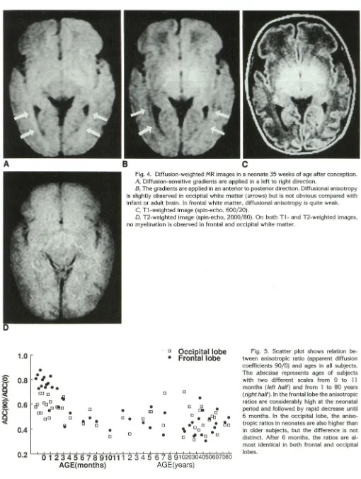
Fig. 4. Diffusion-weighted MR images in a neonate 35 weeks of age after conception.
A, Diffusion-sensitive gradients are applied in a left to right direction.
B, The gradients are applied in an anterior to posterior direction. Diffusional anisotropy
is slightly observed in occipital white matter (arrows) but is not obvious compared with
infant or adult brain. In frontal white matter, diffusional anisotropy is quite weak.
C, T1-weighted image (spin-echo, 600/20).
D, T2-weighted image (spin-echo, 2000/80). On both T1-and T2-weighted images,
no myelination is observed in frontal and occipital white matter.
•
IIJ0
•
0
•
Occipital lobe
Frontal lobe
0
•
0 0
•
De 0
0 • 0 0
•
~fb•
Fig. 5. Scatter plot shows relation be-tween anisotropic ratio (apparent diffusion
coefficients 90/0) and ages in all subjects .
The abscissa represents ages of subjects
with two different scales from 0 to 11
months (left half) and from 1 to 80 years
(right half). In the frontal lobe the anisotropic
ratios are considerably high at the neonatal
period and followed by rapid decrease until
6 months. In the occipital lobe, the aniso
-tropic ratios in neonates are also higher than in older subjects, but the difference is not distinct. After 6 months, the ratios are al
-most identical in both frontal and occipital lobes.
•
0•
•
0•
~0•
\ Do•
•o
0
•
0 # •o0
•
0 1 2 3 4 56 7 8 910111
2 3
4
5 6 7 8
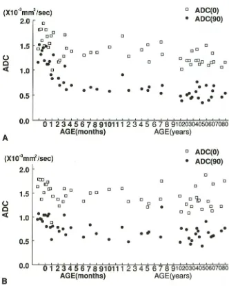
91020304050607080 [image:5.612.52.562.66.741.2]ADC(O)
tween absolute values of apparent diffusion coefficient and ages in the frontal (A) and occipital lobe (B). In the frontal lobe, appar-ent diffusion coefficient (90) reveals a rapid
decrease until 6 months, whereas the appar -ent diffusion coeffici-ent (0) shows a slight fall. In the occipital lobe, both apparent dif
-fusion coefficient (90) and apparent diffusion coefficient (0) remain fairly constant throughout the subjects.
(X10
.
mm
/
sec)
2.0
0•
ADC(90)
llJO0 0
oo 0
0 0
1.5
~~
0 0 0 00 oD
0
0 0 0
•
•
0 00
0 0• •J
!9 0c
0 rno cooD
r::fP
0<t:
•
01.0
0 0•
•
•
•
••
• •••
•
•
•
•
• • •
•
•
•
•
•
•
0.5
•
.
..
,
.
.
•
•
•
•
•
0.0
0 1 2 3 4
56
7 8 910111
2
3 4 5 6 7 8 91020304050607080A
AGE( months)
AG
E(yea
r
s
)
0
ADC(O)
(X10"
3mnt
/
sec)
•
ADC
(
90)
2.0
0
rm.B
0 00 0
0 llJ 0 0 0 0
1.5
0 0 00 0 0
otjb
0 oo 0
0 0 0 0
0
0 0 ooc
0•
0 00 0 0 0
<t:
1.0
.
.,
•
~
...
0. . .
•
•
•
••
,
••
•
•
•
•'
•
~•
•
•
•
•• •
0.5
•
• • •
o.o
L...L.."*o-:::l1,...2~.3~4"5~6~7-=a--::9:'-::1-:-07-11::-:1';--;!;-2
"'*3~4~5~6-=7;-;8~91~o~2o~3~o4';'!;o~5o::;-;:6:';:;:o7~o-:;:;;;;8o8
rest
r
icted diffusion will occur if the range of scale
length is less than 14 to
1
6
Jlm
.
However
,
the
role of the myelin sheath on diffusional anisotropy
is not fully understood
.
Le Bihan et al measured
restricted diffusion in white matter using different
diffusion times and demonstrated that no real
AGE(months)
AGE
(
years)
restrictive diffusion pattern could be found in
white matter (12)
.
Thus, the reduced value of the
[image:6.612.228.560.226.640.2]AJNR: 15, February 1994
sheaths in the developing brain could influence
the degree of restricted diffusion.
Water content of the brain is another important
factor affecting diffusion coefficient in white
mat-ter. The brain is about 90% water at birth and
decreases to 82%
~83
%
at 6 months (14). The
ratio
of
intracellular
water with restriction
to
ex-tracellular
water without
restriction
changes
dur-ing these 6 months
.
As shown in our study,
apparent diffusion coefficient
(0) in
neonatal brain
was slightly higher than
that
in adults. Reduction
of apparent diffusion coefficient (0) during first 6
months may reflect
the
change of tissue water
content in the extracellular space. The decrease
of extracellular water will enhance the diffusional
anisotropy in white matter
.
The anisotropic
ratios in
frontal white matter
was significantly larger
than
those of occipital
white matter at neonatal periods, but
the
differ-ence between them became less significant
in
infancy,
especially after 6 months
.
This fact
sug-gests that the development of myelination starts
earlier in the occipital lobe than
the
frontal lobe.
The anisotropic ratio at 6 months after birth is
already very close to that in adult
in
frontal and
occipital white matter. X-ray computed
tomog-raphy and conventional T1- and T2-weighted MR
images
have been used for the assessment of
brain maturation. Quencer assessed myelination
of neonatal brain using computed
tomography
numbers and pointed out that two phases of
maturation are identified
:
a rapid phase
(first
8-12 weeks) and a gradual phase
(after 12
weeks)
(15)
.
T1- and T2-weighted
images
also
reflect
development of myelination and maturation of
brain (16, 17). On T2-weighted
images,
the
change of signal intensity in white matter
contin-ues after 6 months, and T2-weighted images are
more useful in monitoring brain development in
this
period (18). The signal intensity changes
mainly
relate to
slow decrease of
water
content
in
the
brain after 6 to 8
months (14).
On the other
hand,
T1-weighted images are useful
in
monitor-ing brain development
in the first
6 months
(18).
As is shown
in
the figures
in this
study and
in
our
previous paper (6), the
changes
in diffusional
anisotropy precede
the changes in the signal
intensity on T1- and
T2-weighted images. Thus,
diffusion imaging is expected
to
be
clinically
im-portant because
it
enables earlier assessment
of
not
only
neonatal
brain development
but also
white matter
demyelinating
diseases
compared
with routine MR images
or other
diagnostic
meth-ods
.
DIFFUSIONAL ANISOTROPY
237
It
has been believed
that the diffusion-sensitive
gradient applied
on the phase-encoding axis or
the section-selecting axis is likely to induce more
severe
eddy current
problems than
on
the readout
axis.
As reported in our
previous
paper
(6), the
apparent diffusion
coefficients
of
the water
phan-tom are
fairly constant even when the direction
of
the
diffusion-sensitive
gradient is changed on
X, Y,
and Z axes.
We believe this is attributable
to
the use of active
shielded gradient coils. Based
on
this result, the data of apparent diffusion
coefficient in the
present study were thought to
be highly reliable
.
The
artifacts
caused by motion
may be a problem in
the clinical
application
of
this technique;
however
,
those
artifacts
caused
by vascular pulsations can be
reduced
by
cardiac
or peripheral gating
.
Involuntary
movement of
the head, on the other
hand, is
occasionally
difficult to suppress
even with sedation or some
head-holding devices.
Echo-planar
diffusion-weighted imaging
will
be
the solution
for the
motion
artifact problems.
In conclusion
,
diffusion-weighted
images
dem-onstrated that
the
myelination process
starts
ear-lier in the occipital lobe than
in
the
frontal lobe.
The changes of diffusional anisotropy
in white
matter are completed
within
6
months after birth.
Diffusion-weighted
imaging provides
an
earlier
detection of brain
myelination than the
conven-tional
T1- and
T2-weighted images.
References
1. Le Bihan D, Breton E, Lallemand D, et al. MR imaging of incoherent
motion: application to diffusion and perfusion in neurologic disorders. Radiology 1986; 161:401-407
2. Le Bihan D, Breton E, Lallemand D, et al. Separation of diffusion and
perfusion in intravoxel incoherent motion imaging. Radiology
1989; 168:497-505
3. Le Bihan D, Turner R. Jntravoxel incoherent motion imaging using spin echoes. Magn Reson Med 1991;19:221-227
4. Moseley ME, Cohen Y, Kucharczyk J, et al. Diffusion-weighted MR
imaging of anisotropic water diffusion in cat central nervous system.
Radiology 1990; 176:439-445
5. Chenevert TL, Brunberg JA, Pipe JG. Anisotropic diffusion in human
white matter: demonstration with MR techniques in vivo. Radiology 1990;177:401-405
6. Sakuma H, Nomura Y, Takeda K, et a!. Adult and neonatal human brain: Diffusional anisotropy and myelination with diffusion-weighted
MR imaging. Radiology 1991;180:229-233
7. Duncan DB. Multiple range and multiple F tests. Biometrics
1955;11:1-42
8. Rutherford MA, Cowan FM, Manzur A Y, et a!. MR imaging of anisotropically restricted diffusion in the brain of neonates and infants. J Comput Assist Tomogr 1991 ;15:188-198
9. Hajnal JV, Doran M, Hall AS, et al. MR imaging of anisotropically