1917
Superselective Continuous Arterial Infusion
Chemotherapy through the Superficial Temporal Artery
for Oral Cavity Tumors
Tatsuhiko Nakasato, Kenichi Katoh, Miyuki Sone, Shigeru Ehara, Yoshiharu Tamakawa, Hideki Hoshi, and Saburoh Sekiyama
BACKGROUND AND PURPOSE: High-dose intraarterial chemotherapy with repeated one-shot infusion may be useful for treating head and neck tumors. We evaluated the efficacy of superselective continuous arterial infusion chemotherapy administered via a coaxial catheter system and compared the results with those of subselective catheterization for treatment of oral cavity tumors.
METHODS: Forty-nine consecutive patients with tumors of the oral cavity (clinical stage I, 12 cases; stage II, 19 cases; stage III, six cases; stage IV, 12 cases) were treated by arterial infusion chemotherapy. After a guiding catheter was advanced into the superficial temporal artery, superselective catheterization was performed using a coaxial system microcatheter. Su-perselective catheterization was accomplished in 34 cases, and was unsuccessful in 15, owing to difficulties in performing catheterization or to multiple feeding arteries. In the latter cases, the tip of the catheter was placed near the origin of the feeding arteries (subselective catheterization).
RESULTS: Thirty (88%) of 34 patients had a complete response to superselective arterial infusion chemotherapy and two (6%) had a partial response. Twelve (80%) of 15 patients had a complete response to subselective arterial infusion chemotherapy and three (20%) had a partial response. Local recurrence was more frequent after subselective treatment (13%) than after superselective (6%) treatment.
CONCLUSION: Superselective continuous arterial infusion chemotherapy may be suitable for local control of oral cavity tumors, with a low rate of recurrence.
Intraarterial infusion chemotherapy has the advan-tage of delivering a high concentration of chemo-therapeutic agents into the tumor bed with fewer systemic toxic effects than seen with systemic che-motherapy. Intraarterial chemotherapy delivered into the external carotid artery (ECA) is an estab-lished procedure (1–3). Current improvements in catheter design, including introduction of the co-axial system, have enabled the performance of ret-rograde selective catheterization into the main tu-mor feeders arising from the ECA, such as the superficial temporal artery (STA). Continuous in-fusion chemotherapy by use of a selective
tech-Received January 21, 2000; accepted after revision May 10. From the Department of Radiology, Iwate Medical Univer-sity School of Medicine (T.N., K.K., M.S., S.E., Y.T.); and the Department of Oral Surgery II, Iwate Medical University School of Dentistry (H.H., S.S.), Morioka, Japan.
Address reprint requests to Tatsuhiko Nakasato, MD, De-partment of Radiology, Iwate Medical University School of Medicine, 19–1 Uchimaru, Morioka 020–8505, Japan.
qAmerican Society of Neuroradiology
nique is considered an ideal method for treating malignant neoplasms of the head and neck (4). However, superselective catheterization is occa-sionally made difficult by complex anatomy or multiple feeding arteries. The purpose of this study was to assess the efficacy of superselective contin-uous arterial infusion chemotherapy using a coaxial catheter system and to compare its efficacy with that of the subselective technique.
Methods
Exten-FIG1. Superselective catheterization in a 62-year-old man with tongue cancer (T1N0M0).
A–D, The guiding catheter was placed near the origin of the target vessel by using a guidewire (A). A vascular mapping study was done of the ECA (arrow, LA; B), after which a coaxial microcatheter was ad-vanced through the guiding catheter into the feeding artery (C), and superselective lingual arteriography was performed (D). ICA, internal carotid artery; ECA, stem of external carotid artery; LA, lingual artery; FA, facial artery; IMA, internal maxillary artery.
sion of the lesion into the surrounding tissue and cervical node was evaluated by CT, MR imaging, and sonography.
After inducing local anesthesia of the temporal region, a guiding catheter (UK-II catheter 16G, Unitica, Hyogo, Japan) was advanced into the STA, after which a main feeding artery was selectively catheterized using a coaxial system containing a 2.5F microcatheter (Fastracker-18, Target Therapeutics, Bos-ton Scientific; Galway, Ireland) and a 0.014-inch Transend wire (Boston Scientific, Watertown, MA) or a 0.016-inch torquable guidewire (Radifocus guidewire M, GT wire angle, or double-angle type; Terumo, Tokyo, Japan) (Fig 1). For catheterization of the STA under fluoroscopic guidance, a 0.035-inch guide-wire (Radifocus, angle type) was used to prevent angiospasm or dissection. Systemic heparinization was not performed dur-ing catheterization.
Superselective catheterization by microcatheter was con-firmed with digital subtraction angiography after infusion of
contrast material and was found to be successful in 34 cases and unsuccessful in 15 (Table 1). In such cases, the tip of the microcatheter was placed near the origin of the feeding arteries (subselective catheterization). Unsuccessful catheterization was attributed to anatomic variations and to multiple feeding arteries.
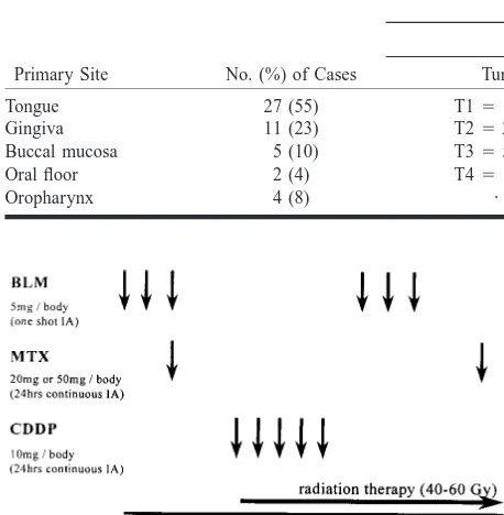
TABLE 1: Patient Data
No. (%)
Primary Site No. (%) of Cases
TNM (M0)
Tumor Node Stage
Tongue Gingiva Buccal mucosa Oral floor Oropharynx
27 (55) 11 (23) 5 (10) 2 (4) 4 (8)
T1514 (29) T2520 (41) T355 (10) T4510 (20)
· · ·
N0542 (86) N154 (8) N2b52 (4) N2c51 (2)
· · ·
I512 (24) II519 (40) III56 (12) IV512 (24)
· · ·
FIG2. Protocols of chemotherapy and radiation therapy.BLM, bleomycin;MTX, methotrexate; CDDP, cisplatin.
FIG3. A and B, Photographs show infusion apparatus attached to catheter system (A) and a patient with a catheter placed by means of a coaxial system (B).
3). Although such treatment may be performed on an outpa-tient basis, we treated our paoutpa-tients in the hospital, in accor-dance with our current clinical practice guidelines.
The position of the catheter and the patency of the vessel were checked by injection of indigo carmine on the first day of each course of chemotherapy. The skin and the catheter were sterilized every day and the catheter was filled with 2000 U of heparin to prevent coagulation. No additional antiplatelet medications were given.
Treatment results were assessed according to World Heath Organization criteria. Results were classified as 1) complete response (disappearance of all tumor masses for at least 1 month); 2) partial response (a decrease of 50% or more in the product of the largest diameter and the perpendicular diameter of the measurable tumor for at least 1 month); 3) no change (a decrease of less than 50% or an increase of less than 25% in the diameter product of the lesion); or 4) progressive disease (an increase of more than 25% in the diameter product or
de-velopment of a new lesion). The Kaplan-Meier method was used to estimate survival rates.
Results
Among the 34 patients who had superselective arterial infusion chemotherapy, 30 (88%) had a complete response and two (6%) had a partial re-sponse, including four patients with a T3 tumor and six with a T4 tumor (Fig 4). Among the 15 patients who had subselective arterial infusion chemother-apy, 12 (80%) had a complete response and three (20%) had a partial response. Local recurrence was more frequent among those who were treated with the subselective technique: two (13%) from this group had a complete response versus two (6%) from the group who underwent the superselective procedure. After therapy, total dissection of the neck was performed in four patients who had a complete response and in three who had a partial response. Nodal metastases were observed in six patients who had a complete response (four from the superselective group and two from the subse-lective group).
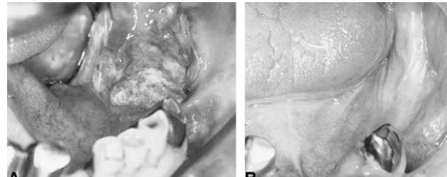
[image:3.612.62.539.557.720.2]FIG4. 64-year-old man with gingival can-cer (T2N0M0, stage II). The tumor in the lower gingiva of the molar region disap-peared after treatment.
A, Before treatment.
[image:4.612.212.528.63.188.2]B, After two courses of chemotherapy with superselective catheterization and ra-diation therapy (40 Gy).
TABLE 2: Results of superselective and subselective arterial in-fusion chemotherapy Superselective, No. (%) Subselective, No. (%) Complete response Partial response No change Progressive disease Local recurrence Nodal metastasis 5-year survival T3 plus T4
30 (88) 2 (6) 1 (3) 1 (3) 2 (6) 4 (12) 82% 30% 12 (80) 3 (20) · · · · · · 2 (13) 2 (13) 87%* 34% * Not statistically significant.
[image:4.612.298.527.235.306.2]FIG5. Overall survival rate of patients with cancer of the oral cavity.
TABLE 3: Staging of patients with complete response to arterial infusion chemotherapy
Stage
Superselective (n530 of 34)
[image:4.612.47.276.235.345.2]Subselective (n512 of 15) I II III IV 8/9 (89) 13/13 (100) 5/5 (100) 4/7 (57) 3/3 (100) 5/6 (83) 1/1 (100) 3/5 (60)
TABLE 4: Results of superselective and subselective arterial in-fusion chemotherapy in patients with tongue cancer
Superselective, No. (%) Subselective, No. (%) Complete response Partial response 5-year survival T3 plus T4
19 (95) 1 (5) 80% 5 (25) 4 (57) 3 (43) 88%* 1 (14) * Not statistically significant.
both the superselective and subselective groups (Table 3). For patients with tongue cancer (n527), the complete response rate was 95% after the su-perselective technique, including three (25%) with T3 tumors and two with T4 tumors. After the sub-selective technique, the complete response rate was 57% among those with tongue cancer, including one patient (14%) with a T4 tumor (Table 4 and Fig 6). The cumulative 5-year survival rate for pa-tients with tongue cancer was 80% in the
super-selective group and 88% in the subsuper-selective group (no statistical difference) (Fig 7).
No major neurologic or systemic complications were noted, except in three patients in whom pul-monary fibrosis developed. Minor side effects, such as nausea, vomiting, and reactive mucositis, were controlled by medication. The position of the cath-eter was confirmed by injection of indigo carmine, and repositioning was performed immediately un-der fluoroscopic guidance in one case.
Discussion
Recent advancements in coaxial microcatheter systems using various types of wires (ie, reshaped or double-angle type) have made them suitable for superselective catheterization of tortuous vessels; however, selective catheterization may still be dif-ficult in cases of a sharply angled feeding artery (Fig 8). Sometimes, anatomic variations, such as a lingual artery with the appearance of a linguofacial trunk, seen in approximately 17% to 18% of pa-tients (5, 6), also create difficulties in performing superselective catheterization. A good knowledge of vascular anatomy is important for determining which artery to catheterize, particularly in patients with gingival cancer.
[image:4.612.47.275.539.612.2]gin-FIG 6. 72-year-old woman with tongue cancer (T1N1M0, stage III). The tumor in the lateral aspect of tongue was not de-tected after treatment.
A, Before treatment.
B, After two courses of chemotherapy with superselective catheterization and ra-diation therapy (60 Gy).
FIG7. Overall survival rate of patients with tongue cancer.
FIG8. Superselective catheterization of the sharply angled feeding artery in a 44-year-old man with tongue cancer (T1N0M0). A–C, The lingual artery is sharply angled (arrow, A). A coaxial microcatheter was introduced initially into the internal carotid (or occipital) artery to make the tip of the catheter J-shaped (B), then the catheter tip was turned around and pulled back, resulting in superselective catheterization into the feeding artery (C). ICA, internal carotid artery; ECA, stem of external carotid artery; LA, lingual artery;FA, facial artery; IMA, internal maxillary artery.
giva, the sublingual artery is the main feeder. These feeders and their anastomoses create a variety of vascular networks between the lingual and the fa-cial arterial system. Therefore, confirmation of the exact feeding artery by injection of a small amount of indigo carmine is important.
In recent years, intravenous chemotherapy using a cisplatin-based regimen has been advocated as a form of induction therapy (7). In describing their work with patients with advanced squamous cell carcinoma of the head and neck, Jacobs et al (8) reported that the use of induction chemotherapy (cisplatin and bleomycin) plus standard surgery,
ra-diation, and maintenance chemotherapy with monthly cisplatin (80 mg/m2) for 6 months resulted
in a significantly lower metastatic rate. This intra-venous maintenance chemotherapy with cisplatin also produced a significantly improved 3-year dis-ease-free survival rate (67%) over that achieved with standard (49%) or induction (44%) treatment protocols in patients with cancer of the oral cavity. In a study of patients with advanced head and neck cancer, Ervin et al (9) reported a 3-year disease-free survival rate of 88% with the use of random-ized adjuvant therapy with cisplatin, bleomycin, and methotrexate as compared with 57% in the control group.
transfemoral approach. Korogi et al (13) reported an overall response rate of 92%, with a complete response rate of 38%, with the use of transfemoral catheterization in patients with advanced carcinoma of the mouth. Their technique consisted of the one-shot slow infusion method repeated several times. On the other hand, the continuous infusion tech-nique used in our series produced an overall re-sponse rate of 94% and 100% among those treated with the superselective and subselective techniques, respectively. The complete response rate after su-perselective catheterization in patients with tongue cancer was also particularly high (95%) relative to that reported in previous series using the same tech-nique (4), although an exact comparison may be difficult owing to differences in patient selection, chemotherapeutic regimen, and combined radiation therapy.
After superselective catheterization, the patients who had a complete response had a lower recur-rence rate; however, no statistical differecur-rence was identified in the 5-year survival rates between the two techniques for any type of oral cancer. Long-term follow-up with a larger patient population will be required to address this issue; however, for pa-tients with tongue cancer, the survival rate seems to be better after superselective catheterization, in that our patients with more advanced disease (T3 plus T4) had a 25% 5-year survival rate after su-perselective catheterization versus a 14% rate after subselective treatment. Because of our low com-plication rate, we believe that the superselective procedure helps improve the quality of life for pa-tients with cancer of the oral cavity.
Conclusion
Complete response and survival rates among pa-tients with oral tumors treated with superselective or subselective continuous arterial infusion che-motherapy were relatively high, with no statistical significance between the two techniques. Further investigation is needed to identify which is the best
technique, as superselective catheterization proba-bly resulted in better survival rates in our study because of the patient group. Superselective con-tinuous infusion chemotherapy through the STA combined with radiation therapy may be suitable for local control of malignant tumors of the oral cavity, with a low recurrence rate.
References
1. Ramsden CH, Duff JK. Continuous arterial infusion of head
and neck tumors. Cancer 1963;16:133–135
2. Cheung DK, Regan J, Savin M, et al. A pilot study of
intraar-terial chemotherapy with cisplatin in locally advanced head and neck cancer. Cancer 1988;61:903–908
3. Claudio F, Cacace F, Comella G, et al. Intraarterial
chemother-apy through carotid transposition in advanced head and neck cancer. Cancer 1990;65:1465–1471
4. Simunek A, Krajina A, Hlava A. Selective intraarterial
chemo-therapy of tumors in the lingual artery territory by a new approach. Cardiovasc Intervent Radiol 1993;16:392–395
5. Kozielec T, Jozwa H. Variation in the course of the facial artery
in the prenatal period in man. Folia Morphol (Warsz) 1997;36:
55–61
6. Adachi B. Das Arteriensystem der Japaner. Bd. 2. Kyoto: Ma-ruzen; 1928:58–96
7. Robbins KT, Vicario D, Seagren S, et al. A targeted supradose
cisplatin chemoradiation protocol for advanced head and neck cancer. Am J Surg 1994;168:419–422
8. Jacobs C, Makuch R. Efficacy of adjuvant chemotherapy for
patients with resectable head and neck cancer: a subset anal-ysis of the head and neck contracts program. J Clin Oncol
1990;8:838–847
9. Ervin TJ, Clardk JR, Weichselbaum RR, et al. An analysis of
induction and adjuvant chemotherapy in the multidisciplinary treatment of squamous-cell carcinoma of the head and neck.
J Clin Oncol 1987;5:10–20
10. Campbell TN, Howell SB, Pfeifle CE, Wung WE, Bookstein J.
Clinical pharmacokinetics of intraarterial cisplatin in humans.
J Clin Oncol 1983;1:755–762
11. Los G, Blommaert FA, Barton R, Heath DD, et al. Selective
intra-arterial infusion of high-dose cisplatin in patients with ad-vanced head and neck cancer results in high tumor platinum concentrations and cisplatin-DNA adduct formation. Cancer
Chemother Pharmacol 1995;37:150–154
12. Imai S, Kajihara Y, Munemori O, et al. Superselective cisplatin
(CDDP)-carboplatin (CBDCA) combined infusion for head and neck cancers. Eur J Radiol 1995;21:94–99
13. Korogi Y, Hirai T, Nishimura R, et al. Superselective
intraarte-rial infusion of cisplatin for squamous cell carcinoma of the mouth: preliminary clinical experience. AJR Am J Roentgenol