Adrenaline and noradrenaline are the primary catecholamines secreted by teleosts in response to severe acute stress (Randall and Perry, 1992; Wendelaar Bonga, 1997; Reid et al. 1998). Typical stressors include hypoxia (Boutilier et al. 1988; Ristori and Laurent, 1989; Fievet et al. 1990; Perry et al. 1991), hypercapnia (Perry et al. 1987, 1989), exposure to air (Walhqvist and Nilsson, 1980), physical disturbance (Nakano and Tomlinson, 1967; Ristori and Laurent, 1985) and exhaustive exercise (Primmett et al. 1986). Upon release into the blood, catecholamines are involved in minimising the detrimental effects of acute stress on blood oxygen transport and are also believed to influence several other physiological processes (for reviews, see Perry and Wood, 1989; Thomas and Perry, 1992; Randall and Perry, 1992; Wendelaar Bonga, 1997).
In teleosts, the principal sources of circulating catecholamines are the chromaffin cells associated with the posterior cardinal vein and the anterior portion of the head kidney (Nandi, 1961; Nakano and Tomlinson, 1967; Nilsson, 1983). These cells are innervated by preganglionic nerve fibres of the sympathetic nervous system that release the
neurotransmitter acetylcholine (Nilsson, 1983). Upon activation of cholinergic receptors (Reid and Perry, 1995; Al-Kharrat et al. 1997), chromaffin cells release catecholamines via exocytosis in a Ca2+-dependent process (Furimsky et al. 1996).
Owing to the nature of the stressors that elicit catecholamine release in fish (see above), the chemical composition of the blood is generally profoundly altered during the secretory event. In particular, the blood is usually hypoxic and/or acidotic (see Randall and Perry, 1982). Only a few studies, however, have directly assessed the effects of altered blood oxygen status (Perry et al. 1991) or acid–base status (Dashow and Epple, 1985; Perry et al. 1993) on catecholamine secretion in fish. Although blood acidosis does not directly stimulate catecholamine secretion in vivo in rainbow trout (Perry et al. 1989), there is indirect evidence to suggest that lowered pH may enhance the secretion of catecholamines during hypoxaemia (Thomas et al. 1994). To date, however, no studies have directly examined a possible role of blood acidosis in modulating catecholamine secretion during acute stress in fish.
JEB1677
The direct and modulating effects of acidosis on catecholamine secretion in rainbow trout (Oncorhynchus mykiss) were assessed in vivo using cannulated fish and in situ using a perfused cardinal vein preparation. In situ, acidosis (a reduction in perfusate pH from 7.9 to 7.4) did not elicit catecholamine release or modulate the secretion of catecholamines evoked by the non-specific cholinergic receptor agonist carbachol (3×10⁻⁷to 10−5mol kg−1) or the muscarinic receptor agonist pilocarpine (10−7mol kg−1). Acidosis, however, significantly increased the secretion rates of noradrenaline and adrenaline in response to nicotine (10−8 to 10−7mol kg−1). In vivo, intra-arterial injections of nicotine (300–600 nmol kg−1) into normocapnic or moderately hypercapnic fish (water PCO∑=5 mmHg or 0.67 kPa) caused a dose-dependent elevation of circulating catecholamine levels. At the highest dose of nicotine, the rise in plasma catecholamine levels was significantly enhanced in the hypercapnic fish.
Acute hypoxia in vivo caused an abrupt release of catecholamines when arterial haemoglobin O2-saturation was reduced to approximately 55–60 %; this catecholamine release threshold during hypoxia was unaltered in hypercapnic fish. However, the hypoxia-induced catecholamine release was significantly greater in hypercapnic fish than in normocapnic fish.
The results of this study suggest that blood acid–base status, while not influencing catecholamine secretion directly or influencing the blood O2content threshold for catecholamine release during hypoxia, may modulate the secretory process specifically in response to nicotinic receptor stimulation of chromaffin cells.
Key words: adrenaline, noradrenaline, chromaffin cell, stress, hypoxia, hypercapnia, acidosis, catecholamine secretion, rainbow trout, Oncorhynchus mykiss.
Summary
Introduction
DOES BLOOD ACID–BASE STATUS MODULATE CATECHOLAMINE SECRETION IN
THE RAINBOW TROUT (ONCORHYNCHUS MYKISS)?
ALEJANDRA E. JULIO, COLIN J. MONTPETIT ANDSTEVE F. PERRY*
Department of Biology, University of Ottawa, 30 Marie Curie, Ottawa, Ontario, Canada K1N 6N5
*Author for correspondence (e-mail: [email protected])
The goals of this study, therefore, were to evaluate the short-term effects of acidosis on catecholamine secretion as well as to determine whether lowered blood pH (as might occur during acute stress) is able to modulate the response of chromaffin cells to cholinergic stimulation. The experiments were performed on rainbow trout in situ using a perfused posterior cardinal vein preparation (Fritsche et al. 1993) and in vivo using either serial blood-sampling techniques or an extracorporeal arterial blood shunt (Thomas, 1994).
Materials and methods Experimental animals
Rainbow trout Oncorhynchus mykiss (Walbaum) of both sexes for in situ perfusion experiments (mass 290.6±9.5 g; mean ± S.E.M., N=116) were obtained from Thistle Springs Trout Farm. Additional fish for in vivo experiments (mean mass 825.1±35 g; N=12) and longer-term perfusion experiments (mean mass 261.3±17 g; N=12) were obtained from Linwood Acres Trout Farm. All fish were kept in large fibreglass tanks supplied with flowing, aerated and dechlorinated, city of Ottawa tap water (15 °C). Fish were maintained on a 12 h:12 h light:dark photoperiod and were fed ad libitum on alternate days using a commercial pelleted fish diet.
In situ experiments
Fish were killed by anaesthetic overdose using 2 g l−1 ethyl-m-aminobenzoate (MS-222, Sigma) buffered with 4 g l−1 NaHCO3. After 10–15 s in the anaesthetic solution, the fish was placed ventral side up on a bed of ice. An incision was made along the length of the animal, beginning at the anus and ending just anterior to the pectoral girdle. The ventricle and bulbus arteriosus were exposed using blunt dissection and a small incision was made in the bulbus arteriosus. A cannula (Clay-Adams PE 160 polyethylene tubing) was inserted through the bulbus into the ventricle and secured using surgical silk at the junction between the two chambers. This served as the outflow for the perfusate, while a posterior cardinal vein cannula (Clay-Adams PE 160 tubing pre-filled with heparinized saline, 25 i.u. ml−1) served as the inflow (Fritsche et al. 1993).
To study the effects of perfusate pH on catecholamine secretion, two beakers were filled with Cortland saline (Wolf, 1963). The control saline was gassed with a mixture of 0.3 % CO2 in air supplied by a Cameron gas-mixing flowmeter (model GF-3/MP) while the experimental saline was gassed with 1.0 % CO2in air supplied by a Wösthoff gas-mixing pump (model M301a/f). The control and experimental saline solutions were left to equilibrate for 3 h; the mean final pH values of the control and acidic salines were 7.9±0.04 and 7.4±0.02, respectively (means ±S.E.M.). The preparation was pre-perfused with control saline for 20 min using a positive pressure differences (approximately 15 cm difference between the surface of the saline and the cannula) to maintain a constant
flow (1.5 ml min−1) through the posterior cardinal vein. After 20 min, four pre-samples of perfusate were taken at 1 min intervals. Flow was then switched to a second beaker containing either control or acidic saline. For short-term perfusions, samples were collected for 10 min after the switch. For the longer-term perfusions, blood samples were collected at 5 min intervals for 30 min.
To determine whether acidosis was capable of modulating the effects of cholinergic stimulation on the chromaffin cells, preparations were perfused with either control (pH 7.9) or experimental (pH 7.4) saline. After the pre-treatment samples had been taken, a bolus injection of carbachol (carbamylcholine chloride; Sigma), nicotine (nicotine-di-D -tartrate; Sigma) or the muscarinic receptor agonist pilocarpine (hydrochloride; Sigma) was delivered through the inflow cannula into different preparations. The doses of carbachol used were 3×10⁻⁷, 10−6, 3×10⁻⁶ and 10−5mol kg−1body mass; the doses of nicotine used were 10−8, 3×10⁻⁸and 10−7mol kg−1; the dose of pilocarpine used was 10−7mol kg−1. The drugs were injected in 0.3 ml of saline. The doses administered are expressed as mol kg−1 because the final concentration of agonist bathing the chromaffin cells cannot be determined. Samples were taken at 1 min intervals for 5 min.
In vivo experiments
Fish were anaesthetised using 0.1 g l−1MS-222 adjusted to approximately pH 7.0 with 0.2 g l−1NaHCO3. Fish were placed on a surgical table where their gills were irrigated continuously with anaesthetic solution. An indwelling polyethylene cannula (Clay-Adams, PE 50) was inserted into the dorsal aorta according to the method described by Soivio et al. (1975). The caudal vein and the caudal artery were cannulated at the level of the caudal peduncle using standard surgical procedures (see Axelsson and Fritsche, 1994). After surgery, the fish were placed into individual opaque acrylic boxes supplied with flowing, aerated fresh water, where they were allowed to recover for 24 h before experimentation.
shown that hypercapnia, if severe enough, can elicit catecholamine release in trout (see Randall and Perry, 1992). Thus, the levels of hypercapnia used in this experiment were selected on the basis of pilot experiments that showed stable plasma catecholamine levels during 20 min of exposure to 1.3 % CO2. After 20 min, a second blood sample was
withdrawn. At this point, graded hypoxia was induced in both the control and experimental groups; this was accomplished by substituting N2 for air. Blood samples were taken at
10 mmHg (1.3 kPa) (PaO∑) intervals until 35 mmHg (4.7 kPa)
was reached, then every 5 mmHg (0.7 kPa) until catecholamine release was presumed to have occurred (indicators are struggling, a sudden lowering of pHa and a pronounced increase in blood pressure). The duration of the hypoxic exposures therefore varied among fish.
In vivo nicotine experiments
Dorsal aortic cannulae were placed into fish weighing between 200 and 450 g, as described above, and the fish were allowed to recover for 24 h before experimentation. Three blood samples (0.5 ml) were taken from each fish; an initial sample followed by samples at 2 and 5 min post-injection. Doses of nicotine of 300, 450 and 600 nmol kg−1(1 ml kg−1)
plus a saline injection (control) were administered to separate groups of fish. Experimental fish were exposed to 20 min of hypercapnia before sampling; %CO2 and total flow from a
Cameron flowmeter were adjusted to give a final PwCO∑ of
approximately 5 mmHg (0.7 kPa). Pilot experiments demonstrated that this level and duration of hypercapnia would not cause plasma catecholamine levels to increase.
Analytical techniques
In the extracorporeal shunt experiments, blood pHa, PaCO∑
and PaO∑ were monitored using Radiometer (CO2, O2) and
Metrohm (pH) electrodes housed in temperature-controlled cuvettes and connected to a Radiometer PHM 73 meter. PwO∑
and PwCO∑ were measured using additional Radiometer O2
and CO2 electrodes connected to a dual-channel O2/CO2
meter (Cameron Instruments). A continuous flow of water across the water-recording electrodes was achieved by siphon. The O2electrodes were calibrated by pumping (using
the peristaltic pump of the extracorporeal shunt) a zero solution [2 % (w/v) sodium sulphite] or air-saturated water continuously through the electrode sample compartments until stable readings were obtained. The CO2electrode was
calibrated in a similar manner using mixtures of 0.5 % and 1.0 % CO2in air provided by a Cameron gas flowmeter. The
pH electrode was calibrated using Radiometer precision buffers. The CO2, O2and pH electrodes were calibrated prior
to each experiment.
Blood pressure was monitored by connecting the dorsal aortic cannula to a pressure transducer (UFI model 1050 BP; UFI Morro Bay, CA, USA) linked to an amplifier (model MP100) and integrated with the data-acquisition system. The pressure transducer was calibrated daily against a static column of water.
In vivo arterial blood samples (40µl) were analysed in
triplicate for oxygen content (CaO∑) using an Oxycon blood
oxygen content analyser (Cameron Instruments). Total CO2
(CaCO∑) was analysed using true plasma (50µl) with a Corning
965 carbon dioxide analyser. Haemoglobin concentration was determined in duplicate on 20µl blood samples using a commercial spectrophotometric haemoglobin assay kit (Sigma).
All blood samples collected for measurements of catecholamines were centrifuged immediately (12 000 g for 1 min), and the plasma was placed in liquid N2and then stored
at −80 °C until subsequent analysis. Plasma samples were subjected to alumina extraction and then analysed by high-performance liquid chromatography (HPLC) with electrochemical detection (Woodward, 1982). 3,4-Dihydroxybenzylamine was used as an internal reference standard in all analyses. Detection limits for adrenaline and noradrenaline were 0.1 nmol l−1. Inter-assay variations for
noradrenaline and adrenaline were 6 and 3 %, respectively; intra-assay variations were 5 and 8 % for noradrenaline and adrenaline, respectively.
Construction of oxygen equilibrium curves
Oxygen specifically bound to haemoglobin (Hb) (mol O2mol−1Hb) was calculated after subtraction of
physically dissolved O2 in the plasma; O2 capacitance
coefficients for human plasma were obtained from Boutilier et
al. (1984). [O2]/[Hb] was plotted against PaO∑
(pre-hypercapnia samples were excluded for the experimental fish), and a sigmoid curve was fitted to the data. 100 % O2
haemoglobin-saturation values were obtained from the normocapnic and hypercapnic curves, and this allowed O2
equilibrium curves to be expressed as a function of percentage haemoglobin O2-saturation. P50 values (PO∑ at 50 %
haemoglobin O2-saturation) were determined automatically
during the curve-fitting procedure using a commercial graphics software package (Sigmaplot 4.0).
Statistical analyses
Results In situ experiments Effects of acidosis on catecholamine secretion
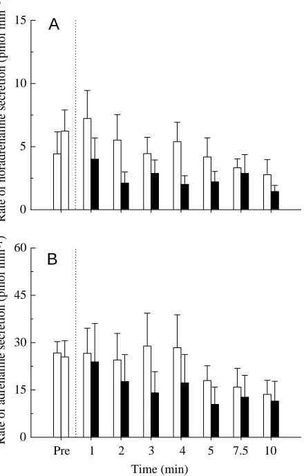
Lowering the perfusate pH from 7.9 to 7.4 was without effect on the secretion rates of adrenaline or noradrenaline during a 10 min period of perfusion (Fig. 1). Although the rate of catecholamine secretion appeared to decline with time in both the control and acidotic groups, the changes were not statistically significant. Indeed, there were no significant differences observed within or between the two groups at any time during the perfusion period.
A longer-term (30 min) perfusion study was conducted to determine whether acidosis might cause a delayed effect on catecholamine secretion. The results demonstrated that acidosis had no significant effects on catecholamine secretion over a 30 min perfusion period (Table 1).
Effects of acidosis on cholinergic-agonist-evoked catecholamine secretion
Carbachol. Maximal catecholamine secretion rates are illustrated in Fig. 2. Both the control (pH 7.9) and experimental (pH 7.4) treatments evoked dose-dependent release of catecholamines upon bolus injection of the non-specific cholinoceptor agonist carbachol. The highest doses (3×10⁻⁶
and 10−5mol kg−1for noradrenaline; 10−6to 10−5mol kg−1for
adrenaline) induced a significant release compared with pre-injection values. However, at any dose, carbachol-evoked catecholamine secretion rate was unaffected by perfusate acid–base status (Fig. 2).
Nicotine. Nicotine-evoked secretion rates for noradrenaline and adrenaline are depicted in Fig. 3. Control preparations (pH 7.9) did not exhibit significant changes in the rates of noradrenaline release at any of the three doses tested (10−8to
10−7mol kg−1); significant changes in the adrenaline secretion
[image:4.609.60.280.314.658.2]rate occurred only at the highest nicotine dose (10−7mol kg−1).
Fig. 1. The effects of perfusate pH on the secretion rates of (A) noradrenaline and (B) adrenaline in situ using a perfused posterior cardinal vein preparation of rainbow trout. The dotted line indicates a switch from control (Pre) saline (pH 7.9; open columns) to experimental saline (pH 7.4; filled columns; N=8). In the control group (N=12), pH was kept constant at 7.9 for the duration of the experiment. Values are presented as means + 1 S.E.M.
Time (min)
Pre 1 2 3 4 5 7.5 10 0
15 30 45 60
Rate of noradrenaline secretion (pmol min )
-1
0 5 10
15
A
B
Rate of adrenaline secretion (pmol min )
-1
Rate of noradrenaline secretion
(p
mol min
-1 )
0 100 200 300 400
Pre 3×10 10 3×10 10
Rate of adrenaline secretion
(pmol min
-1 )
0 500 1000 1500 2000
A
B
‡
‡ ‡ ‡
‡ ‡
‡
‡ ‡
‡
Carbachol dose (mol kg )-1
-7 -6 -6 -5
Fig. 2. The effects of carbachol on the maximal secretion rates of (A) noradrenaline and (B) adrenaline in situ using a perfused posterior cardinal vein preparation of rainbow trout. Open columns indicate preparations continuously perfused with control saline (pH 7.9; N=7–9 for each dose of carbachol); filled columns indicate preparations continuously perfused with acidic saline (pH 7.4;
N=7–9 for each dose of carbachol). ‡ denotes a statistically
[image:4.609.322.542.319.626.2]Acidotic preparations, however, displayed significant increases in the rate of noradrenaline release (compared with the pre-injection sample) and significantly increased secretion rates compared with the control group for doses of 10−8 and
3×10⁻⁸mol kg−1. At the higher doses (3×10⁻⁸ and
10−7mol kg−1), the preparations perfused with acidic saline
displayed significantly greater rates of adrenaline secretion (Fig. 3B).
[image:5.609.328.553.74.379.2]Pilocarpine. Noradrenaline and adrenaline secretion rates after a bolus injection of the specific muscarinic receptor agonist pilocarpine are shown in Fig. 4. Pilocarpine, when added to the control (pH 7.9) and acidotic (pH 7.4) preparations, stimulated noradrenaline secretion (Fig. 4A) equally. Adrenaline secretion rate (Fig. 4B) was statistically elevated by pilocarpine only in the acidotic preparations; statistical significance in the control group was prevented by a high degree of variance in the data set.
Table 1. The effects of perfusate pH on the secretion rates of noradrenaline (NADR) and adrenaline (ADR) in situ following a switch from control saline (pH 7.9) to acidic
saline (pH 7.4)
Control Experimental
NADR ADR NADR ADR
Sample (min) (pmol min−1)(pmol min−1) (pmol min−1)(pmol min−1)
Pre-acidosis 7.6±4.0 48.1±8.3 7.1±2.5 36.8±17.9
5 9.2±3.4 30.3±7.3 4.9±2.8 29.2±11.7
10 10.4±3.4 16.7±7.9 6.0±3.2 20.7±7.6
15 5.7±2.5 17.5±3.4 6.7±3.6 20.2±6.6
20 3.7±1.9 15.3±5.3 6.5±3.5 7.8±5.9
25 5.2±3.7 14.0±4.3 4.6±3.1 6.8±3.3
30 3.1±1.6 5.0±2.0 5.0±3.4 1.5±1.1
In the control group, pH was kept constant at 7.9 for the duration of the experiment.
[image:5.609.48.298.120.246.2]Values are mean secretion rates ± 1 S.E.M., N=6 animals.
Table 2. The effects of short-term (20 min) hypercapnia on selected arterial blood respiratory variables in rainbow trout
Hypercapnic group Control group
Pre 20 min Pre 20 min
PaCO2(mmHg) 2.08±0.09 5.83±0.44*,† 2.64±0.41 2.51±0.41
pHa 7.80±0.06 7.52±0.05*,† 7.71±0.06 7.73±0.06
PaO2(mmHg) 109.8±6.5 135.1±5.3*,† 105.4±5.3 110.4±2.7
CaO2(mmol l−1) 4.19±0.24 3.54±0.26*,† 4.87±0.21 4.88±0.27
[Hb] (mmol l−1) 1.23±0.08 1.26±0.09 1.36±0.09 1.32±0.07
[Hb-O2] (mol O2mol−1Hb) 3.25±0.06 2.61±0.07*,† 3.49±0.14 3.57±0.11
[image:5.609.60.567.539.666.2][Noradrenaline] (nmol l−1) 0.36±0.15 ND 1.43±1.10 ND
[Adrenaline] (nmol l−1) 1.67±0.72 2.38±1.07 9.75±0.41 2.89±1.05
* represents a statistically significant difference from the corresponding pre-treatment value (Pre) (P<0.05; paired t-test); † represents a statistically significant difference from the corresponding value in the control group (P<0.05; unpaired t-test).
ND, not detectable.
Values are means ±S.E.M., N=6 animals. 1 mmHg=0.1333 kPa.
[Hb-O2], the concentration of oxygen bound specifically to haemoglobin (Hb).
Nicotine dose (mol kg ) Pre 10 3×10 10
Rate of adrenaline secretion
(p
mol
min
)
0 250 500 750 1000
Rate of noradrenaline secretion
(p
mol
min
)
0 20 40 60
*
*
*
*
A
B
‡,
‡,
‡, ‡,
-8 -8 -7
-1
-1
-1
‡
In vivo experiments
The effects of hypercapnic acidosis on catecholamine release during acute hypoxia
The effects of external hypercapnia on blood respiratory variables and plasma catecholamine concentrations are summarised in Table 2. Predictably, external hypercapnia caused a marked respiratory acidosis (decreased pHa owing to an increase in PaCO∑) and reductions in CaO∑ and O2
specifically bound to haemoglobin. The increase in PaO∑in the
hypercapnic fish occurred concurrently with an apparent increase in the amplitude of opercular movements (measured by impedance changes; data not shown). Plasma catecholamine levels were unaltered by 20 min of hypercapnia.
Fig. 5 summarises the dynamics of catecholamine release during acute hypoxia under normocapnic (Fig. 5A) or hypercapnic (Fig. 5B) conditions. In each case, plasma catecholamine levels remained virtually constant at baseline levels during most of the period of exposure to graded hypoxia. However, upon reaching a critical PaO∑ threshold, circulating
catecholamine levels increased markedly and abruptly. The
estimated critical PaO∑ thresholds for catecholamine release
varied greatly between the normocapnic and hypercapnic fish. For normocapnic fish, the onset of catecholamine release occurred at a PaO∑ of approximately 15 mmHg (Fig. 5A),
whereas in the hypercapnic fish, catecholamine release occurred at a PaO∑ of approximately 33 mmHg (Fig. 5B). Hypercapnia
had a pronounced influence on haemoglobin O2-binding affinity
as calculated from the in vivo O2equilibrium curves (Fig. 5).
[image:6.609.329.531.208.578.2]The P50values calculated for normocapnic and hypercapnic fish
Fig. 4. The effects of the muscarinic receptor agonist pilocarpine on the maximal secretion rates of (A) noradrenaline and (B) adrenaline in situ using a perfused posterior cardinal vein preparation of rainbow trout. Open columns indicate preparations continuously perfused with control saline (pH 7.9; N=7); filled columns indicate preparations continuously perfused with acidic saline (pH 7.4; N=6). ‡ denotes a statistically significant difference (P<0.05) from the pre-injection (Pre) value. Values are represented as means +1 S.E.M.
Pilocarpine dose (mol kg )-1
Rate of noradrenaline
secretion (pmol min
)
0 10 20 30 40 50 60 70
Pre 10
Rate of adrenaline
secretion (pmol min
)
0 40 80 120 160
‡
‡ ‡
A
B
-7
-1
[image:6.609.59.276.345.644.2]-1
Fig. 5. Plasma total catecholamine (adrenaline plus noradrenaline) levels (filled circles) during acute hypoxia in vivo under (A) normocapnic or (B) hypercapnic conditions. In each panel, the catecholamine data are superimposed upon in vivo oxygen equilibrium curves (open circles). Sigmoid curves were fitted to the data, and P50values of 11.3 and 28.3 mmHg were calculated for the
normocapnic (N=6) and hypercapnic (N=6) fish, respectively. In each panel, the shaded square represents the mean plasma catecholamine concentration ±1 S.E.M. for all PaO∑ values below 20 mmHg
(normocapnic fish) and 37 mmHg (hypercapnic fish). * indicates a statistically significant difference in the mean plasma catecholamine level between the normocapnic and hypercapnic fish (t-test; P<0.05). Note the different plasma catecholamine y-axis scales in A and B. 1 mmHg=0.133 kPa.
0 20 40 60 80 100 120 140 0
25 50 75 100
0 50 100 150 200 250 300 350 400
A
B
*
Plasma [total catecholamines] (nmol l
-1 )
0 200 400 600 800 1000 2000 3000
0 20 40 60 80 100 120 140 0
25 50 75 100
Haemoglobin O
2
-saturation (%)
were 11.3 and 28.3 mmHg, respectively. Thus, owing to the effects of hypercapnia on lowering the affinity of haemoglobin O2-binding, catecholamine release in both groups of fish
corresponded with a decline in haemoglobin O2-saturation to
55–60 %. It is noteworthy that the difference in the PaO∑
catecholamine release thresholds for the normocapnic and hypercapnic fish (18 mmHg) was approximately equal to the difference in the P50values (17 mmHg).
Hypercapnia in vivo significantly increased the levels of plasma catecholamines achieved during acute hypoxia. The mean total catecholamine concentration during normocapnic hypoxia was 117.6±19.1 nmol l−1for all values corresponding
with PaO∑values below 20 mmHg. The mean catecholamine
concentration during hypercapnic hypoxia was 444.1±182.6 nmol l−1 for all values corresponding with Pa
O∑
values below 37 mmHg (the value of 37 mmHg was chosen because it differs from the normocapnic value, 20 mmHg, by the difference in P50values, 17 mmHg, between the two groups
of fish).
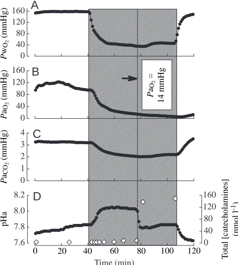
Representative recordings for normocapnic and hypercapnic fish are shown in Figs 6 and 7, respectively. The onset of catecholamine release for the representative normocapnic fish occurred at a PaO∑ of approximately 14 mmHg, and the
maximal plasma catecholamine level reached was 156 nmol l−1
(Fig. 6D). Note that the abrupt increase in plasma catecholamine levels was associated with the sudden development of metabolic acidosis superimposed upon respiratory alkalosis (Fig. 6C,D). The respiratory alkalosis is caused principally by hyperventilation, whereas the metabolic acidosis is believed to reflect the adrenergic activation of red blood cell Na+/H+ exchange (Fievet et al. 1990). The
representative hypercapnic fish exhibited abrupt catecholamine release at a PaO∑of approximately 40 mmHg and achieved a
maximal circulating catecholamine level of 814 nmol l−1
(Fig. 7D).
The effects of hypercapnia on nicotine-induced release of catecholamines
Intra-arterial injections of nicotine elicited significant noradrenaline (Fig. 8A) and adrenaline (Fig. 8B) release in both normocapnic and hypercapnic fish at doses of 450 nmol kg−1 and 600 nmol kg−1. At the highest dose of
[image:7.609.319.565.372.645.2]nicotine, plasma catecholamine levels increased to significantly higher levels in the hypercapnic fish than in normocapnic fish. Values for plasma adrenaline levels in the pre-treatment and saline-injected fish were below the detection limit of the HPLC (0.1 nmol l−1).
Fig. 6. Representative continuous in vivo recordings from a single normocapnic fish illustrating the effects of acute graded hypoxia on (A) PwO∑, (B) PaO∑, (C) PaCO∑and (D) pHa and total catecholamine
levels (open diamonds). The shaded area represents the period of exposure to graded hypoxia. The solid vertical line represents the abrupt onset of catecholamine release; the corresponding PaO∑ is
indicated in B. 1 mmHg=0.133 kPa.
Time (min) 7.6 7.8 8.0 8.2 0 40 80 120 160 0 40 80 120 160 0 40 80 120 160 0 1 2 3 4
A
B
C
D
P wO 2 (mmHg) P aO 2 (mmHg) P aCO 2 (mmHg) pHa0 20 40 60 80 100 120
[image:7.609.51.295.385.656.2]T otal [catecholamines] (nmol l -1) P aO 2 = 14 mmHg
Fig. 7. Representative continuous in vivo recordings from a single hypercapnic fish illustrating the effects of acute graded hypoxia on (A) PwO∑, (B) PaO∑, (C) PaCO∑and (D) pHa and total catecholamine
levels (open diamonds). The shaded area represents the period of exposure to graded hypoxia that was preceded by a 20 min interval of hypercapnia. The solid vertical line represents the abrupt onset of catecholamine release; the corresponding PaO∑ is indicated in B.
1 mmHg=0.133 kPa.
0 40 80 120 160 0 40 80 120 160 0 2 4 6 8 10 Time (min) pHa 7.2 7.6 8.0 0 100 200 300 400 800
A
B
C
D
P wO 2 (mmHg) P aO 2 (mmHg) P aCO 2 (mmHg)0 20 40 60 80
Discussion
Acute stress is often associated with marked changes in the chemical composition of the blood. In particular, physical (exhaustive exercise) and environmental (hypoxia, hypercapnia) stressors, if severe enough, may cause hypoxaemia and/or acidosis. Thus, secretion of catecholamines into the circulation at such times may occur when the chromaffin cells are exposed to acidic/hypoxic extracellular fluid. Therefore, changes in blood chemistry at the moment of catecholamine release could potentially influence secretion by a direct effect on the chromaffin tissue or indirectly by modulating the neurone-mediated reflex release pathway(s).
This is the first study to assess directly the involvement of blood acid–base status in catecholamine secretion in a teleost fish. The results demonstrated that, in rainbow trout, blood acidosis (in the physiological range) does not directly influence catecholamine secretion. However, the data obtained from in situ and in vivo experiments demonstrated that acidosis does
modulate the responsiveness of trout chromaffin cells to cholinergic stimuli.
Absence of any direct effects of acidosis on catecholamine secretion
In situ experiments
The goal of these experiments was to determine whether acidification of the interstitial fluid bathing the chromaffin cells could directly affect catecholamine secretion. Because the chromaffin cells in trout are predominantly localised to the walls of the posterior cardinal vein, this was accomplished by perfusing the posterior cardinal vein with normal (pH 7.9) or acidic (pH 7.4) saline. These values were chosen because they represent typical extracellular pH values in trout at 15 °C under normocapnic conditions and during periods of catecholamine release, respectively. For example, after exhaustive exercise or upon exposure to hypercapnia (i.e. PwCO∑7–8 mmHg), blood
pH is typically reduced by 0.4–0.5 units (Wood and Perry, 1985; Wood, 1991; Perry et al. 1987). The results clearly demonstrated that catecholamine secretion was unaffected by 30 min of perfusion with acidic saline. It seems unlikely, therefore, that acidification of the blood per se during acute stress plays any direct role in the initiation of catecholamine release. However, during periods of acidosis in vivo, there are likely to be significant changes in blood metabolite (e.g. lactate) and hormone (e.g. cortisol) levels. The effects of these other variables were not assessed in the present study and thus we cannot exclude possible contributions from such metabolites and hormones to the response of chromaffin cells to acidosis.
Although similar experiments have not been performed previously on teleost fish, the results are consistent with a previous study on an agnathan, Myxine glutinosa (Perry et al. 1993). In that study, lowering the pH of the perfusate from 8.1 to 7.1 in a perfused heart preparation did not alter rates of catecholamine secretion from the cardiac chromaffin tissue. In contrast, Dashow and Epple (1985) suggested that CO2could
act as a humoral trigger of catecholamine secretion in the lamprey Petromyzon marinus. In that study, however, living fish were exposed to pure (100 %) CO2prior to withdrawing
blood for analysis of catecholamines. Thus, the conclusion of a direct effect of CO2/acidosis on chromaffin tissue in the
lamprey may be premature.
Studies on the effects of alterations in the acid–base status on mammalian chromaffin tissue have produced conflicting results. For example, low pH was reported to stimulate catecholamine release in the perfused adrenal medulla of the rat (Fujiwara et al. 1994). In cultured bovine adrenal chromaffin cells, however, both stimulatory (Kao et al. 1991) and inhibitory (Kruger et al. 1995) effects of acidosis have been documented.
In vivo experiments
In the present study, rainbow trout were exposed to CO2for
a short period (20 min). In one experimental series, the goal was to increase PaCO∑ to approximately 6 mmHg (final
pHa≈7.5; Table 2), whereas in another series, the goal was to
Nicotine dose (nmol kg-1)
Pre Sal 30 450 600 0
100 200 300 400 800 1200 1600
B
‡ ‡
‡
‡,
*
ND ND
0 10 20 30 40 50 200 400 600 800
A
‡ ‡
‡ ‡,
*
[Noradrenaline] (nmol l
[image:8.609.48.285.70.375.2]-1)
[Adrenaline] (nmol l
-1)
achieve a constant level of moderate external hypercapnia (PwCO∑=5 mmHg). These criteria were selected on the basis of
pilot experiments (A. Julio, unpublished data) showing the absence of catecholamine release at these levels of hypercapnia. Indeed, the results clearly demonstrated that plasma catecholamine levels were unaltered during hypercapnia despite the pronounced respiratory acidosis that was elicited. Several previous studies have examined the effects of hypercapnia on the circulating levels of catecholamines in trout (Perry et al. 1987, 1989; Kinkead and Perry, 1991; Kinkead et al. 1993; Thomas et al. 1994; Perry and Gilmour, 1996). The results of these studies are highly variable, with responses ranging from no elevation of catecholamine concentrations (Kinkead and Perry, 1991) to minor increases (Perry et al. 1987, 1989) to large changes (Kinkead et al. 1993; Thomas et al. 1994; Perry and Gilmour, 1996). Owing to the Root effect, a reduction in blood O2
content (or haemoglobin O2-saturation) is believed to be the
trigger for catecholamine release during hypercapnia in trout (Perry et al. 1989). Thus, it is possible that the differences among the studies reflect varying degrees of CO2-induced
hypoxaemia. Although blood O2content was not reported in
every instance, it is noteworthy that Kinkead and Perry (1991) reported no increase in plasma catecholamine levels and also detected no decrease in arterial O2 concentration (CaO∑). In
contrast, the large increase in plasma catecholamine levels (>325 nmol l−1) observed by Kinkead et al. (1993) was
associated with a 33 % reduction in CaO∑. In the present study, CaO∑was reduced by 15 % after 20 min of hypercapnia; under
the present conditions, this degree of hypoxaemia was presumably not severe enough to elicit catecholamine release. Other possible sources of variation between the studies may include the rate at which PwCO∑is brought to equilibrium and
the timing of the blood sampling.
Regardless of the differences among the studies that have examined plasma catecholamine levels in hypercapnic trout, the results of the present study strongly reinforce the notion that blood acidosis, in itself, is not a direct trigger for catecholamine secretion. Thus, in the absence of other accompanying stimuli, such as a threshold level of hypoxaemia (Perry et al. 1989), acidosis does not appear to evoke an acute adrenergic stress response in rainbow trout. Although previous studies (Boutilier et al. 1986; Tang and Boutilier, 1988) have demonstrated significant relationships between blood acidosis and plasma catecholamine levels in trout, it is not possible to differentiate between the effects of acidosis per se and the associated hypoxaemia that is induced by acidosis as a result of the Root effect.
The modulating effects of acidosis on catecholamine secretion In situ experiments
The pH of the perfusion medium did not influence the secretory response of the chromaffin cells to carbachol, a non-specific or dual nicotinic/muscarinic receptor agonist. However, the response of the chromaffin tissue to a selective nicotinic receptor agonist, nicotine, was enhanced markedly by
perfusate acidosis. These results suggested the possibility that acidosis was inhibiting the potential contribution of the muscarinic receptor to catecholamine secretion. Thus, during perfusion with acidic saline, the stimulatory effect on the nicotinic receptor was possibly being counteracted by an inhibitory effect on the muscarinic receptor. The possible involvement of the muscarinic receptor in the control of catecholamine secretion has been investigated in carp Cyprinus carpio (Gfell et al. 1997) and eel Anguilla rostrata (Reid and Perry, 1995; Al-Kharrat et al. 1997) but, prior to the present study, the contribution of the muscarinic receptor to catecholamine secretion from trout chromaffin tissue had not been assessed directly. On the basis of results showing stimulation of catecholamine secretion in the presence of the selective muscarinic receptor agonist pilocarpine, it is clear that a muscarinic receptor is present on trout chromaffin cells. However, the stimulatory effects of pilocarpine on catecholamine secretion were not reduced by acidic saline. Thus, an alternative hypothesis must be sought to explain the lack of any effect of acidosis on carbachol-evoked catecholamine secretion. The current model of catecholamine secretion in fish (for a review, see Reid et al. 1998) advocates that a number of non-cholinergic neurotransmitters and neuromodulators (e.g. vasoactive intestinal peptide, pituitary adenylate cyclase activating polypeptide) are co-released with acetylcholine during neural stimulation of the chromaffin cells. In addition, the chromaffin cells themselves release substances that are capable of eliciting autocrine and paracrine effects (serotonin, adenosine, codeine) (Epple et al. 1994; Bernier and Perry, 1996). Therefore, it is possible that acidosis affects one or more of these non-cholinergic control systems to counter the stimulatory effect on the nicotinic receptor.
The results of the present study suggest a role for the muscarinic receptor in the cholinergic control of catecholamine secretion in trout. To demonstrate conclusively that the muscarinic receptor contributes to catecholamine secretion during neuronal stimulation of the chromaffin tissue, it would be necessary to compare catecholamine secretion during electrical stimulation of the nerves innervating the chromaffin cells in the presence and absence of a selective muscarinic receptor antagonist (e.g. atropine). The preliminary results of such experiments (C. Montpetit and S. F. Perry, unpublished observations) reveal an important stimulatory role of the muscarinic receptor. The stimulatory influence of the muscarinic receptor in trout differs from its function in the American eel Anguilla rostrata, where it is thought to have no role (Reid and Perry, 1995) or an inhibitory role (Al-Kharrat et al. 1997) in catecholamine secretion.
In vivo experiments
Intra-arterial injection of nicotine was used as a tool to evoke catecholamine release in vivo under normocapnic or hypercapnic conditions. Under hypercapnic conditions, when the blood was acidotic, the highest dose of nicotine (600 nmol kg−1) caused a significantly greater release of
the data obtained from the in situ experiments (see above) and further support the notion that catecholamine secretion, evoked by nicotinic receptor stimulation, is enhanced under acidotic conditions. Because detailed dose–response curves for nicotine were not constructed either in situ or in vivo, it is not possible to determine whether acidosis affects the affinity of nicotine-evoked release. However, the increased magnitude of release at the higher doses of nicotine suggests that acidosis up-regulates the nicotinic receptor or one or more downstream components in the signal-transduction pathway leading to catecholamine secretion.
Prior exposure of hypoxic fish to hypercapnia caused a pronounced decrease in the affinity of haemoglobin O2
-binding. Current theory contends that catecholamine release during exposure of trout to acute hypoxia occurs abruptly as PaO∑ falls below a critical threshold corresponding to a
reduction in haemoglobin O2-saturation of 40–50 % (Perry
and Reid, 1992, 1994; Thomas and Perry, 1992; Thomas et al. 1992). Thus, in the absence of any direct effects of acidosis on catecholamine release, it was predicted that hypercapnia would increase the PaO∑threshold for secretion
by an amount equivalent to the increase in the P50value; this
is what was observed. Regardless of the acid–base status of the blood, the threshold for catecholamine release occurred at approximately 55–60 % haemoglobin O2-saturation. Thus,
we conclude that acidosis, in itself, does not alter the threshold for catecholamine release during hypoxia except indirectly by decreasing the affinity of haemoglobin O2
-binding. However, the levels of circulating catecholamines achieved during hypoxia were significantly increased in the hypercapnic fish. These results concur with the previous findings of increased levels and enhanced rates of secretion of plasma catecholamines during acidosis upon nicotinic receptor stimulation in vivo and in situ, respectively. On the basis of these results, it is tempting to speculate that the stimulatory effect of acidosis on nicotinic-receptor-evoked secretion contributed to the enhancement of spontaneous catecholamine release during hypoxia in hypercapnic fish.
Regardless of the exact mechanisms, it is clear that the acid–base status of the blood can markedly influence catecholamine release in the rainbow trout during acute stress. Many types of acute stress are associated with blood acidosis. Thus, the physiological significance of this finding is that, at the moment of catecholamine secretion or shortly thereafter (e.g. if the blood is acidified by adrenergic H+extrusion from
the red blood cells), the chromaffin cells are likely to be sensitised and respond to stimulation by releasing larger quantities of adrenaline and noradrenaline.
This work was financed by Natural Sciences and Engineering Research Council (NSERC) of Canada Research and Equipment grants to S. F. Perry. A. Julio and C. Montpetit would like to thank N. Bernier, M. Furimsky, J. McKendry and P. D. Spencer for all their help and encouragement throughout this project.
References
AL-KHARRAT, H., WEISS, U., TRAN, Q., NIBBIO, B., SCHOLZ, S. AND
EPPLE, A. (1997). Cholinergic control of catecholamine release in the eel. Gen. comp. Endocr. 108, 102–108.
AXELSSON, M. ANDFRITSCHE, R. (1994). Cannulation techniques. In Analytical Techniques (ed. P. W. Hochachka and T. P. Mommsen), pp. 17–36. Amsterdam: Elsevier.
BERNIER, N. J. ANDPERRY, S. F. (1996). Control of catecholamine and
serotonin release from the chromaffin tissue of the Atlantic hagfish. J. exp. Biol. 199, 2485–2497.
BOUTILIER, R. G., DOBSON, G. P., HOEGER, U. ANDRANDALL, D. J. (1988). Acute exposure to graded hypoxia in rainbow trout (Salmo gairdneri): metabolic and respiratory adaptations. Respir. Physiol. 71, 69–82.
BOUTILIER, R. G., HEMING, T. A. AND IWAMA, G. K. (1984). Physiochemical parameters for use in fish respiratory physiology. In Fish Physiology, vol. 10A (ed. W. S. Hoar and D. J. Randall), pp. 403–430. New York: Academic Press.
BOUTILIER, R. G., IWAMA, G. K. AND RANDALL, D. J. (1986). The promotion of catecholamine release in rainbow trout, Salmo gairdneri, by acute acidosis: interaction between red cell pH and haemoglobin oxygen-carrying capacity. J. exp. Biol. 123, 145–157.
DASHOW, L. AND EPPLE, A. (1985). Plasma catecholamines in the
lamprey: intrinsic cardiovascular messengers? Comp. Biochem. Physiol. 82C, 119–122.
EPPLE, A., NIBBIO, B., SPECTOR, S. AND BRINN, J. E. (1994). Endogenous codeine – autocrine regulator of catecholamine release from chromaffin cells. Life Sci. 54, 695–702.
FIEVET, B., CAROFF, J. ANDMOTAIS, R. (1990). Catecholamine release
controlled by blood oxygen tension during deep hypoxia in trout: effect on red blood cell Na+/H+exchanger activity. Respir. Physiol.
79, 81–90.
FRITSCHE, R., REID, S. G., THOMAS, S. AND PERRY, S. F. (1993).
Serotonin-mediated release of catecholamines in rainbow trout Oncorhynchus mykiss. J. exp. Biol. 178, 191–204.
FUJIWARA, N., WARASHINA, A. AND SHIMOJI, K. (1994). Characterisation of low pH-induced catecholamine secretion in the rat adrenal medulla. J. Neurochem. 62, 1809–1815.
FURIMSKY, M., MOON, T. W. AND PERRY, S. F. (1996). Calcium
signalling in isolated single chromaffin cells of the rainbow trout (Oncorhynchus mykiss). J. comp. Physiol. 166, 396–404.
GFELL, B., KLOAS, W. ANDHANKE, W. (1997). Neuroendocrine effects of adrenal hormone secretion in carp (Cyprinus carpio). Gen. comp. Endocr. 106, 310–319.
KAO, L. S., HO, M. Y. ANDCRAGOE, E. J. J. (1991). Intracellular pH
and catecholamine secretion from bovine adrenal chromaffin cells. J. Neurochem. 57, 1656–1660.
KINKEAD, R., AOTA, S., PERRY, S. F. AND RANDALL, D. J. (1993). Propranolol impairs the hyperventilatory response to acute hypercapnia in rainbow trout. J. exp. Biol. 175, 115–126. KINKEAD, R. ANDPERRY, S. F. (1991). The effects of catecholamines
on ventilation in rainbow trout during external hypoxia or hypercapnia. Respir. Physiol. 84, 77–92.
KRUGER, C., HAUNSTETTER, A., GERBER, S., SERF, C., KAUFMANN, A., KUBLER, W. ANDHAAS, M. (1995). Nicotine-induced exocytotic
norepinephrine release in guinea-pig heart, human atrium and bovine adrenal chromaffin cells: modulation by single components of ischaemia. J. molec. cell. Cardiol. 27, 1491–1506.
NAKANO, T. AND TOMLINSON, N. (1967). Catecholamine and
relation to physical disturbances. J. Fish. Res. Bd Can. 24, 1701–1715.
NANDI, J. (1961). New arrangement of interrenal and chromaffin tissues of teleost fishes. Science 134, 389–390.
NILSSON, S. (1983). Autonomic nerve function in vertebrates. In Zoophysiology, vol. 13 (ed. D. S. Farner, B. Heinrich, K. Johansen, H. Langer, G. Neuweiler and D. J. Randall), pp. 1–253. Berlin: Springer-Verlag.
PERRY, S. F., FRITSCHE, R., KINKEAD, R. ANDNILSSON, S. (1991). Control of catecholamine release in vivo and in situ in the Atlantic cod (Gadus morhua) during hypoxia. J. exp. Biol. 155, 549–566. PERRY, S. F., FRITSCHE, R. AND THOMAS, S. (1993). Storage and
release of catecholamines from the chromaffin tissue of the Atlantic hagfish (Myxine glutinosa). J. exp. Biol. 183, 165–184.
PERRY, S. F. AND GILMOUR, K. (1996). Consequences of catecholamine release on ventilation and blood oxygen transport during hypoxia and hypercapnia in an elasmobranch (Squalus acanthias) and a teleost (Oncorhynchus mykiss). J. exp. Biol. 199, 2105–2118.
PERRY, S. F., KINKEAD, R., GALLAUGHER, P. AND RANDALL, D. J.
(1989). Evidence that hypoxemia promotes catecholamine release during hypercapnic acidosis in rainbow trout (Salmo gairdneri). Respir. Physiol. 77, 351–364.
PERRY, S. F., MALONE, S. AND EWING, D. (1987). Hypercapnic
acidosis in the rainbow trout (Salmo gairdneri). I. Branchial ionic fluxes and blood acid–base status. Can. J. Zool. 65, 888–895. PERRY, S. F. ANDREID, S. D. (1992). Relationships between blood
oxygen content and catecholamine levels during hypoxia in rainbow trout and American eel. Am. J. Physiol. 263, R240–R249. PERRY, S. F. AND REID, S. G. (1994). The effects of acclimation
temperature on the dynamics of catecholamine release during acute hypoxia in the rainbow trout Oncorhynchus mykiss. J. exp. Biol.
186, 289–307.
PERRY, S. F. ANDWOOD, C. M. (1989). Control and co-ordination of
gas transfer in fishes. Can. J. Zool. 67, 2961–2970.
PRIMMETT, D. R. N., RANDALL, D. J., MAZEAUD, M. ANDBOUTILIER,
R. G. (1986). The role of catecholamines in erythrocyte pH regulation and oxygen transport in rainbow trout (Salmo gairdneri) during exercise. J. exp. Biol. 122, 139–148.
RANDALL, D. J. AND PERRY, S. F. (1992). Catecholamines. In Fish
Physiology, vol. 12B, The Cardiovascular System (ed. D. J. Randall and W. S. Hoar), pp. 255–300. New York: Academic Press. REID, S. G., BERNIER, N. AND PERRY, S. F. (1998). The adrenergic
stress response in fish: control of catecholamine storage and release. Comp. Biochem. Physiol. (in press).
REID, S. G. AND PERRY, S. F. (1995). Cholinergic release of
catecholamines from chromaffin cells in the American eel (Anguilla rostrata) is mediated exclusively through the nicotinic receptor. J. comp. Physiol. B 165, 464–470.
RISTORI, M. T. ANDLAURENT, P. (1985). Plasma catecholamines and
glucose during moderate exercise in the trout: comparisons with bursts of violent activity. J. exp. Biol. 44, 247–253.
RISTORI, M. T. ANDLAURENT, P. (1989). Plasma catecholamines in rainbow trout (Salmo gairdneri) during hypoxia. Exp. Biol. 48, 285–290.
SOIVIO, A., NYHOLM, K. AND WESTMAN (1975). A technique for
repeated blood sampling of the blood of individual resting fish. J. exp. Biol. 62, 207–217.
TANG, Y. AND BOUTILIER, R. G. (1988). Correlation between catecholamine release and degree of acidotic stress in rainbow trout, Salmo gairdneri. Am. J. Physiol. 255, R395–R399.
THOMAS, S. (1994). Extracorporeal circulation. In Analytical
Techniques (ed. P. W. Hochachka and T. P. Mommsen), pp. 161–167. Amsterdam: Elsevier.
THOMAS, S., FRITSCHE, R. ANDPERRY, S. F. (1994). Pre- and post-branchial blood respiratory status during acute hypercapnia or hypoxia in rainbow trout (Oncorhynchus mykiss). J. comp. Physiol
164, 451–458.
THOMAS, S. AND PERRY, S. F. (1992). Control and consequences of adrenergic activation of red blood cell Na+/H+exchange on
blood oxygen and carbon dioxide transport. J. exp. Zool. 263, 160–175.
THOMAS, S., PERRY, S. F., PENNEC, Y. AND MAXIME, V. (1992). Metabolic alkalosis and the response of the trout, Salmo fario, to acute severe hypoxia. Respir. Physiol. 87, 91–104.
WAHLQVIST, I. AND NILSSON, S. (1980). Adrenergic control of the
cardiovascular system of the Atlantic cod, Gadus morhua during ‘stress’. J. comp. Physiol. B 137, 145–150.
WENDELAARBONGA, S. E. (1997). The stress response in fish. Physiol. Rev. 77, 591–625.
WOLF, K. (1963). Physiological salines for freshwater teleosts. Progve Fish-Cult. 25, 135–140.
WOOD, C. M. (1991). Acid–base and ion balance, metabolism and their interactions after exhaustive exercise in fish. J. exp. Biol. 160, 285–308.
WOOD, C. M. ANDPERRY, S. F. (1985). Respiratory, circulatory and
metabolic adjustments to exercise in fish. In Circulation, Respiration, Metabolism (ed. R. Gilles), pp. 2–22. Berlin: Springer-Verlag.
WOODWARD, J. J. (1982). Plasma catecholamines in resting rainbow