/. exp. Biol. 153, 261-276 (1990) 2 6 1
Printed in Great Britain © The Company of Biologists Limited 1990
THE INFLUENCE OF ACTION POTENTIALS ON THE
DEVELOPMENT OF THE CENTRAL VISUAL PATHWAY IN
MAMMALS
BY RONALD E. KALIL
Center for Neuroscience and Department of Ophthalmology, University of Wisconsin, 1300 University Avenue, Madison, WI53706, USA
Summary
The development of the mammalian visual system begins prenatally at distributed sites, where cells generated at different embryonic ages are destined to interconnect and form the visual pathways, and ends postnatally with the functional tuning of neuronal receptive-field properties. It is reasonable to assume that the earliest stages in this developmental sequence are completed prior to the onset of neural activity, and also that activity may play only a minor role or even none at all in primary axon outgrowth and pathway finding (Harris, 1981; Harris and Holt, 1990). However, recent evidence indicates that subsequent events in development, such as the sorting of axons at their targets, the cellular differen-tiation of target cells and the formation of synaptic contacts by developing axons, are all influenced by action potentials.
Action potentials in the developing retino-geniculo-cortical pathway can be eliminated by blocking the voltage-gated sodium channel with tetrodotoxin. Prenatal blockade prevents the laminar segregation of retinogeniculate axons. Postnatal blockade interrupts the formation of retinogeniculate synaptogenesis, slows the cytoarchitectonic differentiation of the lateral geniculate nucleus and produces abnormalities in the responses of lateral geniculate neurons. In the visual cortex, the development of cells and synapses is retarded and the eye-specific separation of geniculocortical axons is halted, thereby blocking the formation of ocular dominance columns. While the cellular mechanisms underlying these effects are not understood, a partial restoration of normal development can be produced by stimulating blocked axonal pathways electrically.
Introduction
It is now well established that the proper development of many pathways in the mammalian brain is influenced by experience. This is especially apparent in the major sensory systems, where it has been demonstrated in numerous experiments that appropriate stimulation is essential for establishing correct neuronal connec-tions.
262 R. E. KALIL
Studies of the developing visual system have played a prominent part in helping to define the role of sensory experience in the formation of neural pathways. To a great extent, the impressive volume of work in this area is due to an experimental convenience; namely, the relative ease of controlling visual stimulation during development. Thus, in comparison with audition or somesthesis, which are difficult to regulate experimentally because of the opportunity for self-generated stimulation, the nature and extent of visual input can be controlled in developing animals with reasonable precision.
Much of the modern work on the development of the mammalian visual system can be traced to the series of experiments of Hubel and Wiesel initiated during the 1960s (Hubel, 1982; Wiesel, 1982). These pioneering works triggered a subsequent cascade of research emphasizing the importance of suitable visual stimulation during specific periods of development in realizing normal maturation of the central visual pathways. Also emerging from these early experiments, in particular those of Guillery (1972), was an appreciation of the role played by synchronized binocular activity in forming the adult pattern of visual connections.
By 1980, after more than 15 years of intensive effort on the part of many investigators (see Sherman and Spear, 1982, for a review), it had been established beyond doubt that the morphological and physiological development of the visual pathways in mammals is dependent on the presence of patterned binocular stimulation during well-defined 'critical periods'. However, no experimental evidence had yet been adduced that might allow one to decide whether visual stimulation or, more explicitly, its correlate in the form of neural activity, is essential for development per se or serves primarily to ensure that development unfolds correctly.
The reason that this issue could not be settled with the evidence available at the time was that all of it had been collected under conditions that altered the patterns of action potential activity in the visual pathways but did not abolish them. Although deprivation experiments had clearly demonstrated that seemingly major changes in neural connectivity can result from atypical neuronal activity during development, no form of deprivation, long-term dark-rearing included, is suf-ficiently powerful to silence retinal ganglion cells. Indeed, action potentials are initiated in the retina and transmitted centrally in prenatal animals, well before vision is possible (Galli and Maffei, 1988), and ganglion cell discharge persists in postnatal animals, perhaps indefinitely, when they are placed under conditions that preclude vision, such as complete darkness (Rodieck and Smith, 1966; Burke and Hayhow, 1968).
Role of neural activity in visual development 263
topographic maps be considered, since this topic has also been the subject of recent reviews (Fawcett and O'Leary, 1985; Udin and Fawcett, 1988).
Instead, this chapter will focus on studies in mammals that have used tetrodotoxin (TTX) to block voltage-gated sodium channels and thereby prevent the generation of impulse activity. Although extremely toxic (e.g. LD50 of TTX for mice is 10 (i% kg"l intravenously), the action of TTX in blocking sodium channels is highly specific (Narahashi et al. 1964; Catterall, 1980) and appears not to disturb other aspects of neuronal function significantly. Particular attention will be paid to the results of experiments that have used TTX to block retinal ganglion cell activity in order to investigate the role played by action potentials in the morphological development of the retino-geniculo-cortical pathway.
Retinogeniculate axon segregation
In most higher mammals the dorsal lateral geniculate nucleus (LGN) is the primary target of axons from the retina. In species with well-developed binocular vision, the cells of the LGN are arranged in layers. Each cell layer is eye-specific in that it receives axons from one eye or the other but not both. As a result, binocularly driven neurons in the LGN are very uncommon (Bishop et al. 1962; Sanderson, 1971).
The laminar organization of retinal input to the LGN can be demonstrated clearly by injecting one eye with a tracer substance, such as a radioactive amino acid, that will be transported anterogradely along retinogeniculate axons and thereby reveal their projection. The results obtained by injecting the right eye of an adult cat with tritiated proline are shown in Fig. 1. Retinogeniculate axons from the nasal hemiretina of the injected eye cross in the optic chiasm (not illustrated) and enter the left LGN via the optic tract to terminate dorsally in layer A and ventrally in the C layers (C and C2). Fibers from the temporal retina of the injected eye do not cross in the optic chiasm, but instead terminate in the ipsilateral (right) LGN, primarily in layer A l , which lies directly beneath layer A.
The eye-specific laminar organization of the retinogeniculate pathway in the adult cat is not present initially during development. Indeed, when axons from the retina arrive at the LGN at approximately embryonic day 32 (E32) (Shatz, 1983), the nucleus is a relatively homogeneous mass of cells with no apparent lamination. It is not until late in gestation, close to the time of birth at approximately E63, that the cellular layers of the LGN are nearly completely formed (Kalil, 1978) and retinal afferents become almost fully segregated according to their eye of origin (Richards and Kalil, 1974; Shatz, 1983; Sretavan and Shatz, 1986).
264 R. E. KALIL
period E42-E56. This procedure was intended to block the conduction of action potentials in the optic tracts and may also have affected the activity of developing neurons in the LGN.
When retinal projections in the TTX-treated cats at age E57 were compared to those of controls, which had received infusions of citrate buffer, it was evident that treated retinogeniculate axons were not distributed normally. The contralateral projection appeared to fill the entire LGN, whereas the ipsilateral projection had expanded into a region of the LGN (cell layers have yet to be formed at E57) usually occupied only by fibers from the contralateral eye.
By bulk-filling the optic tract with horseradish peroxidase (HRP), Sretavan etal. (1988) were able to isolate individual retinogeniculate axons in fetal cats that had received intracranial infusion of TTX from E42 to E58. Reconstructed, HRP-filled axons from these fetuses differed significantly from normal in having unusual trajectories, elaborate branching patterns and wider terminal arborizations. Treatment with TTX did not retard axon growth but, instead, appeared to promote it nonspecifically. As a result, the normal segregation of retinogeniculate axons was disrupted.
Lateral geniculate nucleus differentiation
Retinogeniculate axons are segregated at birth in tree shrews, but the cellular layers of the LGN are poorly developed. Casagrande and Condo (1988) investi-gated whether retinal ganglion cell activity is important in the formation of LGN cell layers by blocking ganglion cell activity with intraocular injections of TTX during the first 2 weeks after birth. They observed that retinal blockade does not prevent the formation of the cell-free interlaminar zones that define LGN cell layers. However, in comparison with age-matched controls, the widths of the interlaminar zones, the areas of LGN cell somas and the overall size of the LGN
tsmaasmsmm
Role of neural activity in visual development 265
were reduced significantly in animals treated with TTX. Interestingly, no regression of retinogeniculate axon segregation was noted.
On balance, these results indicate that postnatal retinal blockade retards the cytoarchitectonic differentiation of the LGN but does not prevent it. In agreement are results from Golgi studies of developing LGN neurons in the rat, showing that spine number is reduced when action potentials in the retinogeniculate pathway are eliminated during the first 3 weeks after birth (Riccio and Matthews, 19856). Similarly, in the cat, retinal ganglion cell blockade disturbs the dendritic differentiation (Hsiao etal. 1987) of LGN neurons (Fig. 2) and the development of normal cell body size (Kuppermann and Kasamatsu, 1983). In view of the effects of action potential blockade on the growth of cells postsynaptic to silenced retinogeniculate axons, it would be most interesting to know whether the growth and elaboration of retinogeniculate axon arborizations, which occurs mainly after eye-specific segregation, is also influenced by the elimination of impulse activity.
Retinogeniculate synaptic connections Normal morphology and development
The synaptic connections of retinogeniculate axons in the cat are formed mainly after birth during the first 2 postnatal months (Kalil and Scott, 1979; Mason, 1982; Kalil et al. 1986). Axons from the retina end in the LGN as large terminals containing round synaptic vesicles and pale mitochondria. Guillery (1969) named these terminals 'RLP', coining an acronym that takes into account vesicle shape, terminal size and mitochondrial density. Two classes of postsynaptic targets are contacted by RLP terminals: (a) conventional dendrites of relay cells, identifiable by the presence of ribosomes, and (b) specialized dendritic appendages, which contain synaptic vesicles but not ribosomes and are thought to arise from the dendrites of LGN interneurons (Famiglietti and Peters, 1972; Sterling and Davis, 1980; Fitzpatrick etal. 1984; Hamos etal. 1985; Montero, 1986). Since the synaptic vesicles in these dendritic appendages often appear to be flat, they are called F profiles (Guillery, 1969).
In the normal adult cat about 30-40 % of all RLP terminals in the LGN are located in complex synaptic zones, which are sequestered from the surrounding neuropil by astrocytic glial processes (Kalil etal. 1986). These synaptic zones are often composed of a centrally located RLP terminal surrounded by postsynaptic relay cell dendrites and F profiles. Generally, RLP terminals in complex zones contact both F profiles and dendrites, while RLP terminals outside complex zones typically make contacts only with dendrites or occasionally with F profiles (Rapisardi and Miles, 1984).
266 R. E . KALIL
Role of neural activity in visual development 267
(Fig. 3A). By the end of the second postnatal month, synaptic contact length has shortened by 30 %, RLP terminal cross-sectional area has more than doubled, and the number of synaptic contacts made by individual RLP terminals has increased by more than 25 %. In each of these respects, RLP terminals are mature.
In concert with the growth of RLP terminals, two major changes occur in the pattern of retinogeniculate connections during development. These changes begin near the end of the first postnatal month and are largely completed by the end of the second. One major event is the formation of RLP synapses with F profiles. During the first 3 weeks after birth, connections between RLP terminals and F profiles are rare but thereafter these synapses develop rapidly. In fact, quantitative studies show that the increase in the number of synaptic contacts made by developing RLP terminals can be accounted for almost entirely by the addition of synaptic contacts with F profiles. The second major event is the establishment of complex synaptic zones. These zones begin to emerge during the fourth postnatal week and are fully formed, complete with astroglial encapsulation, by the end of the second month (Fig. 3C).
Development with retinal blockade
When retinal ganglion cell action potentials are blocked from birth for 8 weeks postnatally, by making monocular or binocular injections of 11X, the morphologi-cal development of retinogeniculate connections is also blocked (Kalil et al. 1986). In 8-week-old cats, RLP terminals from the TTX-injected eye are essentially identical to those in normal newborn animals (Fig. 3B), while those from the untreated eye or from eyes that received control injections of citrate buffer appear mature in all respects, as would be expected in a normally reared cat at 2 months of age. These results are summarized in Fig. 4.
Although these experiments demonstrate that eliminating action potentials in the retinogeniculate pathway halts synaptic development, the results do not exclude the possibility that synapse formation has been suspended only tempor-arily and might resume if impulse activity were reinstated. To investigate this we raised two groups of cats: one group received monocular injections of TTX from birth for 4 weeks (4/R cats) followed by up to 20 weeks of normal vision; the other group received injections from birth for 10 weeks (10/R cats) followed by 34 weeks of recovery.
Delayed visual experience in 10/R cats promoted a 50 % increase in the size of RLP terminals and a reduction in synaptic contact length to the normal adult value. This experience, however, was inadequate to support the formation of RLP
268 R. E. KALIL
Fig. 3. (A) Retinogeniculate terminal (asterisk) in a newborn kitten making a simple axodendritic synaptic contact. (B) Retinogeniculate terminal in an 8-week-old kitten reared from birth with retinal blockade produced by intraocular injections of tetrodotoxin. This terminal also makes an axodendritic contact. (C) Retinogeniculate terminal in a normal 8-week-old kitten. This terminal lies in an encapsulated synaptic zone and makes synaptic contacts with both dendrites (d) and F profiles (F). Compare the lengths of the synaptic contacts (arrowheads) in C with those in A and B. Scale bar,
[image:10.451.48.416.60.557.2]Role of neural activity in visual development
269
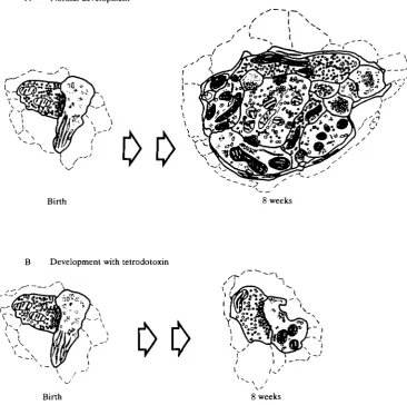
Normal development
Birth
B Development with tetrodotoxin
[image:11.451.44.411.82.446.2]Birth
Fig. 4. The diagram in A summarizes normal retinogeniculate synaptic development during the first 2 months after birth in the cat. Initially, almost all RLP terminals make simple axodendritic contacts. By 8 weeks after birth, RLP terminals have increased in size and synaptic contact lengths are decreased. At this age, many RLP terminals are found in complex synaptic zones in contact with relay cell dendrites and F profiles. (B) Retinogeniculate synaptic development is essentially 'frozen' when ganglion cell action potentials are blocked by tetrodotoxin from birth for 8 weeks.
synapses with F profiles or to establish encapsulated complex synaptic zones. By contrast, 4/R cats displayed nearly normal retinogeniculate synaptic develop-ment. The only significant abnormality was a marked reduction in the number of complex synaptic zones (Kalil and Dubin, 1988).
270 R . E . K A L I L
RLP synaptic development is hardly different, apart from a 20% reduction in synaptic contact length, from that of cats with continuous action potential blockade during the first 8 weeks. In cats given visual experience during the first postnatal month and TTX injections during the second, RLP terminal cross-sectional area is about 65 % greater than in newborns. However, in all other respects, RLP synaptic development with 4 weeks of ganglion cell activity prior to the onset of TTX appears similar to that in cats with only 2 weeks of early stimulation (Kalil and Dubin, 1988).
Collectively, the results from the three sets of experiments described above indicate that action potentials are required throughout the first 2 postnatal months for the normal development of retinogeniculate synaptic connections and their encapsulation in complex synaptic zones by glial lamellae. This conclusion raises an interesting question. Is the presence of action potentials alone sufficient for normal synaptic development or must the temporal pattern of impulse activity also be specified?
One approach to this problem is to replace retinal ganglion cell action potentials during development with controlled electrical stimulation of the retinogeniculate pathway. This procedure maintains activity in developing RLP synapses but natural impulse patterns are not preserved. Results have been collected from three 8-week-old kittens raised from birth with monocular retinal blockade and electrical stimulation of the optic tract during the second postnatal month (R. E. Kalil and M. W. Dubin, unpublished results). While moving freely about inside a large cylinder, the kittens received approximately 5 h of constant-current pulse stimulation each day. Retinogeniculate connections from the blocked eye, which had been stimulated electrically for 4 weeks, differed in two respects from those in cats raised with 8 weeks of uninterrupted blockade. First, stimulated RLP terminals were about 25 % larger in cross-sectional area, and second, synaptic contact lengths were reduced to adult size. These morphological changes confirm that the electrical stimulation was effective, but only partially. Artificial stimu-lation was not adequate to foster synaptic contacts between RLP terminals and F profiles, nor did it produce the normal formation of complex synaptic zones.
Lateral geniculate neuron response properties
Role of neural activity in visual development 271
development. This indicates, therefore, that retinal blockade with 1IX has induced a functional error in wiring that otherwise would not have occurred.
Other abnormalities in the responses of LGN cells in TTX-treated cats include: (a) greater than normal numbers of neurons that receive both X- and Y-cell retinal input, and (b) a large increase in the percentage of cells located at laminar borders that can be driven by both eyes. Both of these properties represent anomalies of degree rather than kind, and can be considered exaggerations of neuronal responses present in normal cats and kittens. Thus 10-15 % of LGN neurons in normal adult cats receive mixed X- and Y-cell input (Cleland et al. 1971) and about 50% of cells at laminar borders are activated binocularly in newborn kittens (Shatz and Kirkwood, 1984). The retention of unusually large numbers of neurons in each response category suggests that retinal blockade is responsible for these wiring errors by interfering with their elimination during development, rather than by creating them de novo.
Geniculocortical axons and visual cortex
Retinal blockade between postnatal days 5 and 21 inhibits the formation of dendritic spines (Riccio and Matthews, 1985c) and synapses (Riccio and Mat-thews, 1985J) in the developing visual cortex of the rat. In comparison with controls, the number of spines on the apical dendrites of layer V pyramidal cells is reduced by approximately 25 % in TTX-injected animals. The reduction is greatest in that region of the apical dendrite that passes through cortical layers III and IV. Similarly, the reduction in the number of synapses in the visual cortex of TTX-treated rats is also most prominent in layers III and IV, being approximately 25 % at 21 days after birth.
In the adult cat, axons from the LGN arborize in layer IV of the primary visual cortex in alternating eye-specific patches that form the morphological substrate for ocular dominance columns (Shatz et al. 1977). However, in the young kitten, geniculocortical afferents representing each eye are intermixed in layer IV of the cortex, and do not appear to begin segregation until the third postnatal week (LeVay et al. 1978). Normally, the segregation process is completed by the second or third postnatal month (LeVay et al. 1978) but, in cats reared with binocular eyelid suture (Kalil, 1982; Stryker and Harris, 1986) or in the dark (Swindale, 1981; Kalil, 1982; Mower et al. 1985; Stryker and Harris, 1986), only partial segregation of geniculocortical afferents takes place. In the absence of pattern vision, spontaneously generated action potentials in the geniculocortical pathway are apparently sufficient to initiate afferent segregation but are inadequate to sustain it to completion.
272 R. E . K A L I L
of these neurons is functionally equivalent to that of normal 2-week-old kittens. This suggests that action potential blockade stopped ocular dominance segre-gation at the stage at which it existed when the binocular blockade was introduced.
In an elegant experiment, Stryker and Strickland (1984) produced evidence that the blockade may have been effective in preventing geniculocortical segregation primarily because it eliminated interocular differences in the patterning of afferent activity, and not simply the activity itself. To demonstrate this point, two groups of kittens were raised with TTX-induced binocular blockade. While retinal ganglion cells were silenced from the second to the eighth postnatal week, one group of kittens received asynchronous electrical stimulation of the optic nerves and the other group received synchronous stimulation. Therefore, in both groups action potential activity was produced artificially in the geniculocortical pathway during development. However, only the kittens that received asynchonous stimulation demonstrated physiological segregation of geniculocortical afferents. In fact, the degree of monocularity of cortical cells that were stimulated asynchronously exceeded that of normally reared 8-week-old kittens (Stryker, 1986).
Concluding remarks
The experiments considered here provide compelling evidence that impulse activity is essential for the normal development of the visual pathways in mammals. This is not to say, however, that activity plays the same role at all stages in development or even that it plays a singular role at any one stage. Indeed, since successful neural development entails solving multiple overlapping problems, it would not be surprising to learn that activity is put to many different uses.
For example, the eye-specific segregation of afferent axons requires that a complex recognition/matching problem be solved; a problem that is similar whether it is confronted prenatally in the retinogeniculate pathway or postnatally in the geniculocortical pathway. When retinogeniculate action potentials are eliminated prenatally, the resulting superfluous and inappropriate axonal growth effectively disorders axon segregation. In contrast, interrupting geniculocortical action potentials postnatally blocks axon segregation by preventing the growth and remodeling of axon arborizations. In summary, abolishing action potentials will preclude axon segregation whether it occurs relatively early or late in develop-ment, but this disruption is caused by promoting excessive axon growth in the first instance and blocking necessary growth in the latter.
Role of neural activity in visual development 273
control mechanism is defeated, allowing axons to grow beyond the limits that would normally obtain.
By contrast, when impulse activity is blocked in the geniculocortical pathway at 2 weeks after birth, synapse formation is well underway and presumably developing contacts are already participating functionally in the mechanisms that will lead to correct, stable patterns of connections. This requires the breaking of incorrect connections and the forming of new appropriate ones; a process that is guided and consolidated by functional synaptic activity (see W. Singer, 1990). When this activity is abolished, the information necessary for making or breaking synaptic contacts is also eliminated, and with it the relevant cues for action. Under this condition, stasis is not an improper response, and appears to be the one adopted, at least temporarily, by developing geniculocortical axons.
Whether the speculations above are correct or not, the evidence on the effects of action potential blockade clearly highlights the need for caution in any attempt to provide a unifying explanation. As illustrated by the retinogeniculate pathway, the development of synaptic connections is often multifaceted and the influence of neural activity is correspondingly diverse. For example, it appears that certain aspects of developing synapses are afforded greater freedom in reaching maturity than others. While synaptic contact length is under the control of impulse activity, action potentials are effective in adjusting contact length whether they are present early or late in development. Similarly, the growth of immature RLP terminals can be advanced by the presence of action potentials during a wide temporal window. In contrast, action potentials must be available during a specific period, the second postnatal month, if RLP terminals are to make synaptic contacts with F profiles. Even more constrained is the relationship between neural activity and the elaboration of astroglial processes. Thus, the glial investment of complex synaptic zones requires action potentials throughout the first 2 postnatal months. Impulse activity during only part of this period or after it does not promote glial encapsulation.
Clearly, the temporal coupling between impulse activity and synaptic develop-ment is not fixed. For those aspects of synaptogenesis that are intrinsic to axon endings, such as terminal growth and the adjustment of contact length, the temporal requirements for impulse activity appear to be defined relatively loosely. Specified with considerably greater precision is the activity-dependence of interactive components of synaptic development, such as the formation of contacts between axon terminals and postsynaptic cells. Perhaps the most rigidly prescribed relationship is that involving neural activity and astroglial development. The results presented here point to a dependent relationship, while the recent work of Miiller and Best (1989) allows for its being cooperative.
274 R. E. KALIL
experiments in the rat (Riccio and Matthews, 1985a) show that blocking action potentials in the developing optic nerve produces a small decrement in the amount of rapidly transported [3H]fucose-labeled glycoproteins. However, inhibition of axoplasmic transport with colchicine in the developing retinogeniculate pathway does not block synaptic development (Matthews etal. 1982). Therefore, although it is possible that a reduction in axonally transported materials is implicated in some of the consequences of impulse blockade during development, it seems unlikely that a decrement of such broad proportions can account for the many diverse and specific effects that result from the elimination of action potentials.
Based on current results, work in the future is logically directed towards the presumptive linkages between neural activity and the intracellular messengers responsible for cytoskeletal development and modification. It will be exciting to discover how electrical messages are decoded in building the nervous system and, in particular, whether this task is executed primarily by presynaptic cells or also involves retrograde feedback from postsynaptic neurons (Williams et al. 1989).
References
BISHOP, P. O., KOZAK, W., LEVICK, W. R. AND VAKKUR, G. J. (1962). The determination of the projection of the visual field on to the lateral geniculate nucleus in the cat. J. Physiol, Lond. 163, 503-539.
BURKE, W. AND HAYHOW, W. R. (1968). Disuse in the lateral geniculate nucleus of the cat. /. Physiol., Lond. 194, 495-520.
CASAGRANDE, V. A. AND CONTJO, G. J. (1988). The effect of altered neuronal activity on the development of layers in the lateral geniculate nucleus. /. Neurosci. 8, 395—416.
CATTERALL, W. A. (1980). Neurotoxins that act on voltage-sensitive sodium channels in excitable membranes. A. Rev. Pharmac. Toxicol. 20, 15-43.
CLELAND, B. G., DUBIN, M. W. AND LEVICK, W. R. (1971). Sustained and transient neurones in the cat's retina and lateral geniculate nucleus. /. Physiol., Lond. 217, 473-4%.
CONSTANTINE-PATON, M., CLINE, H. T. AND DEBSH, E. A. (1989). Neural activity, synaptic convergence, and synapse stabilization in the developing central nervous system. In The Assembly of the Nervous System (ed. L. T. Landmesser), pp. 279-300. New York: Alan R. Liss, Inc.
CONSTANTINE-PATON, M., CLINE, H. T. AND DEBSH, E. (1990). Patterned activity, synaptic convergence, and the NMDA receptor in developing visual pathways. A. Rev. Neurosci. 13, 129-154.
DUBIN, M. W., STARK, L. A. AND ARCHER, S. M. (1986). A role for action-potential activity in the development of neuronal connections in the kitten retinogeniculate pathway. /. Neurosci. 6, 1021-1036.
EDWARDS, D. L. AND GRAJSTEIN, B. (1984). Intraocular injection of tetrodotoxin in goldfish decreases fast axonal transport of [ HJglucosamine-labeled materials in optic axons. Brain Res. 299, 190-194.
EDWARDS, D. L. AND GRAFSTEIN, B. (1986). Intraocular tetrodotoxin reduces axonal transport and transcellular transfer of adenosine and other nucleosides in the visual system of goldfish. Brain Res. 364, 258-267.
FAMIGUETTI, E. V. AND PETERS, A. (1972). The synaptic glomerulus and the intrinsic neuron in the dorsal lateral geniculate nucleus of the cat. /. comp. Neurol. 144, 285-334.
FAWCETT, J. W. AND O'LEARY, D. D. M. (1985). The role of electrical activity in the formation of topographic maps in the nervous system. Trends Neurosci. 8, 201-206.
Role of neural activity in visual development 275
GALLI, L. AND MAFFEI, L. (1988). Spontaneous impulse activity of rat retinal ganglion cells in prenatal life. Science 242, 90-91.
GULLLERY, R. W. (1966). A study of Golgi preparations from the dorsal lateral geniculate nucleus of the cat. J. comp. Neurol. 128, 21-50.
GUILLERY, R. W. (1969). The organization of synaptic inter-connections in the dorsal lateral geniculate nucleus of the cat. Z. Zellforsch. mikrosk. Anat. 96,1-38.
GUILLERY, R. W. (1972). Binocular competition in the control of geniculate cell growth. J comp.
Neurol. 144, 117-130.
HAMOS, J. E., VAN HORN, S. C , RACZKOWSH, D., ULRICH, D. J. AND SHERMAN, S. M. (1985). Synaptic connectivity of a local circuit neurone in the lateral geniculate nucleus of the cat.
Nature 317, 618-621.
HARRIS, W. A. (1981). Neural activity and development. A. Rev. Physiol. 43, 689-710. HARRIS, W. A. AND HOLT, C. E. (1990). Early events in the embryogenesis of the vertebrate
visual system: cellular determination and pathfinding. A. Rev. Neurosci. 13, 155-169. HSIAO, C. F., DUBIN, M. W. AND KAJJL, R. E. (1987). Rearing cats with TTX-blockade of
retinal ganglion cell (RGC) activity affects the morphological development of LGN neurons.
Invest. Ophthal. vis. Sci. (Suppl). 28, 21.
HUBEL, D. H. (1982). Exploration of the primary visual cortex. Nature 299, 515-524.
KALJL, R. E. (1978). Development of the dorsal lateral geniculate nucleus in the cat. /. comp.
Neurol. 182, 265-292.
KALJL, R. E. (1982). Development of ocular dominance columns in cats reared with binocular deprivation or strabismus. Soc. Neurosci. Abstr. 8, 4.
KALIL, R. E. AND DUBIN, M. W. (1988). The role of action potentials in the morphological development of retinogeniculate connections in the cat. In Cellular Thalamic Mechanisms (ed. M. Bentivoglio and R. Spreafico), pp. 479-488. Amsterdam: Elsevier.
KALIL, R. E., DUBIN, M. W., SCOTT, G. L. AND STARK, L. A. (1986). Elimination of action potentials blocks the structural development of retinogeniculate synapses. Nature 323, 156-158.
KALIL, R. E. AND SCOTT, G. L. (1979). Development of retinogeniculate synapses in the dorsal lateral geniculate nucleus of the cat. Soc. Neurosci. Abstr. 5, 791.
KUPPERMANN, B. D. AND KASAMATSU, T. (1983). Changes in geniculate cell size following brief monocular blockade of retinal activity in kittens. Nature 306, 465-468.
LEVAY, S., STRYKER, M. P. AND SHATZ, C. J. (1978). Ocular dominance columns and their development in layer IV of the cat's visual cortex: a quantitative study. /. comp. Neurol. 179, 223-244.
MASON, C. A. (1982). Development of terminal arbors of retinogeniculate axons in the kitten. II. Electron microscopical observations. Neurosci. 7, 561-582.
MATTHEWS, M. A., NARAYANAN, C. H., NARAYANAN, Y. AND SIEGENTHALER-MATTHEWS, D. J. (1982). Inhibition of axoplasmic transport in the developing visual system of the rat. III. Electron microscopy and Golgi studies of retino-fugal synapses and post-synaptic neurons in the dorsal lateral geniculate nucleus. Neurosci. 7, 405-422.
MAYER, M. L. AND WESTBROOK, G. L. (1987). The physiology of excitatory amino acids in the vertebrate central nervous system. Prog. Neurobiol. 28, 197-276.
MONTERO, V. M. (1986). Localization of y-aminobutyric acid (GABA) in type 3 cells and demonstration of their source to F2 terminals in the cat lateral geniculate nucleus: a Golgi-electron-microscopic GABA-immunocytochemical study. J. comp. Neurol. 254, 228-245.
MOWER, G. D., CAPLAN, C. J., CHRISTEN, W. G. AND DUFFY, F. H. (1985). Dark rearing prolongs physiological but not anatomical plasticity of the cat visual cortex. J. comp. Neurol. 235, 448-466.
MULLER, C. M. AND BEST, J. (1989). Ocular dominance plasticity in adult cat visual cortex after transplantation of cultured astrocytes. Nature 342, 427-430.
NARAHASHI, T., MOORE, J. W. AND SCOTT, W. R. (1964). Tetrodotoxin blockage of sodium conductance increase lobster giant axons. /. gen. Physiol. 47, 965-974.
RAPISARDI, S. C. AND MILES, T. P. (1984). Synaptology of retinal terminals in the dorsal lateral ^ geniculate nucleus of the cat. J. comp. Neurol. 225, 515-534.
276 R. E. KALIL
potentials: less-active inputs become dominant when kitten visual cortical cells are pharmacologically inhibited. Proc. natn. Acad. Sci. U.S.A. 85, 3623-3627.
RICCIO, R. V. AND MATTHEWS, M. A. (1985a). The effect of intraocular injection of tetrodotoxin on fast axonal transport of [3H]proline- and [3H]fucose-labeled materials in the developing rat optic nerve. Neurosci. 16, 1027-1039.
RJCCIO, R. V. AND MATTHEWS, M. A. (1985£>). Effects of intraocular tetrodotoxin on the postnatal development of the dorsal lateral geniculate nucleus of the rat: a Golgi analysis.
J. Neurosci. Res. 17, 440-451.
RJCCIO, R. V. AND MATTHEWS, M. A. (1985c). Effects of intraocular tetrodotoxin on dendritic spines in the developing rat visual cortex: a Golgi analysis. Devi Brain Res. 19, 173-182. RICCIO, R. V. AND MATTHEWS, M. A. (1985<f). The postnatal development of the rat primary
visual cortex during optic nerve impulse blockade by intraocular tetrodotoxin: a quantitative electron microscopic analysis. Devi Brain Res. 20, 55-68.
RICHARDS, W. AND KALIL, R. (1974). Dissociation of retinal fibers by degeneration rate. Brain
Res. 72, 288-293.
RODIECK, R. W. AND SMITH, P. S. (1966). Slow dark discharge rhythms of cat retinal ganglion cells. J. Neurophysiol. 29, 942-953.
SANDERSON, K. J. (1971). The projection of the visual field to the lateral geniculate and medial interlaminar nuclei in the cat. /. comp. Neurol. 143, 101-118.
SHATZ, C. J. (1983). The prenatal development of the cat's retinogeniculate pathway.
J. Neurosci. 3, 482-499.
SHATZ, C. J. AND KIRKWOOD, P. A. (1984). Prenatal development of functional connections in the cat's retinogeniculate pathway. /. Neurosci. 4, 1378-1397.
SHATZ, C. J., LINDSTROM, S. H. AND WIESEL, T. N. (1977). The distribution of afferents representing the right and left eyes in the cat's visual cortex. Brain Res. 131, 103-116. SHATZ, C. J. AND STRYKER, M. P. (1988). Prenatal tetrodotoxin infusion blocks segregation of
retinogeniculate afferents. Science 242, 87-89.
SHERMAN, S. M. AND SPEAR, P. D. (1982). Organization of visual pathways in normal and visually deprived cats. Physiol. Rev. 62, 740-855.
SINGER, W. (1990). The formation of cooperative cell assemblies in the visual cortex. J. exp.
Biol. 153, 177-197.
SRETAVAN, D. W. AND SHATZ, C. J. (1986). Prenatal development of retinal ganglion cell axons: segregation into eye-specific layers within the cat's lateral geniculate nucleus. J. Neurosci. 6, 234-251.
SRETAVAN, E. W., SHATZ, C. J. AND STRYKER, M. P. (1988). Modification of retinal ganglion cell axon morphology by prenatal infusion of tetrodotoxin. Nature 336, 468-471.
STARK, L. A. AND DUBIN, M. W. (1986). Response properties of neurons in the lateral geniculate nucleus of neonatal kittens. Vision Res. 26, 409-414.
STERLING, P. AND DAVIS, T. L. (1980). Neurons in cat lateral geniculate nucleus that accumulate exogenous [3H]-y-aminobutyric acid (GABA). J. comp. Neurol. 192, 737-749.
STRYKER, M. P. (1986). The role of neural activity in rearranging connections in the central visual system. In The Biology of Change in Otolaryngology (ed. R. W. Ruben, T. R. Van De Water and E. W. Rubel), pp. 211-224. Amsterdam: Elsevier.
STRYKER, M. P. AND HARRIS, W. A. (1986). Binocular impulse blockade prevents the formation of ocular dominance columns in cat visual cortex. /. Neurosci. 6, 2117-2133.
STRYKER, M. P. AND STRICKLAND, S. L. (1984). Physiological segregation of ocular dominance columns depends on the pattern of afferent electrical activity. Invest. Ophthal. vis. Sci. (Suppl.) 25, 278.
SWINDALE, N. V. (1981). Absence of ocular dominance patches in dark-reared cats. Nature 290, 332-333.
UDIN, S. B. AND FAWCETT, J. W. (1988). Formation of topographic maps. A. Rev. Neurosci. 11, 289-327.
WIESEL, T. N. (1982). Postnatal development of the visual cortex and the influence of the environment. Nature 299, 583-591.