Posterior Fossa Malformations
Nolan R. Altman, 1
Thomas P. Naidich, and Bruce H. Braffman
From the Miami Children's Hospital (NAA), the Baptist Hospital of Miami (TPN), and the Memorial Hospital, Hollywood, FL (BHB)
Posterior fossa malformations are best classi-fied in terms of their embryogenesis from the rhombencephalon ( 1-7). The diverse manifesta-tions of the dysgeneses can be related to the stages at which development of the cerebellum became deranged. The cerebellar malformations can also be classified by their effect on the fourth ventricle and cisterna magna, and by whether any cystic spaces represent expansion of the rhombencephalic vesicle or secondary atrophy of the parenchyma. Since the size of the posterior fossa depends largely on the size of the rhomben-cephalic vesicle at the time that the mesenchyme condenses into the bony-dural walls of the pos-terior fossa (see article by MeLone in this issue), identification of the size of the posterior fossa often proves to be more helpful in differentiating among these malformations than the simple pres-ence, or abspres-ence, of a cyst. In this discussion, the Chiari malformations have been specifically ex-cluded since they have been addressed previously (8-17).
Definitions
The normal mature cerebellum is divided into three lobes: anterior, posterior, and flocculono-dular (Fig. 1).
A. The anterior lobe consists of the first three lobules of vermis with their lateral extension into the hemi-spheres, ie,
vermis
1. lingula,
and
with
hemispheres
no hemispheric por-tion in man
1
Address reprint requests to Dr Altman, Department of Radiology, Miami Children's Hospital, 6125 SW 31st Street, Miami, FL 33155.
Index terms: Posterior fossa, abnormalities and anomalies; Pediatric neuroradiology
AJNR 13:691-724, Mar/Apr 1992 0195-6108/92/1302-0691 © American Society of Neuroradiology
691
2. central lob- with ule
the two alae, and
3. culmen with the (anterior) quadran-gular lobule.
The anterior lobe is the most rostral portion of the cerebellum and is separated from the more caudal portions of the cerebellum by the primary fissure.
B. The posterior lobe consists of the next five lobules of the vermis with their hemispheric connections, ie,
4.
5.
6.
7.
8.
vermis and hemispheres
declive with the lobulus simplex folium with the superior semilunar
lobule
tuber inferior semilunar,
and with the gracile and pyramis biventral lobules uvula with the cerebellar tonsils.
The posterior lobe is separated from the more caudal cerebellum by the posterolateral (flocculonodular) fis-sure. The posterior lobe undergoes the greatest growth in man receiving the influx of the pontocere-bellar fibers (2).
C. The flocculonodular lobe if formed by
vermis and hemispheres
9. nodulus with the flocculi.
692
A
Fig. 1. Anatomy of the mature cerebellum.
A, Midsagittal section of the cerebellum after removal of
the brain stem.
8-D, Surface features of the cerebellum as seen from 8,
superior; C, posterior; and D, posteroinferior. The pia arach-noid has been removed and the fissures have been spread to
emphasize their shapes and relationships. Note the fastigium (open white arrow) that is the superior midline point of the
fourth ventricle and the white matter of the vermis (desig-nated the arbor vitae) that appears to radiate outward from
the fastigium like a starburst. The first major division (curved black arrow) of the arbor vitae indicates the division of the
cerebellum into the anterior lobe and posterior lobe at the primary fissure (single arrowheads). The division to the flocculonodular lobe is not shown. The nine lobules of vermis
B
are directly continuous laterally with the lobules of the
cerebellum. The culmen is the largest lobule of vermis. The
specific branching pattern of the arbor vitae helps to identify C
each lobule. The vermis is well defined and can be
distin-guished from the cerebellar hemispheres by paired
paraver-mian grooves and by the abrupt angulation of the folia and fissures as they change from the horizontal orientation of the vermis to the oblique orientation of each hemisphere. Note
the relationships of the labeled fissures to the adjacent
lobules: primary fissure (single arrowheads), superior poste-rior fissure (single short arrows), great horizontal fissure (single long arrows), ansoparamedian fissure (double arrow-heads), prebiventral fissure (double short arrows), intrabiven-tral fissure (double long arrows), and secondary fissure (triple arrowheads). The posterolateral fissure (crossed arrow) that
separates the posterior from the flocculonodular lobes is
shown only at the vermis. Vermis: I= lingula, ce = central,
cu =culmen, d= declive, f= folium, t= tuber, p =pyramis,
u = uvula, n = nodulus. Hemispheres: AQ = (anterior) D
quadrigeminal lobule, LS = lobulus simplex, 55 = superior
semilunar lobule, IS = inferior semilunar lobule, G = gracile
lobule, 8 = biventral lobule, T = tonsils. The flocculi are not
shown.
[image:2.612.200.553.82.731.2]AJNR: 13, March/Apri11992
In recognition of this temporal sequence of development and in accord with the sequential evaluation of these structures in phylogeny, the portions of the cerebellum may also be desig-nated the archicerebellum, paleocerebellum, and neocerebellum.
Archicerebellum
The term archicerebellum designates the floc-culonodular lobe (2, 3). It is the first portion of the cerebellum to differentiate and is believed to be the most primitive. Because it is concerned with the vestibular nuclei, it is also designated the
vestibular cerebellum.
Paleocerebellum
The term paleocerebellum designates the floc-culi of the hemispheres plus the entire vermis (2). This appears to be a useful distinction, because the vermis of the anterior and posterior lobes forms before the corresponding hemispheres, so the flocculonodular lobe and the vermis coexist before the anterior and posterior lobes of the cerebellar hemispheres are well formed.
Neocerebellum
The term neocerebellum designates the cere-bellar hemispheres except for the flocculi (2, 3).
The cerebellar dysplasias can be classified in these terms as archicerebellar, paleocerebellar,
and neocerebellar dysplasias. Paleocerebellar dys-plasias have vermian defects as key features. These tend to include subtle malformations of the medulla, especially the inferior olives and the pyramidal tracts (2). Neocerebellar dysplasias in-volve the cerebellar hemispheres with relative sparing of the vermis and flocculi.
Normal embryogenesis of the cerebellum
There is general agreement among the author-ities as the broad order and the sequence of timing of the crucial events in embryogenesis ( 1-7, 18). The authorities differ substantially in the precise times at which they state specific events occur (1, 2, 18). Therefore, although every effort has been made to be accurate, the following discussion is intended more to provide an over-view of cerebellar embryogenesis than to give exact dates for each single event.
693
The cerebellum arises by a series of steps best understood in terms of the Carnegie Stages of Development (1).
Stage 10
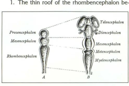
At approximately 22 days, before the neural tube closes, two constrictions in the cephalic portion of the neural tube divide it into the pros-encephalon, mespros-encephalon, and rhombence-phalon (Fig. 2). These constrictions become more evident during stages 11 and 12 (approximately 24-27 days). The rostral neuropore closes during stage 11 (19). When the brain becomes a closed tube, the three divisions of the neural tube are designated the primary brain vesicles. The
pro-sencephalon and rhombencephalon each
promptly divides into two secondary vesicles. The subdivisions of the rhombencephalon are desig-nated the metencephalon (future pons and cere-bellum) and the myelencephalon (future medulla oblongata). A constricted region, the isthmus,
unites the mesencephalon with the metence-phalon (5).
Stages 13-16
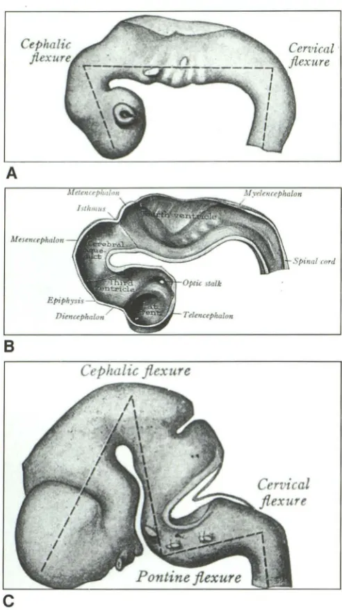
From approximately 28-37 days, the rhom-bencephalon flexes increasingly, convex ven-trally, to form the pontine flexure (Fig. 3). As a consequence:
1. The thin roof of the rhombencephalon
be----
Telencephalon ---ProsencephalonMesencephalon
Rhombe11cephalon
A
Fig. 2. Embryology; diagrammatic representation of the pri-mary and secondary brain vesicles.
A, Initially the neural tube constricts to form the three primary brain vesicles. These define the hindbrain (rhombencephalon), the midbrain (mesencephalon), and the forebrain (prosencephalon).
[image:3.614.314.561.454.619.2]694
A
Afese11crphalou
cnrd
8
Cephalic flexure
c
Fig. 3. Embryology; diagrammatic representation of the
flex-ing neural tube. A, 6-mm stage; B, 11-mm stage (hemisection);
C, 14-mm stage. (Reprinted with permission from Arey (5), p.
431, Figs. 406 and 407 .)
comes folded and forms a transverse crease (the plica choroidea) that runs perpendicular to the long axis of the neural tube. The transverse crease is approximately in line with the future plane of the foramina of Luschka (Figs. 4 and 5). The choroid plexus of the fourth ventricle will develop within the transverse crease by apposition of mesoderm to ependyma (1).
2. As the pons flexes, the alar plates that form the sides of the neural tube flare laterally, so the lumen of the rhombencephalon at the level of the crease becomes the widest portion of the neural
AJNR: 13, March/April1992
tube (Fig. 4) (1). Because of the flaring, the side walls of the rhombencephalon assume a diamond shape (as seen from posterior). From their widest separation at the transverse crease, the lips angle obliquely back toward the midline both cephali-cally (toward the mesencephalon) and caudally (toward the myelencephalon). A series of tran-sient, transversely oriented rhombic grooves be-comes evident along the floor of the rhomben-cephalic cavity from stages 10-15 (approxi-mately 22-34 days) (Fig. 4A). These identify the
intervening rhombomeres (synonym:
neurom-eres) that are related to the anlagen for cranial nerves 5-10. The rostral-most groove lies at the level of the pontine flexure and is related to the trigeminal nerve. The other grooves lie caudal to the pontine flexure. The first rhombencephalic segment caudal to the isthmus may be designated
rhombomere 1.
Stages 13-18
From approximately 28-44 days, the cerebel-lum will arise from the alar plate of the rhomben-cephalon rostral to the plica choroidea (Figs. 48, 4C, and 5) (1). That portion of the alar plate rostral to the developing trigeminal sensory nerve will form the major portion of the cerebellum. Intense neuroblastic activity causes the lateral aspects of tlie alar plates to hypertrophy into thick rhombic lips (1). Although the first indica-tion of the developing cerebellar hemispheres can be seen histologically at stages 13 or 14 (6), gross morphologic development of the cerebellar hemi-spheres is not appreciated until stage 18 (7). At this stage, the external cerebellar bulge first ap-pears along the caudal margin of the cerebellum as a transversely oriented strip of tissue that is separated from the rest of the cerebellum by a transverse groove (Figs. 4 and 5) (7). The trans-verse groove will become the posterolateral (floc-culonodular) groove and the transverse strip will form the flocculonodular lobe. Also at this stage the internal cerebellar bulge first appears as a protrusion into the developing fourth ventricle. The internal bulge is derived from the alar plates of both rhombomere 1 and the isthmic segment (7). It will form the anterior and posterior lobes of the cerebellum.
[image:4.614.55.301.77.516.2]AJNR: 13, March/Aprill992 695
Fig. 4. Embryology; diagrammatic representation of future development of the hindbrain, as seen from behind.
A, 5 weeks. The neuromeres are visible along the floor of the diamond-shaped rhombencephalic vesicle. The alar plates angle
medially from the widest point of the diamond toward the isthmus and toward the myelencephalon.
B, 9 weeks. Note the alignment of the lateral recesses and the midpoint of the fourth ventricle in a transverse plane that corresponds to the plica choroidalis. The cerebellum develops predominantly from the alar plates rostral to the plica choroidalis.
C, 15 weeks. Note the differentiation of the lobules of the vermis and hemispheres. Reprinted with permission from Arey (5), p. 435, Fig. 409.)
Stages 18-19
During stages 18 and 19 (approximately 44
days and 48 days) the flocculi, superior cerebellar
peduncles, dentate, and olivary nuclei form. The first choroid plexus becomes recognizable within the fourth ventricle ( 19).
The primary fissure forms at about 73 days. The individual vermian lobules can be identified
by approximately 4 months gestation (Figs. 5C and 6) (1). The entire vermis is formed by the end of the 15th week (18). The hemispheres tend to lag 30-60 days behind the vermis. Eventually,
hemispheric growth obscures the vermis (1). More rapid proliferation of the midregions of the
cerebellum creates the typical mushroom shape (1). Marked proliferation of the cerebellar cortex
and relative inactivity of the roof of the fourth ventricle causes the fastigium to assume the shape of an inverted "V" when viewed sagittally. The nodulus thus becomes tucked in and comes to lie adjacent to the apex of the fastigium (1). At birth, the human cerebellum is morphologically identical to the adult except for size. However,
cellular differentiation and migration continue. (See "Embryogenis of the Cerebellar Cortex" on page 719 in this article).
The medullary vela and the choroid plexus are believed to form as follows (20): The cavity of the rhombencephalon-the rhombencephalic
vesicle-expands greatly, especially anteriorly just behind the mesencephalon, to become the fourth ventricle. In the normal brain, the pontine flexure creates the transversely oriented plica choroidea that runs across the roof ofthe
rhom-bencephalic vesicle (Figs. 3 and 7). This divides the roof into two membranous areas: 1) the area membranacea superior that is situated between the caudal edge of the cerebellar anlage and the plica choroidea, and 2) the area membranacea inferior that lies caudal to the plica choroidea. The area membranacea superior normally disap-pears by becoming incorporated into the devel-oping choroid plexus. As a result, the choroid plexus comes to lie in direct relation to the caudal edge of the cerebellum. The area membranacea inferior will later become permeable at the fora-men to Magendie.
As overgrowth of the midportion of cerebellum
occurs creating the "mushroom shape" of the cerebellum: 1) the lateral edges of the choroid remain at the lateral recesses of the fourth ven-tricle at the site of the pontine flexure and the
originally widest part of the rhombencephalic vesicle, and 2) the medial portions of the choroid
plexi are displaced caudally and then rostrally by the "tucking in" of the nodulus. Thus, the choroid
plexus that originally developed caudal to the
696
Lateral recess
Rhombic
A
(:uadrif!,eminal plate
f>allium
l'ermis
Hem1'sphere
B
.\1 edulla
oblongata
AJNR: 13, March/ April 1992
c
[)
Fig. 5. Embryology; diagrammatic representation of cerebellar development.
A, 6 weeks; note the orientation of the lateral recesses to the pontine flexure.
8, 2 months; note the early formation of vermis and hemisphere.
C, 4 months and D, 5 months; note the progressive differentiation of the lobules of the cerebellum with caudal position of the flocculonodular lobe and progressive displacement of that lobe by the "mushroom" growth of the anterior and posterior lobes cephalic to it.
Reprinted with permission from Arey (5), p 441, Fig. 415.)
infant, the choroid plexus can be seen to course from the lateral recesses of the fourth ventricle (the derivation of the widest portion of the rhom-bic roof at the pontine flexure) toward the midline at the fastigium and then swing caudally to run into cisterna magna via the foramen of Magendie on the remnant of the posterior medullary velum
(1). The foramina of Luschka appears late in the
fourth month of gestation ( 195 mm crown-rump length) (1, 20). The time of opening of the fora-men of Magendie is not established (1, 20).
Cerebellar Pathology
Complete Cerebellar Aplasia
Total cerebellar aplasia is exceedingly uncom-mon. Stewart (21) summarized two cases from
the literature. In the first, the cerebral hemi-spheres were normal or slightly large. The
tento-rium was intact. In the space normally occupied by cerebellum, there was a gelatinous membrane connected to the medulla by two membranous pedicles. Two detached pea-like masses of white matter were observed in the neighborhood of the pedicles. The quadrigeminal plate was intact. The aqueduct was seen within softened tissue. This softening encroached on the inferior cerebellar peduncles and the olives. No fourth ventricle and no pons could be found. The medullary pyramids diverged directly into the crura cerebri.
[image:6.615.66.554.79.439.2]AJNR: 13, March/April 1992
E
E.J::. bD
c:
Q) ...J
A
200.----.--~---~~~
150
100
70
50
30
Basal plate
1
Q+----+-:-;;-;;--I'-F--~
C
-
,a
-
n•a
l
1
fl
e•u'<
I~
~ CerebellumI
...
~-7'\---,~hombenceoha"c '001 Cerv•cal flexure
5 4---,l~-1---- nt•ne flexure
Cran1al flexure I
I
Cerebellum
Rhombencephalic roof
2~~-4~~--r-+-~rT4---~
20 30 40 60 100 150
Gestational age (days)
8
697
18 24 30
days
2 3 5 7 9
months Gestational age
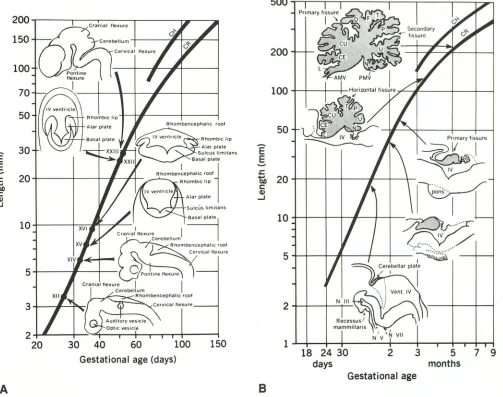
Fig. 6. A, and 8, Sequential development of the cerebellum. Please note the progressive evolution of the size and configuration of the cerebellum. The precise time of each stage cannot be stated exactly and is presently under continuing reevaluation. (Reprinted with permission from Lemire et al (1).)
medullary pyramids ran up to the quadrigeminal plate. A posterior fossa was present, but there was no tentorium. The cerebrum was markedly thinned by distension of the ventricles.
In this condition, the posterior columns of the spinal cord may be separated throughout their length by an ependyma-lined cleft. The uncrossed pyramidal tracts may extend inferiorly as promi-nent bundles in the anterior columns of the cord
(22).
Embryologically, these cases appear to result from destruction of the cerebellum. The presence of a posterior fossa in case two suggests the prior development of a hindbrain that is no longer present.
Paleocerebellar Dysgenesis
The majority of cerebellar dysgeneses are pa-leocerebellar dysgeneses. In these, the dysgenesis of the vermis may be partial or complete. The major entities in this group are Dandy-Walker complex, Joubert syndrome, tectocerebellar dys
-raphia, and rhombencephalosynapsis.
Dandy-Walker Complex
The conditions called Dandy-Walker malfor-mation, Dandy-Walker variant, and mega cisterna magna are believed to represent a continuum of
developmental anomalies designated the Dandy
[image:7.612.57.560.89.486.2]698 AJNR: 13, March/ April 1992
A.
.
~B(f
Cmbel/um fo'm;ng
;::::;~b,.;n
_
_;~
Foramen of Magendie opens
c.
Ant. membranous area
~
Post. membranous? area
'•
''
~
D-Delayed opening of Foramen of Magendie
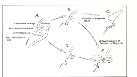
Fig. 7. Embryology; diagrammatic representation of the developing choroid plexus and roof of the fourth ventricle. Midsagittal
section, cephalic is to the readers right.
A,-C, In the normal brain, the area membranacea superior (labeled Ant. membranous area) becomes incorporated into the developing
choroid plexus, so the choroid comes to lie at the caudal edge of the cerebellum. The area membranacea inferior (labeled Post,
membranous area) will be the site of the foramen of Magendie.
D and £, Failure to incorporate the area membranacea superior into the choroid plexus could lead to progressive ballooning of a
thin-walled fourth ventricular cyst caudal to the vermis and cerebellum, but cephalic to the choroid plexus. This is the proposed
embryogenesis of the Dandy-Walker malformation.
Dandy-Walker Malformation
The Dandy-Walker malformation was first
re-ported by Dandy and Blackfan (1914) who
de-scribed marked dilatation of the fourth ventricle
and anterior displacement of the vermis, which they attributed to primary atresia of the cerebellar foramina (22). A further review by Taggart and Walker (1942) (23) led Benda (1954) to suggest
the name Dandy-Walker malformation (24).
Benda emphasized that atresia of the outlet
fo-ramina of the fourth ventricle was not essential
to this malformation.
The typical Dandy-Walker malformation is
characterized by a triad of 1) complete or partial
agenesis of the vermis, 2) cystic dilatation of the
fourth ventricle, and 3) an enlarged posterior
fossa with upward displacement of the lateral
sinuses, tentorium, and torcular (Figs. 8-1 0) (2).
Clinical Findings
The Dandy-Walker malformation occurs in 1
per 25,000-30,000 births (25). There is a slight, statistically insignificant, female predilection (25).
Dandy-Walker malformation accounts for 2%
-4% of hydrocephalus and 14% of cystic posterior
fossa malformations (26). The disorder most commonly is an isolated malformation with a low
risk of recurrence in later siblings (1 %-5%) (27).
However, the risk of recurrence may be high when the Dandy-Walker malformation is associ-ated with 1) autosomal recessive Mendelian con-ditions such as Warburg syndrome, Coffin-Siris syndrome, Frasier cryptophthalmus and Meckel-Gruber syndrome; 2) the dominant X-linked Acardi syndrome; or 3) diverse chromosomal anomalies such as duplications of 5p, 8p, and 8q;
trisomy of chromosomes 9, 13, and 18; and
triploidy (27). Rubella, cytomegalovirus,
toxo-plasmosis, coumadin, and alcohol have also been
reported to cause Dandy-Walker malformation.
Many Dandy-Walker patients show develop-mental delay. However, the degree of intellectual impairment may have been overestimated in the past. Hirsch et al found an intelligence quotient
greater than 80 in 60% of patients (25). Golden
et al found normal mental development in 75%
of patients (28). Specific review of intellectual/
[image:8.612.78.558.68.340.2]AJNR: 13, March/April 1992
A B
c
699
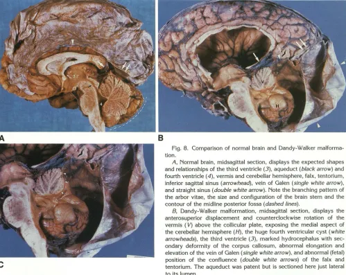
Fig. 8. Comparison of normal brain and Dandy-Walker malforma-tion.
A, Normal brain, midsagittal section, displays the expected shapes
and relationships of the third ventricle (3), aqueduct (black arrow) and
fourth ventricle ( 4), vermis and cerebellar hemisphere, falx, tentorium, inferior sagittal sinus (arrowhead), vein of Galen (single white arrow), and straight sinus (double white arrow). Note the branching pattern of the arbor vitae, the size and configuration of the brain stem and the
contour of the midline posterior fossa (dashed lines).
8, Dandy-Walker malformation, midsagittal section, displays the anterosuperior displacement and counterclockwise rotation of the
vermis ( V) above the collicular plate, exposing the medial aspect of the cerebellar hemisphere (H), the huge fourth ventricular cyst (white arrowheads), the third ventricle (3), marked hydrocephalus with sec -ondary deformity of the corpus callosum, abnormal elongation and elevation of the vein of Galen (single white arrow), and abnormal (fetal) position of the confluence (double white arrows) of the falx and tentorium. The aqueduct was patent but is sectioned here just lateral to its lumen.
C, Magnified view of the hindbrain. The branching pattern of the arbor vitae shows that many of the verminal lobules are intact, but hypoplastic. The anterior lobe is intact. The posterior lobe is intact, at least to pyramis. The tissue that should have formed the uvula and nodulus appears to form a thin band of distorted folia (white arrowheads) along the everting margin of the cyst (see also Fig. 1 OB).
(Figs. 8 and C reprinted with permission from Masdeu et al (67), through the kindness of Dr Joseph C. Masdeu, New York.)
syndrome documents that 47% have normal in-tellect with little or no motor deficit; an additional 26% have normal intellect and little to no motor deficit but require special help with specific
learn-ing disabilities; 11% have moderate
develop-mental delay with little to no motor deficit; and 16% have severe delay with spastic cerebral palsy
(29).
Greater than 80% of Dandy-Walker patients
present with hydrocephalus early in life (2).
How-ever, the hydrocephalus is often not evident at
birth; it develops postnatally and presents by 3
months of age in 75% of patients. This delay of
onset of hydrocephalus may be explained by the
secondary opening of the foramina of Luschka
that occurs later, or by bleeding from
unsup-ported, easily torn vessels along the wall of the
large posterior fossa cyst at the time of delivery.
The possibility that this bleeding may occur dur
-ing labor lends support to those advocating
Cae-sarian section for Dandy-Walker patients
diag-nosed prenatally (25).
These patients usually require surgery for
cor-rection of hydrocephalus. Cystoperitoneal and/or
ventriculoperitoneal shunting are now the proce
-dures of choice, although controversy persists as
[image:9.612.57.556.83.480.2]mortal-700
A
B
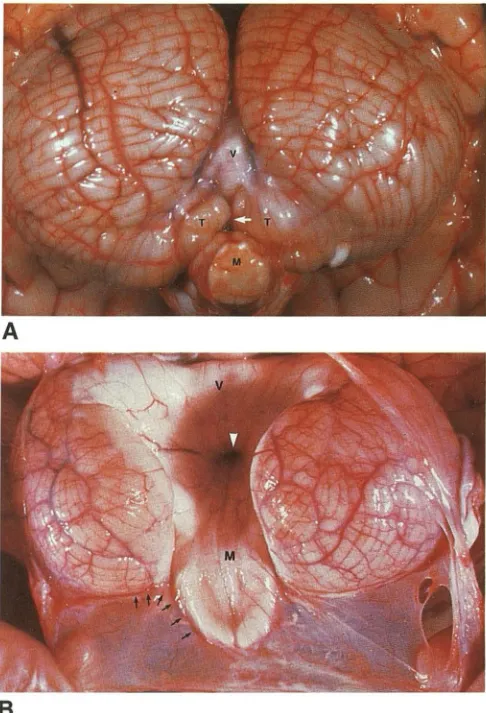
Fig. 9. Comparison of normal brain and Dandy-Walker mal-formation.
A, Normal unfixed cerebellum viewed from behind and below displays the folia and fissures of the hemispheres, the inferior vermis ( V), the tonsils ( 7), and the medulla (M). The vallecula
(white arrow) leads between the tonsils to the fourth ventricle (covered by the inferior vermis).
8, Dandy-Walker malformation. Unfixed specimen viewed from behind and below. The cerebellar hemispheres are hypoplastic and rotated laterally. The vermis is hypoplastic and rotated an ter-osuperiorly. The thin-walled fourth ventricular cyst attaches to the brain stem along the tela choroidea and expands behind and above the cerebellar hemispheres where it appears to attach along the posterolateral margin of the hemispheres (but see Fig. 13B).
Consequently, one can look directly from the cyst through the fourth ventricle to the ventricular surface of the superior vermis ( V), the ventricular end of the aqueduct (white arrowhead), and the ventricular surface of the medulla (M).
ity may result from upward herniation of the posterior fossa cyst following lateral ventri
culo-peritoneal shunting or downward herniation of
the cerebrum and lateral ventricles following
di-rect shunting of the cyst itself.
AJNR: 13, March/April1992
Concurrent systemic anomalies are present in approximately 25% of cases, and include low-set ears, polydactyly, syndactyly, Klippel-Feil syn-drome, Cornelia de Lange syndrome, and cleft palate (Fig. 11) (2, 31 ). Facial angiomas are seen in approximately 10% of patients (25). Golden et
al (31) observed two dissimilar populations with
Dandy-Walker syndrome. Those with concurrent hydrocephalus had a very high incidence of other central nervous system (CNS) malformations
(91%) and a smaller incidence of visceral anom-alies (55%), including one case of secundum atrial septal defect (9% ). Those without hydrocephalus had a very high incidence of complex cardiac malformations (80% ).
Pathologic Changes
Bone and Dura: The head circumference is in-creased with a dolichocephalic configuration (Fig.
11 A). The lambdoid sutures may be widened and separated preferentially. The posterior fossa is markedly expanded (Fig. 11 B). Cerebrospinal fluid (CSF) pulsations and secondary pressure changes cause erosive scalloping of the posterior surfaces of the petrous pyramids and of the occipital squama. Infrequently, marked expan-sion of the posterior fontanelle or a true dehis-cence of the occipital squama is associated with an occipital encephalocele (32, 33). The tento-rium and transverse sinuses lie in high position along the parietal bone (Fig. 8B) (34, 35). The incisura is inclined vertically and is wider than normal. The angle formed by the junction of the straight sinus with the superior sagittal sinus increases from a normal acute angle of 50°-75°
to an obtuse angle of approximately 90°-150°
(18). The falx cerebri is normal. There is no falx cerebelli in the true Dandy-Walker malformation (26).
Cerebellum, Vermis and Brain Stem: The vermis shows a range of deformity (Figs. 11 and 12). It is completely absent in approximately 25% of cases (2). The remainder show partial aplasia of the posterior vermis (2). The superior medullary velum and any residual portion of the superior
vermis are rotated anterosuperiorly and reflected upward. In severe cases, they appear to be re
[image:10.613.55.298.82.439.2]AJNR: 13, March/April1992
A
8
Fig. 10. Dandy-Walker malformation; two specimens. A, View from behind. Opening and reflecting the dura (D) exposes the thin arachnoid external to the unruptured Dand
y-Walker cyst.
B, Fixed specimen opening and reflecting the dura (D), arach
-noid and cyst reveal the size of the cyst and its relationship to the
cerebellum (same specimen as Figs. 88 and 8C). In this case, the
701
9 and 13). They appear to be "winged outward" and displaced anterolaterally against the petrous pyramids. They may be symmetrical or asym -metrical in size and position (35). There may be disorganization and heterotopias of the cerebellar cortex (2, 31 ). The brain stem usually appears small as a result of hypoplasia and of compression by the Dandy-Walker cyst. There can be pontine hypoplasia, subtotal aplasia of the inferior olives, and dysplasias of the other nuclear groups. Het-erotopias of the inferior olivary nuclei and anom-alies of the crossing corticospinal tracts are com-mon (2). The aqueduct may be patent, narrowed, kinked, or occluded.
Dandy-Walker Cyst: The Dandy-Walker cyst is
actually the distended fourth ventricle that bal-loons posteriorly behind the developing cerebellar hemispheres and displaces them laterally and anteriorly (Fig. 9). Conceptually, the cyst wall is formed of three layers (35). The inner layer is ependyma, which is continuous with the epen-dyma lining the wall of the anterior fourth ventri-cle. This layer may show discontinuities where the ependyma is reduced to small ependymal nests (2). The outer layer is pia, which is reflected around the cyst to be continuous with the pia along the medial and posterior surfaces of the cerebellar hemispheres. The intermediate layer is the stretched out neuroglial tissue that would have formed the inferior vermis and medial hemi-spheres. This residual tissue is thickest laterally and superiorly, near to the hemispheres, tonsils, and residual superior vermis. Calcification may be present in this tissue (7%) (31).
The cyst wall is attenuated progressively to-ward the midline where the fourth ventricle ex-panded most widely. Thus, the anterolateral walls of the fourth ventricle-cyst are the smooth white matter of the residual vermis and the cerebellar hemispheres (2) (Figs. 98 and 13). The posterior wall of the fourth ventricle-cyst is a thin, easily ruptured, translucent membrane (2). The cyst wall attaches to the brain stem along the course of the tela choroidea. It appears to be attached to the cerebellar hemispheres posterolaterally, but the reflection of the cyst can often be traced
hemispheres lie more closely together but remain ununited. The
undersurface of vermis ( V), aqueduct (white arrowhead) and floor
of medulla (M) are visible through the midline defect. Note the
vertical orientation of the incisura.
(Reprinted with permission from Masdeu et al (67), through the
[image:11.615.55.300.81.649.2]702
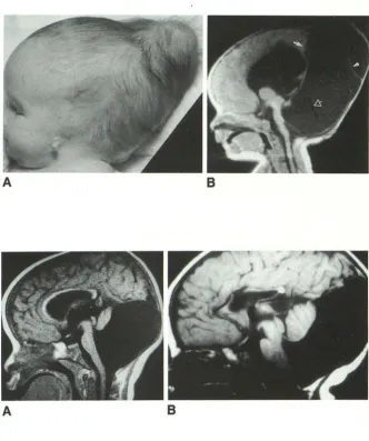
Fig. 11. Dandy-Walker malformation, 1 month-old girl.
A, Lateral patient photograph shows
marked dolichocephaly, bulging posterior
fontanelle, low-set malformed pinna and atresia of the external auditory canal.
8, Midsagittal T1 MR reveals that the
dolichocephaly and bulging fontanelle result
directly from the gross expansion of the
posterior fossa cyst. The tentorium and
straight sinus (closed white arrow) are el e-vated so the bulging fontanelle is purely
infratentorial. The superior vermis is tiny. The brain stem is hypoplastic. The cyst membrane (white arrowhead) has partially collapsed away from the dural wall of the
posterior fossa after shunting. Note flow void
(open white arrow) in shunt.
Fig. 12. Dandy-Walker malformation.
A-8, Midsagittal T1 MR from two patients shows that the vermis is hypoplastic and rotated anterosuperiorly by the cyst in each
case. Together with Figure 11 B, these fi
g-ures show a gradation of vermian hypopl a-sia.
A
A
far medially to where it joins the white matter of the fourth ventricle.
The choroid plexus is displaced far laterally and
inferiorly, and comes to lie within the lateral
recesses and along the caudal insertion of the
cyst wall at the medulla (2). Choroid plexus may be entirely absent from the fourth ventricle at
postmortem in 40% of cases (36). The exit
foram-ina of the fourth ventricle may be patent or occluded. Hart et al documented patency of the foramen of Magendie at postmortem in at least
two of their 28 cases of Dandy-Walker
malfor-mation (7%) and patency of the foramina of
Luschka in 18% (31). The foramina were clearly
closed in 18%. Hirsch et al showed
communica-tion between the fourth ventricle and the
sub-arachnoid space in vivo in more than 80% of
cases (25).
Between the outer pia of the cyst wall and the pia of the cerebellar hemispheres is a thin com
-AJNR: 13, March/ April 1992
B
B
pressed subarachnoid space (Fig. 13) (35).
Exter-nal to that is the normal arachnoid mater of the cisterna magna, the dura that lines the inner table of the occipital bone, and the inner table itself.
The expanding cyst bulges into, expands, and
occupies the space that would have become
cisterna magna. The outer cyst wall bulges
against the midline arachnoid so tightly it almost
"closes" the space. The "normal" subarachnoid space between the Dandy-Walker cyst and the occipital bone becomes visible only when shunt-ing of the cyst collapses the cyst wall away from
the occipital bone (Fig. 13), or when contrast is
introduced into the subarachnoid space (Fig. 14). The cyst occupies most of the enlarged
poste-rior fossa. In 44% of cases, the cyst has a superior
extension or diverticulum that extends superior
to the residual superior vermis. This may bulge into the quadrigeminal plate cistern above the
[image:12.617.228.561.72.469.2]AJNR: 13, March/April 1992
A
8
c
D E
703
Fig. 13. Dandy-Walker malformation; re-lationship of the cerebellar hemispheres and
the fourth ventricular cyst.
A, Untreated Dandy-Walker
malforma-tion, Axial T1 MR. The cerebellar
hemi-spheres are hypoplastic and winged antero-laterally by expansion of the cyst posteriorly. No space is seen between the cyst and the cerebellum or between the cyst and the wall of the posterior fossa. Note that the cyst
stops exactly at the lateral angle of the cerebellar hemispheres (arrows) and does
not extend anterior to them.
B, Shunted Dandy-Walker malformation
with intraventricular hemorrhage. Postmor-tem specimen; 3-month-old girl. The cyst
wall (curved arrow) appears to arise in con-tinuity with the white matter of the cerebellar
hemispheres and to extend posterolaterally behind-but separate from-the posterior
surface of the cerebellar hemispheres to the lateral angle of the hemispheres (black
ar-rows). It is then reflected posteromedially to form the back wall of the cyst.
C, Diagrammatic representation of the Dandy-Walker cyst arising (curved black
ar-rows) medially from the white matter of the cerebellar hemispheres and forming a wall
(black arrows) that is interposed between the cerebellar hemispheres (H) and the bony posterior fossa, but remains separate from
them. The pial lining of the dorsal surface of
the hemispheres is reflected into the anter-olateral portion of the cyst wall leaving a
compressed subarachnoid space between the cyst and the hemispheres and between the cyst and the dorsal dura.
D, Shunted Dandy-Walker cyst, T2 MR. Note the fluid in the subarachnoid space
(white arrowhead) between the dorsal
sur-face of the cerebellar hemispheres and the
origin (curved black arrows) of the cyst along the side wall of the fourth ventricle.
E, Shunted Dandy-Walker malformation
(same patient as Fig. 11). Contrast-enhanced CT shows the expanded posterior fossa and enhancing tentorium. Shunt (open white ar-row) decompression collapses the cyst wall
(white arrowheads), separating it from the
bony and dural walls of the posterior fossa and from the posterior surface of the
cere-bellar hemispheres (H, H). Compare the
origins (curved white arrow) of the cyst wall
[image:13.612.55.507.83.744.2]704
does not communicate with the quadrigeminal cistern or the suprapineal recess but may com-press these structures. Inferiorly, the cyst usually extends downward through the foramen magnum
Fig. 14. Dandy-Walker malformation; axial CT. Contrast placed into the lumbar subarachnoid space and carried cranially delineates the supratentorial cisterns, the cisterns along the cer
e-bellar hemispheres (black arrows), and the thin subarachnoid
cisterns (white arrowheads) between the cyst (C) and the dorsal
surface of the cerebellar hemispheres. Contrast also layers po
s-teriorly against the occipital squama. (Case courtesy of Dr Jeremy Altman; reprinted with permission from Naidich et al (35).)
Fig. 15. Dandy-Walker malformation;
upward herniation following direct shunting of the lateral ventricle only. Axial CT after instillation of contrast via the ventricular
catheter show decompression of the lateral
ventricular hydrocephalus with marked u
p-ward herniation of the noncommunicating,
undecompressed Dandy-Walker cyst. Note
the mild constriction (arrows) of the cyst by the tentorium at the wide incisura.
A
AJNR: 13, March/April1992
behind the cord to the C1-C2 level. In the un-treated state (Fig. 13A) the cyst does not extend anterior to the cerebellar hemispheres, ie, anterior to the lateral angles of the cerebellum (between the anterior cerebellum and the petrous pyramid) (35). If a cyst extends anterior to the lateral angle of the cerebellum in the untreated case, the cyst most likely represents an arachnoid cyst, not the cyst of a Dandy-Walker malformation (Dr Larissa Bilaniuk, Philadelphia, unpublished data) (see Fig. 22).
Ventricles: Patients with Dandy-Walker
malfor-mation almost always develop hydrocephalus (Fig. 88). The degree of hydrocephalus varies from mild to severe and does not correlate with the size of the posterior fossa. The site of CSF obstruction varies: Carmel et al (36) found ague-ductal stenosis in 28% of cases, communication between the third ventricle and the cyst in 72%
of cases, patency of the fourth ventricular foram-ina in 39% of cases, and obstruction of the incisura in 11% of cases. When the aqueduct is occluded, shunting the lateral ventricles may cause upward herniation of the undecompressed fourth ventricular cyst and a characteristic "snow
man" deformity of the posterior fossa cyst (Fig.
15). Conversely, direct shunting of the noncom-municating fourth ventricular cyst may cause downward transincisural herniation of the unde-compressed lateral ventricles (38). In such a case, the portion herniating is usually the medial atrial
[image:14.621.98.264.154.376.2] [image:14.621.226.563.482.745.2]AJNR: 13, March/April1992
wall, forming diverticula that extend into the posterior fossa unilaterally or bilaterally. Approx-imately 25% of Dandy-Walker patients show downward herniation of the medial atrial walls after shunt therapy (Fig. 16). This post-shunting herniation results from a transincisural pressure gradient caused by malfunction of a ventriculo-peritoneal shunt, application of suction to the cystoperitoneal shunt, or increase in supratento-rial pressure for unknown causes (38). Once the herniation occurs, it may persist despite shunt revision and restoration of normal pressure gra-dients (38).
The patency of the CSF pathways is important to determine in Dandy-Walker malformation. Magnetic resonance (MR) demonstration of CSF flow may obviate the need for ventriculography, direct opacification of the cyst, or placement of contrast into the subarachnoid space before ther-apy with diverting shunts. Contrast placed into
705
the subarachnoid space fills the compressed cis-terna magna and percolates into the small sub-arachnoid space between the cyst wall and the posterior surface of the cerebellar hemispheres (35). If the foramen of Luschka is patent, contrast
may also enter the cyst itself.
Vasculature: The anomalies of the vertebral bas-ilar vasculature associated with Dandy-Walker malformation have been described by Wolpert et
al and by La Torre and associates (39, 40). Pa-tients with the Dandy-Walker malformation dem-onstrate high position of the posterior cerebral vessels due to the high tentorium. The superior cerebellar arteries are often displaced anterosu-periorly, above the posterior cerebral arteries, due to the marked enlargement of the fourth ventricle. The posterior-inferior cerebellar arteries are short-ened with superior displacement of the tonsilar loops (35). The inferior vermian branches are often lacking. The venous phase show absent
Fig. 16. Dandy-Walker malformation; downward transincisural herniation post-shunt
-ing; 4 112-year-old boy.
A
B
A, Axial CT following multiple shunt revisions for shunt malfunction show the
decompressed posterior fossa, persistent dilatation of the lateral and third ventricles
despite the lateral ventricular shunt, and bilateral medial atrial diverticula (white arrow-heads) that herniate downward through the incisura of the tentorium (arrowheads).
Band C, Postmortem specimen, same patient.
B, Axial section through the lateral ventricle shows atrial dilatation and the ostia of
the bilateral atrial diverticula (curved arrows).
C, Lateral view of the cerebrum (A = anterior, P = posterior), the tentorium ( 7) and
the cerebellar hemisphere (H) shows the two left (L) and right (R) medial atrial diverticula (arrows) that herniated downward through the incisura before the cerebrum was lifted upward to obtain this picture. ( C reprinted with permission from Naidich et al (38).)
[image:15.614.55.561.346.744.2]706
inferior vermian vein, elevation of the great vein of Galen, and high position of the transverse sinuses and torcular. Arachnoid cysts, in contra-distinction, show a normal complement of vessels that are secondarily displaced and compressed
by the avascular retrocerebellar cyst (39, 40).
Associated CNS Anomalies: The Dandy-Walker
malformation is associated with other CNS
ab-normalities in 68% of cases (30). These include
supratentorial abnormalities such as callosal
dys-genesis (one-sixth of patients), subependymal
neuronal heterotopias polymicrogyria, agyria,
schizencephaly, and lipomas (Fig. 17). Of these,
the callosal dysgenesis is the most frequently
associated supratentorial abnormality (31 ).
En-cephaloceles and lumbosacral meningocele-may
also concur.
Fig. 17. Dandy-Walker malformation; concurrent CNS anomalies.
A, Neuronal heterotopia. Coronal T2 MR
shows subependymal nodules of heterotopic gray matter (arrows) with Dandy-Walker
malformation.
B, Callosal agenesis. Sagittal T1 MR. Note
the position of the straight sinus (arrow) at the vertex. V = vermis; H = cerebellar hem -isphere.
C and D, Axial proton density MR show bilateral schizencephaly, agyria/pachygyria, absent septum pellucidum, and callosal d ys-genesis with collapse of the Dandy-Walker
cyst (C) wall (arrowheads) and bilateral m e-dial atrial diverticula (white arrows) following
shunt decompression of the cyst (same p a-tient as Fig. 11 and Fig. 13E.)
A
AJNR: 13, March/April 1992 Dandy-Walker Variant
The Dandy-Walker variant consists of hypopla-sia of the cerebellar vermis with cystic dilatation
of the fourth ventricle, but no enlargement of the
posterior fossa (41). Hydrocephalus may be
pres-ent or not.
Clinically, these children may present with an
enlarging head due to hydrocephalus or with
developmental delay. The Dandy-Walker variant
is more common than the true Dandy-Walker
malformation and accounts for one third of posterior fossa malformations (26). Shunting is performed only if there is hydrocephalus. The supratentorial anomalies associated with Dandy-Walker variant include agenesis of the corpus
callosum (21 %), malformations of cerebral gyri
and cerebral heterotopias (21% ),
[image:16.613.224.561.323.730.2]AJNR: 13, March/ April 1992
cephaly (10%), diencephalic cysts (10%), and posterior fossa meningoencephalocele ( 10%) (26). Mental deficiency appears to be related to the associated supratentorial anomalies.
Pathologically, the fourth ventricle is large but less dilated and better formed than in true Dandy-Walker malformation (Fig. 18) (35). The tento-rium is high in only 10% of cases. There is a falx cerebelli in 32% of cases, although it may be
displaced (35). There is less severe hypoplasia of the inferior vermis and less severe superoanterior rotation of the superior vermis. The cerebellar hemispheres are hypoplastic. The brain stem usu-ally appears nearly normal (Fig. 19). Rarely, der-mal sinuses and teratoma may concur (Fig. 20).
Mega Cisterna Magna
The term mega cisterna magna (synonyms:
retrocerebellar arachnoid pouch, communicating
arachnoid cyst, and Blake pouch cyst) refers to
a cystic malformation of the posterior fossa
char-acterized by an intact vermis, an enlarged cisterna
magna, and an enlarged bony/ dural posterior
fossa. The size of the mega cisterna magna is variable. The fourth ventricle communicates with the subarachnoid space (Fig. 21). The brain stem
and the cerebellum are usually normal. This con-dition accounts for 54% of the cystic posterior fossa malformations (26).
Clinically, these children present with
hydro-A
707
cephalus and/or developmental delay. The men-tal retardation is due to associated supratentorial anomalies, such as agenesis of the corpus cal-losum (6%), diencephalic cysts (6%), and holo-prosencephaly (6%) (26). Posterior meningoceles are identified in 3% of cases (26). Rarely, there is neuronal heterotopia supratentorially. Posterior meningoceles are also identified in 3% of cases (26).
Pathologically, there is evagination of the tela
choroidea of the fourth ventricle behind the intact vermis and cerebellar hemispheres. The cystic expansion of the tela choroidea may extend far beyond the normal borders of the cisterna magna laterally, posteriorly, and superiorly (35). It may reach the quadrigeminal plate cistern but does not communicate with it (Fig. 21). The cyst may extend superiorly into a focal dehiscence of the falcotentorial junction and, then, is usually asso-ciated with an interruption or fenestration of the straight sinus. The tentorium is elevated in 12.5%
of cases. A falx cerebelli is seen in 62.5% of cases (35). The brain stem is typically normal (18).
Embryogenesis of the Dandy-Walker Complex
Embryologically, the Dandy-Walker malfor-mation is believed to result from a broad insult to the alar plate involving the dorsal fourth
ven-B
Fig. 18. Dandy-Walker variant; postmortem specimen. A, Midsagittal section, and 8, posterior inferior view of the uncut specumen, show mild vermian hypoplasia and communication of the ventricle with the posterior fossa space medial to the cerebellar hemispheres
[image:17.612.52.564.466.691.2]708
Fig. 19. Dandy-Walker variant; twin pa
-tients. A and B, Midsagittal Tl MR, C and
D, corresponding coronal Tl MR. The twins show slightly different degrees of vermian hypoplasia and rotation and slightly different degrees of fourth ventricular dilatation. The cerebellar hemispheres are nearly normal. The posterior fossae are only slightly e n-larged.
A
c
tricle and the rhombic lips (28). The manifesta-tions of the Dandy-Walker malformation would appear to be explained best by failure to incor-porate the area membranacea superior into the developing choroid plexus at the proper time (Fig. 7) (20). Persistence of this membrane between the caudal edge of the developing vermis and the rostral edge of the developing choroid plexus easily explain the large fourth ventricular (rhom-bencephalic) cyst that balloons posterior to the
cerebellar hemispheres, the relation of the cyst
to the vermian remnant anterosuperiorly, the
in-sertion of the cyst into the brain stem along the tela choroidea (but insertion of the cyst into the
white matter of the medial surfaces of the
cere-bellar hemispheres), and the low position of the
choroid plexus. The area membranacea inferior
could persist unopened leading to absence of the
foramen of Magendie, or it could open at the
AJNR: 13, March/April 1992
B
D
appropriate or at a later time, accounting for the
variability observed.
The high position of the tentorium, straight
sinus, and torcular may indicate arrested
devel-opment with the straight sinus remaining at the vertex as it is in the early embryo, with failure to
migrate to its normal deep occipital position (1).
Mechanical interference with such migration
be-cause of the expanding posterior fossa cyst may
also contribute.
The variations in the severity of the
malfor-mation among the true Dandy-Walker, the
Dandy-Walker variant, and the mega cisterna
magna could be explained in terms of the severity
· of the initial insult leading to vermian hypoplasia
and to persistence of the area membranacea
superior. If the insult is sever and diffuse, it may
affect the cerebellar hemispheres, vermis, and
AJNR: 13, March/April 1992
A B
c
E
Dandy-Walker malformation (18). More localized
insult to the developing cerebellum could lead to vermian/ cerebellar hypoplasia with little effect on the fourth ventricular roof. This could result in
the Dandy-Walker variant (18). Greater effect of
the insult on the fourth ventricular roof than on the developing cerebellar primordia could lead to cystic dilatation of the ventricle and little
cere-bellar hypoplasia, as is seen with mega cisterna
magna (18).
Posterior Fossa Arachnoid Cyst
True arachnoid cysts are collections of CSF
enclosed within the layers of the pia-arachnoid
and lined by arachnoid cells and collagen (35).
709
Fig. 20. Dandy-Walker variant with mi-doccipital dermal sinus and teratoma.
A, Midsagittal Tl MR shows vermian hy -poplasia and rotation, enlarged fourth ven -tricle and retrocerebellar cyst, slightly high position of the straight sinus (black arrow-head) and torcular (black arrow), and an infratorcular sinus tract (white arrowhead) that leads to an ovoid mass (white arrow) within the retrocerebellar cyst.
B, Axial Tl MR through the upper cere-bellum shows the transverse extent of the cyst and mass and the heterogeneous con-tent of the mass.
C-E, Surgical photographs.
C, Operative exposure. After suboccipital craniotomy and opening of the dura, the mass (arrows) is seen to lie deep to the arachnoid.
D, External surface of mass.
E, Opening the mass reveals hair, carti -lage, and sebaceous debris of a teratoma.
Clinically, the arachnoid cysts present at vary
-ing ages from the first few months of life until
adulthood. Most lesions appear to be sporadic,
although arachnoid cyst may be associated with
the mucopolysaccharides or with the conditions of dural ectasia such as neurofibromatosis.
The major clinical symptoms result from
hy-drocephalus and compression of the underlying
brain (Fig. 22). Ataxia is frequent, headache and
dysdiadochokinesia less common (18). In
new-borns, arachnoid cysts may present as rapid ex
-pansion of the cyst by hemorrhage due to the
trauma of delivery and rupture of bridging veins
(Fig. 23). Risk of such hemorrhage may indicate
need for a Cesarean section if the diagnosis is
[image:19.613.56.565.83.532.2]710
systemic anomalies are specifically associated
with the retrocerebellar arachnoid cyst.
Increasing use of neuroimaging has led to
in-creased recognition of small, asymptomatic,
ap-parently incidental posterior fossa cysts. Serial
studies will determine whether a proportion of
these ever becomes symptomatic. When there is
hydrocephalus, these cysts may be
decom-pressed successfully with either cystoperitoneal
shunting or fenestration.
Pathologically, one-fourth to one-third of all
arachnoid cysts involve the posterior fossa.
Eleven percent are situated within the
cerebella-Fig. 21. Mega cisterna magna. Midsagittal Tl MR shows a normal vermis, fourth ventricle, and vallecula with a large cisterna magna that extends above the vermis toward the quadrigeminal plate cistern, and posterosuperiorly through a focal deficiency in the falx and tentorium, associated with a posterior fenestration of the straight sinus (arrows). The inner table of the occipital squama is scalloped.
Fig. 22. Posterior fossa arachnoid cyst, sagittal (A) and axial (B) Tl MR images demonstrate a large posterior fossa CSF col -lection that compresses the vermis and di s-places it anteriorly. The occipital bone shows slight scalloping. The straight sinus is slightly elevated. No communication with the fourth ventricle or vallecula is demonstrated. The cyst lies entirely within the midline.
A
AJNR: 13, March/April 1992
pontine a~gle, 1 O% lie in the quadrigeminal plate
cistern, 9% are related to the vermis, and 3% lie
in the prepontine and interpeduncular cisterns (35). The cysts may extend through the incisura or the foramen magnum.
The arachnoid cysts are unilocular, round, oval,
or crescentic, fluid-filled masses that can be
sep-arated from the fourth ventricle and the vallecula
(Fig. 22). The vermis, cerebellar hemispheres, and
brain stem are normal except for associated mass effect. There are no mural nodules or calcifica-tions. Ultrastructural studies have shown certain cells along the interior wall of the cyst contain specialized membranes and enzymes usually as-sociated with secretory activity (42). It may be that CSF accumulates within the cyst as a result of secretion from these cells, rather than by osmotically induced filtration or a ball-valve mechanism (34).
Imaging studies show that the cyst fluid has computed tomography (CT) density or MR signal intensity nearly identical to the CSF of the ven-tricular system (35). There is no evidence of
contrast enhancement. Many arachnoid cysts
ex-tend anterior to the cerebellar hemispheres (Fig. 23), so it is easy to rule out the Dandy-Walker complex. However, those arachnoid cysts that originate in the midline and lie wholly posterior to the cerebellum may be difficult to differentiate from the mega cisterna magna itself, except for their mass effect.
Embryologically, the retrocerebellar arachnoid cyst may represent a persistent diverticulum of the fourth ventricle, which fails to involute (the Blake pouch) (43). It may represent a focal failure
[image:20.617.95.265.266.422.2]AJNR: 13, March/ April 1992 711
c
Fig. 23. Hemorrhagic posterior fossa arachnoid cyst. Midsagittal (A), left parasagittal (B), and axial (C) Tl MR images demonstrate a large asymmetric posterior fossa arachnoid cyst that extends from the posterior midline into the left cerebellar pontine angle anterior
to the cerebellar hemisphere and compresses the neural structures. Increased signal intensity within the cyst represents methemoglobin. There is no hemorrhage within the ventricles or other sign of communication with the fourth ventricle.
to establish free communication throughout the
developing space between the inner and the outer layers of the pia-arachnoid as the mesenchyme
condenses (44).
Joubert Syndrome
This familial syndrome was described by
Joub-ert et al ( 1969) in four siblings, all of whom
presented with neonatal hyperpnea alternating
with apnea, abnormal eye movements, ataxia,
and psychomotor retardation (45). Two of these
children had agenesis of the posterior vermis, one had complete agenesis of the vermis at
postmor-tem, and the fourth had complete agenesis of the
vermis with an occipital meningoencephalocele
(probably a gliocele) (45). The tentorium was
elevated in three of the four siblings (45).
Joubert syndrome is inherited as an autosomal
recessive trait, so genetic counseling of the
par-ents is indicated. Males predominate by more than 2: 1 (2). The children are mentally retarded,
hypotonic, and ataxic with a poor clinical
out-come. Hemifacial spasms, campylodactyly,
syn-dactyly, and cystic kidneys also concur (45). Joubert patients usually present as neonates with breathing abnormalities that improve with
age. In Kendall's series, 44% of children had
respiratory abnormalities ( 46). Visual difficulties
are common. Sixty-seven percent of cases show
nystagmus or abnormalities of supranuclear
con-trol of eye movements (46). Forty-four percent show mild to moderate reduction in visual acuity
and retinal dystrophy (46). These clinical
abnor-malities can be related to dysfunction of the cerebellum and medulla oblongata. The episodic tachypnea and apnea are most likely due to
abnormalities in the solitary fascicle and the nu-clei gracili of the brain stem that control afferent respiratory impulses (42). The oculomotor
dys-functions such as dysmetric saccades, impair
-ment of smooth pursuit, and optokinetic
nystag-mus can be related to abnormalities in the vermis.
Decrease of the velocity of the saccadic eye movement may be associated with brain stem abnormalities ( 42).
Pathologically, Joubert patients show a
vari-able degree of dysplasia of the cerebellar vermis
(Figs. 24 and 25) (46, 47). The vermis may be
completely absent or show partial agenesis/hy
-poplasia that affects either the superior or the
inferior vermis predominantly (46). In Kendall's
series, the posterior vermis was dysplastic in all cases and exhibited a deep midline groove or cleft (46). The superior vermis showed particular
hy-poplasia of the culmen (46). The inferior
cerebel-lar peduncles were small (46). The superior cer-ebellar peduncles were small and appeared to course nearly perpendicular to the brain stem
(46). The fourth ventricle was modestly enlarged
but there was no hydrocephalus or posterior fossa
[image:21.612.58.556.83.273.2]712
Fig. 24. Joubert syndrome. Axial Tl fv\R
sections at the medulla (A) and the pons (B)
show absence of the inferior vermis and
thimble shape of the fourth ventricle. The fourth ventricle communicates directly with the enlarged cisterna magna.
A
Fig. 25. Joubert syndrome in two siblings.
B
A and B, Tl fv\R images of the older child in the sagittal (A) and coronal (B) planes demonstrate marked vermian dysgenesis, hypoplasia of the cerebellar hemispheres, and hypoplasia of the brain stem. The basilar cisterns, fourth ventricle, and cisterna magna are enlarged secondarily. The posterior fossa is not enlarged. The cerebral
hemispheres are atrophic.
C and D, Tl fv\R images of the younger sibling in the sagittal (C) and coronal (D)
planes demonstrate less severe abnormalities, with hypoplasia of the inferior vermis
and the cerebellar hemispheres and secondary enlargement of the cisterna magna in
a posterior fossa of normal size. This case resembles the Dandy-Walker variant.
AJNR: 13, March/ April 1992
c
[image:22.615.60.563.86.746.2]AJNR: 13, March/ April 1992
Multiple foci of heterotopic cerebellar cortex are found in the subcortical white matter (2). Asso-ciated abnormalities of the cervicomedullary junction include complete absence of the pyram-idal decussations, dysplasia of the olivary and para-olivary nuclei, and hypoplasia of the nucleus gracilis, solitary fascicle, and descending trigem-inal tracts (46).
Occipital meningoceles or meningoencephalo-celes may be present in up to one-third of patients (2). Infrequent cerebral abnormalities include vari-able degrees of dysgenesis of the corpus callosum and cerebral atrophy (Fig. 25) (46). In some cases, the findings may resemble those of the Dandy-Walker variant or mega cisterna magna.
The embryologic defect in Joubert syndrome has not been determined. The malformation may result from a genetically determined arrest or derangement of the development of the rostral portions of the rhombic lips, with relative sparing of the rest of the alar plates. Thus the vermis would be affected predominantly with more nearly normal cerebellar hemispheres and fourth ventricle (19).
Tectocerebellar Dysraphia
Tectocerebellar dysraphia refers to the combi-nation of vermian hypoplasia or aplasia, occipital encephalocele, and dorsal traction of the brain stem so that the hypoplastic cerebellar hemi -spheres come to lie ventral and lateral to the brain stem (Fig. 26) ( 48). These cases have previously been termed "inverse cerebellum with occipital
A
B
713
encephalocele" (49). This disorder has been thought to be a link between the Arnold-Chiari and Dandy-Walker malformations (49, 50), b e-cause the dysplasia of the cerebellar vermis, the large fourth ventricle, and the high transverse sinuses seen in this condition are also seen with Dandy-Walker malformation, while the beaked tectum, the kinking of the brain stem, and the position of the cerebellum ventral to the pons seen in this condition are also seen (in milder form) in Chiari II malformation. It should be noted, however, that tecta! beaking and kinking of the brain stem are commonly seen in occipital en -cephaloceles; therefore, the "link" to the Chiari II malformation is, at most, a superficial resem-blance.
Tectocerebellar dysraphia is a rare sporadic malformation that more commonly affects males. The patients all present with occipital e ncepha-loceles, and may be microcephalic (49). The chil-dren usually die within the first year of life, but may survive to age 8 years. Children who survive the neonatal stage usually require shunting for hydrocephalus. Tectocerebellar dysraphia shows similarities to Walker-Warberg syndrome, an au-tosomal recessive disorder, with agyria, hydro-cephalus, and retinal dysplasia.
Pathologically, these infants demonstrate an occipital encephalocele that contains cerebellar cortex. There is associated deformation of the tectum, such that the posterior colliculi form long, thin, peg-shaped processes that project towards the bony defect (Fig. 26). The vermis is absent or severely hypoplastic, with small remnants of
[image:23.612.51.565.514.745.2]714 AJNR: 13, March/ April 1992
A
c
DAJNR: 13, March/April1992
the superior vermis. The cerebellar hemispheres are rotated ventrally and may come to lie ventral to the brain stem due to hypoplasia/absence of the vermis. The posterior fossa is small and shallow. Variably associated supratentorial malformations include fusion of the thalami, anomalous cerebrocortical convolutions termed polymicrogyri by Smith (50), dysplasia of the mammillary bodies, and aplasia of the corpus callosum. The atlas and the occipital squama may be bifid. There is occasional cervical hydromyelia (2). Tectocerebellar dysraphia is distinguished from simple traction deformities secondary to dislocation of cerebellar tissue into an occipital cephalocele by the severity of the deformities, their complexity, and the severe hypoplasia/ apla-sia of the vermis (2).
Embryologically, this disorder is believed to result from an insult early in embryogenesis at the 10- to 30-mm stage. Defects of dorsal induc-tion and neuroschisis cause an encephalocele that pulls the brain stem tightly against the posterior aspect of the calvarium. Consequently, the tec-tum and the cranial nerves become elongated and the cerebellar hemispheres assume a ventral position (50). Most likely, the encephalocele re-sults in separation of the cerebellar hemispheres, preventing their fusion in the midline and thereby blocking formation of the vermis.
Rhombencephalosynapsis
The term rhombencephalosynapsis signifies a hypoplastic single-lobed cerebellum, with ver-mian hypogenesis or agenesis and fusion of the cerebellar hemispheres, cerebellar peduncles, and the dentate nuclei (Fig. 27) ( 19). This disorder was first described by Obersteiner ( 1916) (51) on
Fig. 27. Rhombencephalosynapsis.
715
the postmortem examination of a normal 28-year-old suicide victim (52). DeMorsier named the disorder rhombocephalosynapsis (53), which was changed to rhombencephalosynapsis by Gross (54). To date, 17 cases have been reported
(19).
Clinically, these children present with signs of cerebellar and motor dysfunction (55). Rhomben-cephalosynapsis appears to be one of the few posterior fossa malformations that present with
"cerebellar signs." The intellectual impairment
and life expectancy are variable. Those children with a rudimentary cerebellum appear to do worse. Th