*Present address: Human Performance Laboratory, Faculty of Physical Education, University of Calgary, 2500 University Drive NW, Calgary, Alberta, Canada T2N 1N4.
Key words: mammals, intervertebral joint anatomy, sagittal bending, resistance, compliance.
MAMMALIAN SPINAL BIOMECHANICS
II. INTERVERTEBRAL LESION EXPERIMENTS AND MECHANISMS OF BENDING RESISTANCE
BYJULIANNA M. GÁL*
Department of Pure and Applied Biology, University of Leeds, Leeds, LS2 9JT, UK
Accepted 24 August 1992
Summary
Three-point cyclic bending was applied to intervertebral joint complexes (three vertebrae with two intervertebral discs) of monkey (Macaca fascicularis), wallaby (Wallabia rufogrisea frutica), tiger (Panthera tigris ), jaguar (Panthera onca) and seal (Halichoerus grypus). Force–displacement loops were recorded for intact specimens in both extension and flexion. Reductions in peak forces at given displacements were measured, following lesions of ventral ligaments, superspinous ligaments, interspinous ligaments and muscles, ligamenta flava and the articular capsules. Subsequently, the vertebral arches were removed from the specimens to test the bending resistance of the intervertebral discs alone. The results of these lesion experiments, coupled with details of intervertebral joint anatomy, suggest that extension resistance is ultimately due to articular joint impaction for all species tested. The prominent ligamenta flava of the monkey and wallaby contrast with the robust discs of jaguar and tiger and illustrate two distinct mechanisms for resisting flexion in mammalian intervertebral joints. The conspicuous absence of soft structural elements in seal intervertebral joints contributes to their low bending resistance. The implications of these findings for mammalian locomotion, behaviour and scaling are discussed.
Introduction
dynamic mechanical properties. Differences between mammals are manifest largely by the degree to which such structures are enhanced or diminished.
Relatively few studies have involved successive lesioning of intervertebral structures to identify passive mechanisms of joint resistance. Tencer et al. (1982) performed extensive mechanical tests with cadaveric human intervertebral joints. They found that the intervertebral discs played the most vital role in limiting lateral and anterior shear, axial compression and flexion. The facets played a major role in resisting posterior shear and axial torque. Dumas et al. (1987) conducted tensile tests with human cadaveric intervertebral joints. Intervertebral discs were sectioned, leaving only the ligamentous attachments intact. The latter were sequentially resected and the tensile tests repeated. The most restrictive ligament was ligamentum flavum, followed by the articular, interspinous and superspinous ligaments. Non-human lesion studies are even rarer. Smeathers (1981) conducted Euler buckling tests with rabbit and goat lumbar spines. Lesion experiments showed that flexion was resisted primarily by ligamenta flava in the rabbit and the intervertebral discs in the goat intervertebral joints.
Gál (1993) investigated the static and dynamic bending behaviour of the intervertebral joints of a variety of mammals. Results showed that scaling theory alone could not explain the mobility patterns observed. Statistical comparisons of mathematical parameters describing joint compliances suggested that flexion resistance was controlled by different structures in different species. Mathematical modelling and statistics are indispensible tools for predicting and detecting significant differences between joints, but they fall short of identifying the structures responsible for the observed bending properties. Therefore, a protocol of successive lesion experiments was carried out with intervertebral joint complexes, following the previous dynamic bending and strain energy tests. Extension and flexion bending cycles, within bending plateau ranges, were followed by serial cuts through the interspinous muscles and ligaments and the deeper articular capsules and ligamenta flava. Reductions in peak forces for given actuator displacements were indicative of reductions in joint resistance. The removal of the vertebral arches and associated structures allowed inferences to be made about the resistance capabilities of the intervertebral discs alone. The results of the lesion experiments and intervertebral joint dissections of this study are combined with the intact bending properties described in the previous paper to provide a more complete discussion of the functional morphology of the mammalian vertebral column.
Materials and methods
scalpel and post mortem (PM) knife and bending tests were repeated. Reductions in peak forces for given actuator displacements were measured. The detailed protocol follows.
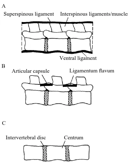
Ventral ligament
The ventral (or anterior) ligament is a ligament that connects the ventral surfaces of adjacent vertebral centra and discs. This continuous strip extends the entire length of the vertebral column. Because the axes of rotation of intervertebral joints presumably run through the middle of the discs and centra, the ventral ligament tends to resist joint extension (see Fig. 1A for a schematic diagram). Following the recording of a force–displacement loop for each intact specimen in extension, the actuator was lowered to expose the ventral surface of the centra (recall that, for these three-point bending tests, extension and flexion meant that the ventral surfaces of the centra and the spinous processes, respectively, rested on the two outer blocks). A scalpel or PM knife was used to sever this ligament midway along the ventral length of each centrum and disc. The actuator was returned to its original level and the extension test was repeated. Since the moment arms were unchanged, reductions in peak forces reflected reductions in resistive moments offered by the joints.
Superspinous ligament and interspinous ligaments and muscles
The superspinous ligament fuses with the spinous processes of adjacent vertebrae (see
Superspinous ligament
Articular capsule
Intervertebral disc
Interspinous ligaments/muscles
Ligamentum flavum
Centrum A
B
C
[image:3.612.141.350.70.334.2]Ventral ligament
Fig. 1A for a schematic diagram). Often it is closely bound to the aponeurosis of the longissimus dorsi muscle and the lumbodorsal fascia, making separation very difficult. Where separation was not possible (particularly in the jaguar and tiger), all three structures were included under the single heading of superspinous ligament. This structure tended to resist joint flexion. Following intact flexion tests (intact except for the severed ventral ligament from the previous extension tests), the actuator was lowered to allow cutting of the superspinous ligament between each joint. The actuator was returned to its initial setting and the bending cycle was repeated. The actuator was lowered several more times to allow cuts of varying depths through the interspinous muscular and connective tissues, effectively deepening the initial superspinous cut. Each cut was followed by the replacement of the actuator to its original level and a repeat of the bending cycle. Changes in peak flexion forces reflected changes in the resistive moments of the joints.
Articular joint capsules and ligamenta flava
Interspinous muscular and connective tissue was carefully dissected from between both joints of each specimen to expose the articular facet joints. These are synovial articulations between pairs of cranial and caudal articular processes of adjacent vertebrae. They can vary in structure from fairly simple areas of overlap to elaborate cradle-like constructions. Ligamentum flavum is a ligament that connects the dorsal aspect of adjacent vertebral arches (see Fig. 1B for a schematic diagram of both the articular joints and ligamentum flavum). It usually contains elastin, a rubber-like protein (Wainwright et al. 1976). Once the interspinous structures are dissected away, ligamentum flavum is usually visible through a gap between successive vertebrae. Sometimes, however, it is hidden by the overlapping bony plates on the floor of adjacent vertebral arches. Exposure of this ligament was sufficient in all test species here to enable lesion. Following dissection, the specimen was remounted in the clamp and reattached to the Instron load cell. A new intact flexion cycle was recorded for each demuscled specimen, based upon the same actuator displacement as in the intact specimens. Successive lesioning through ligamenta flava and up, around and between the articular joints was followed in turn by repositioning of the actuator and repeating the bending cycle. Changes in peak flexion forces were compared to the new maxima of the demuscled specimens for given displacements.
Discs and centra
intervertebral joints, the observed changes in intervertebral angle for discs and centra were compared to the predicted changes in intervertebral angle for the corresponding intact joints. This was done by substituting the observed discs and centra bending moments into the appropriate bending equations for the intact joints (see Gál, 1993, Tables 2–5 inclusive) and calculating predicted changes in intervertebral angle.
Lumbar intervertebral joints from monkey, wallaby, tiger, jaguar and seal were examined by careful dissection and successive boiling and bleaching. This allowed scrutiny of the deeper intervertebral structures. Particular attention was given to the soft structural tissues.
Results
Figs 2–4 illustrate some examples of the responses of wallaby, seal and monkey and tiger joint complexes, respectively, to the lesion experiments described. A summary of the reductions in peak forces is shown in Table 1. Peak extension forces changed very little following lesion of the ventral ligament. The most dramatic reductions in peak flexion forces (for demuscled specimens) followed the lesion of ligamenta flava and the articular capsules of the monkey and wallaby (approximatedly 90 and 80% reduction respectively). Much smaller changes in peak flexion forces were observed for jaguar and tiger complexes (approximately 19 and 10–12% respectively).
Table 2 shows the results of the cyclic bending tests with discs and centra for monkey, jaguar and seal. Wallaby and tiger complexes were also tested but corresponding X-rays
Actuator displacement (mm) 1
[image:5.612.162.332.75.283.2]2
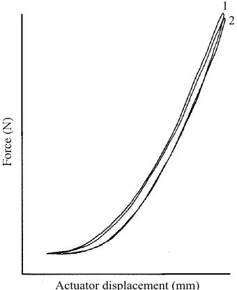
Fig. 2. The force–displacement loops for the intact and lesioned wallaby joint complex L2L3L4, subject to cyclic extension moments, are shown. Maximum changes in intervertebral angle were +2.5˚ for each joint. Maximum specific moments were 0.046 and 0.077 Nm kg21
were not taken. Predicted changes in intervertebral angles for intact monkey joints were much lower than those observed for monkey discs and centra subjected to the same bending moments. In contrast, predicted and observed changes in extension angle were similar in jaguar and seal. A similar, if not more pronounced, pattern was seen in flexion. Predicted changes in intervertebral angles were virtually the same as observed changes in jaguar and seal. Predicted changes for monkey were again much smaller than the observed changes.
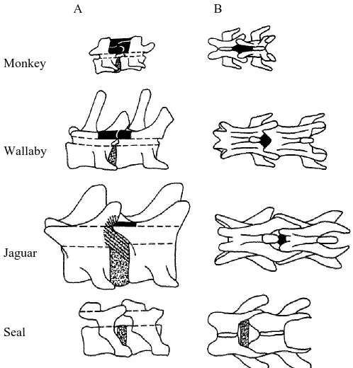
Fig. 5 illustrates mid-lumbar intervertebral joint anatomy of monkey, wallaby, jaguar and seal (in sagittal and dorsal perspectives). These schematic representations emphasize the approximate positions and relative sizes of the intervertebral discs and ligamenta flava in the species.
Monkey
Monkey lumbar joints had rather small intervertebral discs that were thicker in the ventral half than in the dorsal half. Transverse processes were bent approximately half way along their length so that they terminated in a cranial direction in close association with the lateral aspect of each disc. Ligamentum flavum was a prominent yellowish ligament that appeared to extend dorsally from the vertebral arches to span the gap between adjacent spinous processes. It would seem, therefore, to have been an interspinous ligament as well. Articular joints were formed by the superposition of the
Actuator displacement (mm) 1
[image:6.612.170.311.78.292.2]2
Fig. 3. The force–displacement loops for the intact and lesioned seal joint complex L1L2L3, subject to cyclic flexion moments, are shown. Maximum changes in intervertebral angle were 27.5 and 210˚ for L1L2 and L2L3 respectively. Maximum specific moments were 20.0020 and 20.0033 Nm kg21 for L1L2 and L2L3 respectively. Loop 1 represents the
Actuator displacement (mm) Tiger
1 2 3 1
2 Monkey
[image:7.612.142.365.76.318.2]3
Fig. 4. The force–displacement responses of intact and lesioned monkey (L5L6S) and tiger (L3L4L5) joint complexes, subject to cyclic flexion moments, are shown. Maximum changes in intervertebral angle for the monkey joints were 24.5 and 26 ˚ (for L5L6 and L6S respectively) and maximum specific moments were 20.039 and 20.036 Nm kg21(for L5L6
and L6S respectively). For the tiger joints, maximum changes in intervertebral angle were 26.5 and 24 ˚ (for L3L4 and L4L5 respectively) and maximum specific moments were 20.051 and 20.031 Nm kg21 (for L3L4 and L4L5 respectively). Loops 1 represent the
force–displacement responses of the intact demuscled joints. Loops 2 follow lesion of the articular capsules. Loops 3 follow complete cuts through (inactivation of) ligamenta flava and the articular capsules. See text for further explanation.
Table 1. Approximate percentage reductions in peak joint complex forces following lesions of intervertebral structures
Cut superspinous
ligaments, interspinous Cut articular Cut ventral ligaments and capsules and ligament muscles ligamenta flava
Animal (ext) (flex) (flex)
Monkey − 5–18 90
Wallaby 0–8 0–12 80
Jaguar 0 14–22 19
Tiger 0 8–23 10–12
Seal 1 19 19
[image:7.612.57.426.484.603.2]caudal articular processes of the more cranial vertebra with the cranial articular processes of the caudal vertebra of the adjacent pair. Articular joint capsular material fused with ligamentum flavum such that they were very difficult to separate. Spinous processes sloped very slightly cranially and were quite short and blunt.
Wallaby
[image:8.612.63.432.103.370.2]Like the monkey, the wallaby had relatively small, ventrally thickened intervertebral discs. Transverse processes sloped ventrally and cranially to overlap the area of the discs. Ligamentum flavum was a robust yellowish stucture and was very closely associated with the articular joint capsules. Caudal articular processes of the more cranial vertebra of adjacent pairs formed cradle-like supports for the large cranial articular processes of the more caudal vertebra. Spinous processes were large tapering processes with slightly cranial orientations.
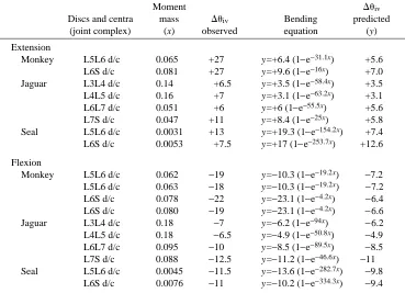
Table 2. Observed and predicted changes in intervertebral angle for discs and centra in extension and flexion
Moment Duiv
Discs and centra mass Duiv Bending predicted
(joint complex) (x) observed equation (y)
Extension
Monkey L5L6 d/c 0.065 +27 y=+6.4 (1−e−31.1x) +5.6
L6S d/c 0.081 +27 y=+9.6 (1−e−16x) +7.0
Jaguar L3L4 d/c 0.14 +6.5 y=+3.5 (1−e−58.4x) +3.5
L4L5 d/c 0.16 +7 y=+3.1 (1−e−63.2x) +3.1
L6L7 d/c 0.051 +6 y=+6 (1−e−55.5x) +5.6
L7S d/c 0.047 +11 y=+8.4 (1−e−25x) +5.8
Seal L5L6 d/c 0.0031 +13 y=+19.3 (1−e−154.2x) +7.4
L6S d/c 0.0053 +7.5 y=+17 (1−e−253.7x) +12.6
Flexion
Monkey L5L6 d/c 0.062 −19 y=−10.3 (1−e−19.2x) −7.2
L5L6 d/c 0.063 −18 y=−10.3 (1−e−19.2x) −7.2
L6S d/c 0.078 −22 y=−23.1 (1−e−4.2x) −6.4
L6S d/c 0.080 −19 y=−23.1 (1−e−4.2x) −6.6
Jaguar L3L4 d/c 0.18 −7 y=−6.2 (1−e−94x) −6.2
L4L5 d/c 0.18 −6.5 y=−4.9 (1−e−50.8x) −4.9
L6L7 d/c 0.095 −10 y=−8.5 (1−e−89.5x) −8.5
L7S d/c 0.088 −12.5 y=−11.2 (1−e−46.6x) −11
Seal L5L6 d/c 0.0045 −11.5 y=−13.6 (1−e−282.7x) −9.8
L6S d/c 0.0076 −11 y=−10.2 (1−e−334.3x) −9.4
The results from the discs and centra bending experiments are summarized. The applied specific bending moments are shown, with the observed changes in intervertebral angle. Duiv(predicted) is the
Jaguar
Jaguar transverse processes were relatively short, with a slight ventral and cranial orientation. Unlike the monkey and wallaby, jaguar intervertebral discs were very prominent structures. Further, white (presumably collagen) fibres, appearing to originate from the dorsal half of the discs, fused with the vertebral centra around the site of the articular joints. The articular joints were also cradle-like in nature, with prominent cranial processes of the more caudal vertebra being supported by the smaller caudal processes of the more cranial vertebra of an adjacent pair. Ligamentum flavum had the yellowy appearance observed in the monkey and wallaby although, relative to the other intervertebral structures, it was much less substantial. Spinous processes were thick short structures, with a slight taper and cranial orientation. Similar features were observed for tiger mid-lumbar joints, which were larger in all respects.
Seal
Transverse processes of the seal again had a slightly ventral and cranial slope, terminating in a space overlapping the intervertebral discs. However, the seal joints
Monkey
Wallaby
Jaguar
Seal
[image:9.612.132.381.74.334.2]A B
differed markedly from those of the other species examined; they were conspicuous in their almost total lack of soft structural tissue. Ligamentum flavum was reduced to a very fine strip of weak connective tissue, such that the intervertebral discs were clearly visible between the centra. Large spaces were evident between the vertebrae, exposing the vertebral canal. Intervertebral discs were reasonably thick structures that had a slightly concave profile, rather than the convex outline of the intervertebral discs of the other species studied. The articular joints had approximately vertical orientations, where caudal processes of the more cranial vertebra lay medial to the cranial processes of the more caudal vertebra of adjacent pairs. Spinous processes were very short, wide and blunt.
Fig. 6 shows schematic diagrams of cross sections through mid-lumbar intervertebral discs of monkey, wallaby, jaguar and seal. In normal healthy discs, two distinct components can be identified: the nucleus pulposus and the annulus fibrosus. The nucleus pulposus is the central gel-like component, composed primarily of water and a diffuse network of collagen fibres and proteoglycan molecules. The annulus fibrosus is made up of several lamellae containing strong, white, collagenous fibres. Collagen fibres are parallel within each lamella. Fibre orientation varies between lamellae, so that the macroscopic pattern of the annulus is a criss-cross weave (Panagiotacopulos et al. 1987a,b,c; Shirazi-Adl, 1989a,b). These two components were distinctly visible in all four species and they looked superficially similar. However, the terrestrial species differed from the seal in that they all had asymmetrically placed nuclei. In the monkey and jaguar and to a slightly lesser extent the wallaby, the ventral half of the annulus was greatly thickened. The nucleus was dorsally displaced. The nucleus pulposus of the seal was more centrally located, with annulus thickness being dorsally, ventrally and laterally symmetrical. The annulus lamellae appeared to permeate through to the nucleus in all species except the monkey, where the annulus phase without the lamellae appeared
Monkey Wallaby
[image:10.612.82.415.72.263.2]Seal Jaguar
opaque-white but distinct from the gel. The lamellar fibres looked similarly white in all species.
Discussion
This study began with an investigation of intervertebral sagittal mobility in a series of mammals differing in size, locomotor mode and taxonomic lineage. Of the seven terrestrial species initially considered, only the monkey, wallaby and tiger were chosen for the dynamic bending tests and lesion experiments presented here. Intervertebral joints from these animals were lacking in measurable neutral zones and flexed more readily than extended. Mathematical modelling of the results of bending tests showed that wallaby and monkey joints had greater flexion capacities and were relatively stiffer than homologous tiger and jaguar joints (Gál, 1993). Here, successive lesion of intervertebral muscular and ligamentous tissue showed that passive resistance to flexion moments was due largely to tension in ligamenta flava and the associated articular capsules in monkey and wallaby intervertebral joints. In contrast, the intervertebral discs of tiger and jaguar joints were the most important structures in resisting passive flexion moments. Passive extension moments were resisted largely by articular joint impaction in monkey and wallaby joints. Intervertebral disc compression and articular joint contact both appeared to play roles in resisting passive extension moments in homologous tiger and jaguar joints.
Successive lesion of intervertebral structures to determine resistance hierarchy has been employed in relatively few studies. Tencer et al. (1982) performed static mechanical tests with human cadaveric lumbar intervertebral joints. Extension and flexion tests were included in their protocol. Changes in intervertebral angle and displacements of joint centres were recorded following lesions of the anterior (ventral) ligament, posterior (interspinous, superspinous and intertransverse) ligaments and anterior discs fibres and facet removal. They found that lesion of the anterior ligament and 50% of the anterior disc fibres did not change any of the position parameters of the joint. They suggested that extension resistance resulted from bulk compression of the disc. Likewise, flexion resistance appeared to be largely the result of disc compression. Only with large displacements were the posterior ligaments and articular capsules shown to play a role.
Smeathers (1981) conducted lesion experiments with rabbit and goat lumbar vertebrae in order to correlate the results of buckling experiments with the intervertebral components. He found that, while flexion resistance in rabbit joints was due primarily to tension in ligamenta flava, resistance to flexion moments was due mainly to intervertebral disc compression and articular joint impaction in homologous goat joints. Articular joint contact was almost entirely responsible for limiting extension in the goat, whereas only in extreme extension did impaction act to limit extension in the rabbit joints.
component for intervertebral flexion is the intervertebral disc. It is suggested that there may be a scale-dependent shift in the emphasis of particular intervertebral structures, based upon the material composition of those structures. In other words, there may be a shift from primarily elastin-containing to primarily collagen-containing structures with increasing animal size.
Elastin is a highly extensible rubber-like protein. Purified samples of ligamentum nuchae (80% elastin with a small amount of collagen and other materials) break at roughly twice their resting length. Its elastic modulus, about 6.03105Nm22, is very
similar to that of other protein rubbers (Wainwright et al. 1976; Gosline, 1980). Collagen is a very stiff material in tension. The Young’s modulus of vertebrate tendons (dry mass about 70–80% collagen, Wainwright et al. 1976) is about 1.53109Nm22(Bennett et al.
1986). However, it has an ultimate strain of about 0.08–0.10 (Wainwright et al. 1976) and is therefore rather inextensible. So, elastin is relatively compliant with high extensibility, while collagen is relatively stiff with low extensibility. The great mobility of seal lumbar joints has been attributed, at least in part, to the lack of soft structural tissue. This is not a viable solution for joint mobility in terrestrial mammals because of gravitational considerations. Incorporating a highly extensible material like elastin would allow considerable changes in intervertebral angle, while maintaining joint integrity. However, since elastin is relatively compliant, the volume of material required to support the relatively greater bending stresses associated with increasing body mass could become prohibitive. Structural proteins are metabolically costly to synthesize and maintain and, therefore, it might be more economical in large animals to switch to a collagenous system for passive spinal resistance in order to keep the required volume of material to a minimum.
To test this idea, it would be useful to conduct similar dynamic bending and lesion experiments with intervertebral joint complexes from taxonomically related mammals of different sizes. The cat family would be a good group to study because gross morphology is conserved despite the wide range of sizes. The working hypothesis might be that flexion resistance would be determined by tension in ligamenta flava in the intervertebral joints of a domestic cat. This would contrast with the observed dependency upon the intervertebral discs in the tiger and jaguar intervertebral joints of this study. The inclusion of a cheetah in the investigation would make it even more interesting, given its superior sprinting capabilities. Another group that would prove useful to investigate would be the bovids. Halpert et al. (1987) studied lumbar vertebral morphology in a range of African bovids. They were concerned with dimensions of vertebral centra and processes and possible scaling relationships. Most of their vertebrae were museum specimens, so morphological comments were limited to those about the vertebral bone. Gross vertebral morphology of the smaller dik-dik was quite different from that of homologous eland vertebrae, particularly with respect to the relative size and orientation of the transverse processes. Smeathers (1981) distinguished between mammals with ‘flexible’ and ‘stiff’ backs, at least partly on the basis of differences in vertebral bony morphology. The results of these studies have demonstrated the importance of soft structural tissues (intervertebral ligaments and discs) in endowing particular stiffnesses to mammalian intervertebral joints. Given the differences in vertebral bone morphology, it would be of interest to examine the role of the soft structural tissues in bovid joints. Smeathers (1981) observed that many small mammals tended to have long arched ‘flexible’ lumbar columns, while many larger mammals tended to have short straight ‘stiff’ lumbar columns. He hypothesized that there was a trade-off between the energetic requirements of eliciting spinal movement and maintaining posture. Small mammals with flexible spines would be able to execute quick bending of the column, but would require constant axial muscle contraction to maintain a particular orientation. He surmised that the energy associated with maintaining posture as size increased would become prohibitive. Thus, large animals with passively stiff columns could maintain posture economically, at the relative expense of mobility.
The marked sagittal compliance of seal lumbar–lumbar and lumbosacral joints first observed and quantified by Gál (1993) was shown to have an anatomical basis. The joints were virtually devoid of soft structural tissue, except the discs (see Fig. 5). Ligamenta flava were reduced to fine transparent strips, loosely associated with adjacent arches. The intervertebral discs could be seen at the floor of the vertebral canal. The articular capsules were weak structures; the articular joints were areas of simple overlap. The near-vertical orientation of the articular joints allowed considerable extension and flexion. Passive resistance would therefore be exerted primarily by the ultimate contact of the bony surfaces and tension in the disc (annulus) fibres. However, impaction of the articular joints appeared to occur only at very high, unlikely physiological angles. From a closer inspection of the X-radiographs, adjacent centra did not appear to make contact with each other. When a beam is bent, the concave and convex surfaces are subject to compressive and tensile forces respectively. If this analogy is extended to bending the intervertebral joints, then bony surfaces verge on impaction while annulus fibre tension is increasing. Therefore, it is difficult to separate these two effects. Seal intervertebral discs differed dramatically from those of terrestrial animals, not only with respect to the placement of the nuclei, but also in gross compressive properties. Adjacent joints could be grasped and squeezed together longitudinally with ease, by several millimetres. Therefore, it is unlikely that the intervertebral discs of the seal could exert substantial passive bending resistance, except perhaps at or near the extremes of joint movement.
Many anecdotal reports describe the grace and expertise of swimming pinnipeds. There are three families in the order Pinnipedia: Phocidae, Otariidae and Odobenidae, the true seals, eared-seals and walruses respectively. The harbour or common seal (Phoca vitulina) is a typical true seal, having sleek hair and no external ear-flaps. They swim almost exclusively with their hindflippers, using their foreflippers for steering. They stretch their hindflippers backwards and put the soles of their feet together in the vertical plane to form a type of vertical tail-fin. The hindflippers and hind end (to a certain extent) are then oscillated laterally in the horizontal plane. When the foreflippers are not being recruited for steering, they are held at the side of the body in depressions, thereby conserving their stream-lined body profile. Eared-seals, like the Cape Fur Seal, have thick fur and scroll-like external earflaps. They use their enlarged foreflippers as hydrofoils, effectively ‘flying’ through the water. With increasing speed, there may be the occasional vertical oscillation of the hind end and hindflippers. More often, however, the hindflippers are used for steering. So that while the eared-seals employ vertical oscillatory movements, true seals employ primarily horizontal oscillations to swim (Slijper, 1946; Burton, 1972; Burton and Burton, 1979; Burn, 1980; Feldkamp, 1987; Fish et al. 1988).
impression of a kind of gallop. The neck acts as an important counterbalance to maintain the balance of the body over the foreflippers. True seals are very awkward on land by comparison. They are unable to raise themselves on their foreflippers and equally unable to flex their hindflippers under their body. Instead, they progress by crawling or ‘humping’, flexing their bodies and taking their weight alternately on chest and pelvis (Slijper, 1946; Burton, 1972; Burton and Burton, 1979; Burn, 1980).
Hebrank (1982) investigated lateral bending and tensile properties of the backbones of Norfolk spot (Leiostomus xanthurus), a subcarangiform swimmer, and skipjack tuna (Katsuwonus pelamis), a thunniform swimmer. Bending properties reflected the mode of propulsion, with lateral mobility distributed throughout the length of the column in the spot and isolated in the caudal peduncle in the tuna. Movement in planes other than those resulting in net thrust generation were prevented by vertebral morphology. Oscillating streamlined bodies suffer greater friction drag than similar static streamlined bodies. Lighthill (1969, 1971) suggested that this phenomenon was due to thinning of the boundary layer. Even those animals that do employ body oscillations to propel themselves use some other strategy to reduce unwanted body oscillations or ‘wagging’. Increasing depth of mid-section, either by adding large dorsal and/or ventral fins, or simply by deepening body profile, increases resistance to the turning moments generated by the oscillating caudal region. Fish et al. (1988) investigated the kinematics of swimming harp and ringed seals and estimated thrust production. They calculated drag coefficients and found them to be greater than those predicted for rigid streamlined bodies of similar size and shape. They commented that the propulsive lateral body and tail oscillations enhanced body drag by the mechanism previously described. They did not comment on the presence or absence of sagittal oscillations. To maintain sagittal rigidity during swimming, a seal would probably have to contract antagonistic axial muscles. This would tend to increase the energetic cost of swimming, relative to what it would be if sagittal rigidity were passively maintained. For these reasons, greatly enhanced sagittal mobility is probably not the greatest asset for an animal that swims by lateral oscillations of the hindflippers.
However, an effectively limbless semi-aquatic mammal, destined to return to the land or pack-ice to breed and moult, might just find that sagittal mobility is a necessity. It is suggested that the enhanced sagittal mobility observed in the common and grey seals (specimens from Gál, 1993, the static and dynamic bending tests respectively) and probably all true seals, is an adaptation of these semi-aquatic mammals to locomotion on land. With the exception of the somewhat laterally displaced iliocostalis muscle, most of the axial musculature of the seal tends to extend and flex the spine. Therefore, true seals are able to make progress over land by a method of locomotion similar to that used by some caterpillars. Extension and flexion of the body axis is coupled with alternate periods of grip and release of the substratum. Terrestrial locomotion is very probably an expensive pursuit for seals, requiring vigorous muscle contraction. Fortunately, gravity plays a relatively trivial role in the life of a true seal, so that static support of muscle bulk is probably not costly in itself. Perhaps it is not surprising that pinnipeds seem to spend a good deal of their terrestrial life dozing.
theory, the product of the transverse diameter (breadth) and the square of the sagittal diameter (height)] of many mammals. He found that the moments of resistance for true seals lay between values for the fully aquatic cetaceans and sirenians and values for the terrestrial carnivores. Clearly, passive joint resistance must also be affected by the presence or absence of soft structural tissue as well as end-point properties. Perhaps modifications in the basically carnivore vertebral column involved the loss of ligaments (especially ligamentum flavum) and the loss of disc integrity. The superspinous and interspinous ligaments in the dog and cat have been studied by Heylings (1980). He described them as weaker than homologous ligaments of human intervertebral joints. He did not comment on the ligamenta flava or the intervertebral discs. Certainly there were marked differences between the jaguar and seal joints studied here (see Fig. 5). The differences between the bending properties of the intervertebral discs were particularly pronounced, despite gross similarities in the appearance of the disc annulus lamellae and nuclei. The material composition and corresponding mechanical properties of these discs warrant further investigation.
This series of studies has demonstrated some of the sagittal bending properties of mammalian lumbar–lumbar and lumbosacral joints. The enhanced compliance of seal joints was suggested to be an adaptation to terrestrial locomotion in true seals. Two distinct mechanisms for resisting flexion moments in the terrestrial species were presented. Considering the available literature on flexion resistance in the intervertebral joints of other species, it was suggested that there may be a scale-dependent shift in material from elastin to collagen in order to accommodate the increasing demands of stiffness and strength, while maintaining some spinal mobility. This idea, appearing to transcend differences in posture, locomotor mode and taxonomic lineage, certainly warrants further study.
The author gratefully acknowledges the financial support of an NSERC PGS4 scholarship and ORS and Tetley-Lupton awards. Special thanks are due to Professor R. McNeill Alexander for his helpful advice on this work. Dr Caroline Pond provided assistance in the acquisition of many of the experimental specimens.
References
BENNETT, M. B., KER, R. F., DIMERY, N. J. ANDALEXANDER, R. MCN. (1986). Mechanical properties of
various mammalian tendons. J. Zool., Lond. A 209, 537–548.
BURN, D. M.(1980). The Complete Encyclopaedia of the Animal World. London: Octopus Books Ltd. BURTON, M.(1972). Encyclopaedia of Animals. London: Octopus Books Ltd.
BURTON, M. ANDBURTON, R.(1979). Encyclopaedia of Mammals. London: Galley Press, Cathay Books.
DIMERY, N. J., ALEXANDER, R. ANDDEYST, K. A. (1985). Mechanics of ligamentum nuchae of some artiodactyls. J. Zool., Lond. A 206, 341–351.
DUMAS, G. A., BEAUDOIN, L. ANDDROUIN, G. (1987). In situ mechanical behaviour of posterior spinal ligaments in the lumbar region. An in vitro study. J. Biomech. 20, 301–310.
FELDKAMP, S. D. (1987). Foreflipper propulsion in the California sealion, Zalophus californianus.
J. Zool., Lond. 212, 43–57.
FISH, F. E., INNES, S. ANDRONALD, K. (1988). Kinematics and estimated thrust production of swimming
GAL, J. M. (1993). Mammalian spinal biomechanics. I. Static and dynamic mechanical properties of
intact intervertebral joints. J. exp. Biol. 174, 247–280.
GOSLINE, J. M.(1980). The elastic properties of rubber-like proteins and highly extensible tissues. In The Mechanical Properties of Biological Materials (ed. J. F. V. Vincent and J. D. Currey), pp. 331–358.
Cambridge: Cambridge University Press.
HALPERT, A. P., JENKINS, F. A. ANDFRANKS, H. (1987). Structure and scaling of the lumbar vertebrae in
African bovids (Mammalia: Artiodactyla). J. Zool., Lond. 211, 239–258.
HEBRANK, M. R. (1982). Mechanical properties of fish backbones in lateral bending and in torsion. J. Biomech. 15, 85–89.
HEYLINGS, D. J. A. (1980). Supraspinous and interspinous ligaments in dog, cat and baboon. J. Anat. 130, 223–228.
LIGHTHILL, M. J. (1969). Hydromechanics of aquatic animal propulsion. A. Rev. Fluid Mech. 1,
413–446.
LIGHTHILL, M. J.(1971). Large-amplitude elongated body theory of fish locomotion. Proc. R. Soc. Lond. B 179, 125–138.
PANAGIOTACOPULOS, N. D., POPE, M. H., KRAG, M. H. ANDBLOCK, R.(1987a). A mechanical model for
the human intervertebral disc. J. Biomech., 20, 839–850.
PANAGIOTACOPULOS, N. D., POPE, M. H., KRAG, M. H. ANDBLOCK, R. (1987b). Water content in human
intervertebral discs. I. Measurement by magnetic resonance imaging. Spine 12, 913–917.
PANAGIOTACOPULOS, N. D., POPE, M. H., KRAG, M. H. ANDBLOCK, R.(1987c). Water content in human
intervertebral discs. II. Spine 12, 918–924.
SHIRAZI-ADL, A. (1989a). On the fibre composite material models of disc annulus: Comparison of
predicted stresses. J. Biomech. 22, 357–365.
SHIRAZI-ADL, A. (1989b). Strain in fibres of a lumbar disc: Analysis of the role of lifting in producing
disc prolapse. Spine 14, 96–103.
SLIJPER, E. J. (1946). Comparative biologic–anatomical investigations on the vertebral column and
spinal musculature of mammals. Verh. Kon. Nederl. Akad. Wetensch. Amsterdam II 42, 1–128. SMEATHERS, J. E. (1981). A Mechanical Analysis of the Mammalian Lumbar Spine. PhD thesis,
Department of Zoology, University of Reading, UK.
TENCER, A. F., AHMED, A. M. ANDBURKE, D. L.(1982). Some static mechanical properties of the lumbar
intervertebral joint, intact and injured. J. Biomech. Eng. 104, 193–201.