Original Article
Clinical and immunological characteristics of severe
asthma with fungal sensitization and allergic
bronchopulmonary aspergillosis: potential
utility for differential diagnosis
Peiyan Zheng1,2*, Shiquan Wu3*, Jiaxing Xie1, Wenting Luo1, Huimin Huang1,2, Nili Wei1,2, Lu Li1,2, Xi Chen1, Qingling Zhang1,2, Baoqing Sun1,2
1Guangzhou Institute of Respiratory Diseases, State Key Laboratory of Respiratory Disease, National Clinical Re-search Center for Respiratory Disease, Department of Allergy and Clinical Immunology, First Affiliated Hospital of
Guangzhou Medical University, Guangzhou 510120, Guangdong, China; 2Sino-French Hoffmann Institute, Guang
-zhou Medical University, Guang-zhou 511436, Guangdong, China; 3Department of Respiration, Hexian Memorial Affiliated Hospital, Southern Medical University, Guangzhou 511400, Guangdong, China. *Equal contributors. Received February 7, 2017; Accepted September 23, 2017; Epub December 15, 2017; Published December 30, 2017
Abstract: Fungal-sensitized asthmatic condition SAFS and the Aspergillus-related asthmatic condition ABPA are the focus of intense research. To investigate the clinical and immunological characteristics of severe asthma with fungal sensitization (SAFS) and allergic bronchopulmonary aspergillosis (ABPA) and provide for the early differential diagnosis and treatment of ABPA. The following parameters were measured in the serum of all recruited patients: total IgE (tIgE) levels, specific IgE (sIgE) and IgG (sIgG) antibodies against Aspergillus fumigatus, and sIgE antibod-ies against mixed fungal cultures (sIgE-M.x). Pulmonary function, fractional exhaled nitric oxide, and induced spu- ta were examined; routine blood tests and computerized tomography of the lungs were performed. For SAFS pati- ents, IgE antibody levels against eight types of fungi were measured. The percentage level of bronchiectasis, ratio of eosinophils in the induced sputa, and total number of blood eosinophils were significantly higher in ABPA than in SAFS. There were no significant differences in lung indicators, (percentage of predicted forced vital capacity [FVC% pred], percentage of predicted forced expiratory volume in 1 s [FEV1% pred], FEV1%, and FVC). SIgE-M.x, sIgE, and sIgG antibodies against A. fumigatus, and tIgE antibodies were significantly higher in ABPA than in SAFS. The clini -cal reference values for total blood eosinophils, sIgE-M.x antibodies, and sIgE as well as sIgG antibodies against A.
fumigatus for differential diagnosis of SAFS and ABPA were 0.41 × 10⁹/L, 3.22 kU/L, 3.86 kU/L, and 28.75 mgA/L, respectively. This panel should be considered for the differential diagnosis of ABPA and SAFS.
Keywords: Bronchial asthma, allergic bronchopulmonary aspergillosis, fungal allergen, specific IgE
Introduction
Fungi are common allergens, known to exacer-bate the severity of asthma in asthmatic pa- tients. Moreover, Aspergillus fumigatus, Peni- cillium chrysogenum, Candida albicans, Alter- naria alternata, and Cladosporium herbarum
are commonly implicated in exacerbating as-
thma [1-3], while Setomelanomma rostrata,
Mucor racemosus, and Botrytis cinerea are less commonly reported. Fungal allergens are
widely found both indoors and outdoors [4].
Long-term exposure to an environment contain-ing fungi is correlated with poor management
of asthma, an increased incidence of acute asthmatic attacks, higher hospitalization rates,
and increased mortality [3, 5-8]. Simon-Nobbe
et al. [9] additionally reported that over half the
patients with asthma were sensitized by fungi.
Denning [10] showed that fungal sensitization
was prevalent in asthmatic patients who re- quired hospitalization and that the severity of asthma was correlated with the degree of
fun-gal sensitization [11]. However, studies of
pa-tients with severe asthma with fungal sensitiza-tion (SAFS) suggest that prolonged antifungal
therapy has beneficial effects in the context of
anti-fungal treatment has been reported to impro- ve clinical symptoms in patients with allergic
bronchopulmonary aspergillosis (ABPA) [12]. In 1952, Hinson [13] described ABPA for the first
time as a potentially fatal lung disease trig-gered by allergic responses to Aspergillus anti-gens in the human bronchi, with inappropriate treatment leading to irreversible pathological damage. SAFS did not reach the diagnostic standard of ABPA, so it is necessary to differ- entiate from ABPA. At present, bronchiectasis mustn’t be recognized in the diagnosis of AB- PA, That may be the early stage of ABPA, called serological ABPA (ABPA-S). SAFS and ABPA-S may have a certain degree of overlap. The
diag-nosis of SAFS patients with mild ABPA inflam -mation is very similar. Thus, it is believed that SAFS and ABPA represent different stages of
the same disease [14, 15]; however, it is diffi -cult to distinguish between them by clinical doctors. Because of the overlapping features
of ABPA with asthma, cystic fibrosis, and
oth-er diseases, this condition often remains un- derdiagnosed and there may be a long delay
(of up to 10 years) between the first
occurren-ce of symptoms and subsequent diagnosis
[16]. Therefore, further understanding of clini -cal and immunologi-cal characteristics of SAFS
and ABPA is necessary. The present findings
should enable clinical standardization and early diagnosis of SAFS and support early treatment of SAFS to prevent its progression to ABPA.
Materials and methods
Research participants
Sixty-seven patients admitted to the First Af-
filiated Hospital of Guangzhou Medical
Uni-versity from 2014 to 2016 were recruited in this study. Of these, 31 patients presented wi- th SAFS, and 36 patients presented with ABPA. Diagnosis of SAFS was carried out as
recom-mended by the 2015 Global Initiative for As-thma [17], wherein a positive reaction in the specific IgE (sIgE) antibody test against certain
fungi (one or more positive results against ei- ght types of fungi) is indicative of SAFS, and in accordance with the diagnostic criteria pro-posed by Agarwal et al. [14]. ABPA diagnosis
was carried out in accordance with the con- sensus set of diagnostic criteria proposed by Agarwal et al. in 2013 [18]. Patients with the
following conditions were excluded: active, acute, or chronic pulmonary diseases, severe
immune diseases, blood diseases, malignant tumors, coronary heart disease, and hyperten-sion. In addition, pregnant and breastfeeding women and individuals prescribed a daily oral medication were excluded. The following infor-mation was recorded for each patient: gender, age, history of asthma and sinusitis, and bron-chiectasis status as indicated by lung com- puterized tomography scanning. Tests were carried out to analyze the levels of sIgE anti- bodies against mixed fungi (sIgE-M.x), sIgE an- tibodies against A. fumigatus (sIgE-A.f), speci-
fic IgG antibodies against A. fumigatus
(sIgG-A.f), serum total IgE (tIgE), blood eosinophil counts, erythrocyte sedimentation rate (ESR), fractional exhaled nitric oxide (FeNO), induced sputum, and pulmonary function.
Assessment of total IgE and allergen-specific IgE and IgG antibodies
Serum samples for biomarker analysis were stored at -80°C until analysis. sIgE-M.x, sIgE antibodies against eight types of fungi (A. fumigatus [sIgE-A.f], C. albicans [sIgE-C.a], P. chrysogenum [sIgE-P.c], C. herbarum [sIgE-C.h],
A. alternata [sIgE-A.a], M. racemosus
[sIgE-M.r], B. cinerea [sIgE-B.c], and S. rostrata [sIgE-S.r]), as well as sIgG-A.f antibodies and serum tIgE, were determined by using a
fluoro-im-munoassay technique using an UniCAP 1000
instrument (Thermo Fisher Scientific, Uppsala,
Sweden) according to the manufacturer’s in-
structions. IgE and IgG measurements were reported in kU/L and mg/L, respectively [19,
20]. A positive sIgE result was set at a value of
≥0.35 kU/L. The detection range was 2-200 mg/L for sIgG. For sIgG levels higher than 200
mg/L, blood samples were diluted 10-fold before testing.
FeNO measurement
For all patients, standard measurements of FeNO (NioxMino, Aerocrine, Sweden) were
car-ried out at a flow rate of 50 mL/s [21] using
signals fed back for the control. This test was performed prior to body plethysmography and bronchial provocation to prevent the distortion of FeNO data that may result from the breath-ing maneuvers involved.
Induced sputum
Sputum induction was carried out according to
Table 2. Clinical characteristics of the two groups
SAFS (n = 31) ABPA (n = 36) P-value
FeNO (ppb) 29 (9-65) 68 (14-92) 0.177
Total leukocytes (109/L) 8.62 (5.80-10.46) 8.09 (6.21-10.65) 0.921
Neutrophils (109/L) 6.00 (3.30-8.50) 4.70 (3.10-7.50) 0.498
Eosinophils (109/L) 0.15 (0.00-0.30) 0.85 (0.30-1.41) 0.001
Induced sputum EOS (%) 11.50 (6.50-45.50) 13.00 (1.50-23.50) 0.047
FVC% pred 84.70 (74.40-99.50) 78.30 (66.00-89.00) 0.408
FEV1% pred 62.80 ± 22.82 55.005 ± 24.41 0.628
FEV1%/FVC 60.84 (48.70-69.00) 53.36 (41.53-74.20) 0.514
FEF25–75% pred 22.50 (15.10-51.65) 14.85 (7.45-41.76) 0.062
Erythrocyte sedimentation rate (mm/h) 9 (4-17) 22 (15-40) 0.005
C-reactive protein (mg/L) 0.75 (0.1-4.3) 0.1 (0.1-5.2) 0.457
Glucocorticoids (mg/day)a 10 (0-25) 15 (10-20) 0.373
Inhaled corticosteroids (µg/day)b 500 (320-640) 320 (320-640) 0.154
1-3-β-d-glucan (pg/mL) 10 (10-17) 10 (10-49) 0.320
Use of glucocorticoids [number of patients (%)] 23 (74.2%) 29 (80.6%) 0.815 Use of antibiotics [number of patients (%)] 12 (38.7%) 21 (58.7%) 0.235 EOS, eosinophil; FeNO, fractional exhaled nitric oxide;FVC% pred, percentage of predicted forced vita capacity;FEV1% pred,
percentage of predicted forced expiratory volume in 1 s. aGlucocorticoid use was assessed via the measurement of prednisone
acetate. bInhaled corticosteroid use was assessed via the measurement of beclomethasone dipropionate.
µg) was inhaled; 15 min later, two
concentra-tions of saline (0.9% and 3%, for 7 min each)
were nebulized and inhaled. Sputum was then
induced, and differential counts of inflammato -ry cells were analyzed in the sputum.
Lung function measurements
Lung function measurements were carried out in asthmatic patients to evaluate the pulmo-nary ventilation function and the lung function index, including the percentage of predicted
forced vital capacity (FVC% pred), percentage
of predicted forced expiratory volume in 1 s
(FEV1% pred), and the ratio FEV1%/FVC, as well
as the percentage of predicted peak expiratory
flow rate (PEF% pred) and forced expiratory flow
(FEF25-75% pred).
Statistical analyses
All analyses were carried out using the IBM SPSS 19.0 software. Results were expressed
[image:3.612.93.523.296.522.2]as the number of patients (%), the mean ± stan -dard deviation for normally distributed vari-ables and the median and interquartile range
Table 1. General characteristics of patients
SAFS (n = 31) ABPA (n = 36) P-value
Female (number of patients [%]) 10 (32.7%) 16 (44.4%) 0.059
Age (years) 46 ± 13 47 ± 15 0.167
Weight (kg) 60.0 (52.0-67.0) 52.5 (48.0-61.2) 0.006
Height (cm) 163 (159-170) 160 (152-168) 0.304
BMI (kg/m2) 22.2 (19.0-24.5) 21.2 (19.1-23.1) 0.510
Smoking history [number of patients (%)] 4 (12.9%) 9 (25%) 0.423
Obesity history [number of patients (%)] 2 (4.7%) 0 (0%) 0.059
Sinusitis [number of patients (%)] 25 (80.6%) 23 (63.9%) 0.491
Bronchiectasis [number of patients (%)] 11 (25.6%) 32 (88.9%) 0.001
Disease duration (years) 10 (4-30) 10 (3-20) 0.871
ABPA, allergic bronchopulmonary aspergillosis; SAFS, severe asthma with fungal sensitization. BMI, body mass index [weight
(kg)/height2 (m2)]; Obesity, BMI values greater than 30 kg/m2; Smoking history, smoking more frequently than once a week,
(IQR) for non-normally distributed data, The dif-ferences in percentages between two groups were analyzed by the X2 test. We compared the non-normally distributed data between groups by using the nonparametric Mann-Whitney U test, and Normally distributed data by the unpaired t-test. Receiver operating characteris-tic (ROC) curves were produced using an empir-ical model. A value of P < 0.05 was considered
statistically significant (Supplementary Data).
Results
General observations
There were no significant differences in basic
indicators such as gender, average age, dis-ease progress, height, body mass index, smok-ing history, obesity, and sinusitis between the
se patients; however, there were no statistical-
ly significant differences between the two
gr-oups (Table 2).
Serum immunological indicators
The sIgE-M.x, sIgE-A.f, sIgG-A.f, and serum tIgE levels were significantly higher in patients with
ABPA than in those with SAFS (all P < 0.001), indicating that the level of sensitization against
A. fumigatus was significantly higher in the
patients with ABPA than in those with SAFS (Figure 1).
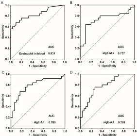
Analysis of ROC curves of laboratory test indicators
ROC curves were plotted for blood eosinophil
counts and sIgE-M.x, sIgE-A.f, and sIgG-A.f lev -Figure 1. Analysis of sIgE-M.x, sIgE-A.f, sIgG-A.f, and total IgE levels in pa
-tients with SAFS and ABPA. ABPA, allergic bronchopulmonary aspergillosis; SAFS, severe asthma with fungal sensitization; sIgE-A.f, specific IgE against
Aspergillus fumigatus; sIgE-M.x, specific IgE against mixed mycosis agents; sIgG-A.f, specific IgG against A. fumigatus. Statistical differences were de-termined by the Mann-Whitney nonparametric test. The scatter plots show that the levels of sIgE-M.x (A), sIgE-A.f (B), sIgG-A.f (C), and total IgE (D) were higher in the patients with ABPA than in those with SAFS, * P < 0.01.
SAFS and ABPA groups. Body
weight was significantly lower,
while the bronchiectasis rate
was significantly higher, in the
ABPA group than in the SAFS group (Table 1). These results indicated that the patients wi- th ABPA experienced bronchi-ectasis at a higher rate than those with SAFS and that the bronchial damage was more severe in the former group.
Clinical characteristics
Induced sputum analysis for the two groups showed that
eosinophilic inflammation was
dominant in both types of pa- tients. The eosinophil
percent-age (EOS%) in the induced spu -tum, blood eosinophil counts,
and ESR were significantly
hig-her in the ABPA group than in the SAFS group. With respect to pulmonary indicators such
as FVC% pred, FEV1% pred, FEV1%/FVC, and FEF25-75%
pr-ed, there were no statistically
significant differences
betwe-en the ABPA and SAFS groups. The FeNO levels were higher than the normal level (25 ppb) in both groups, suggesting an
[image:4.612.91.369.72.387.2]the-els in the patients with SAFS and ABPA (Figure 2). The areas under the ROC curves (AUCs) of total blood eosinophil counts, M.x,
sIgE-A.f, and sIgG-A.f were >0.7, with P < 0.05 (see
Table 3 for details). The sensitivity levels were
>60%; in particular, the sensitivity level of sIgG-A.f was 76.0%. The specificity levels for total
blood eosinophil counts, sIgE-M.x, and sIgE-A.f
were 90.0%, 90.0%, and 93.0%, respectively.
The clinical reference values of total blood
eosinophil counts, sIgE-M.x, sIgE-A.f, and
sIgG-A.f for differential diagnosis of SAFS and ABPA were 0.41 × 10⁹/L, 3.22 kU/L, 3.86 kU/L, and 28.75 mgA/L, respectively. The AUC value for
tIgE was 0.626 and not statistically significant
(P = 0.110). These results demonstrated that
the level of sensitivity of sIgG-A.F as a diagnos -tic tool for ABPA and SAFS was higher than those of the other three indicators, which,
how-ever, showed higher specificity. In particular,
the focus of intense research [18]. ABPA, a
more severe disease with a rapid onset, was
first described in 1952 [20]. The first case of
ABPA in an adult was reported in the US in
1968 [27]. Subsequently, Wen et al. [28]
reported three cases of ABPA in China in 1985, and since then, ABPA has been widely recog-nized as a clinical disorder. ABPA is considered an exacerbated form of Aspergillus
sensitiza-tion, which is likely represents the first step in
the development of ABPA. Agarwal et al. [29]
conducted a meta-analysis of 21 cases of asth-ma and sensitization by A. fumigatus and found that the rate of A. fumigatus-induced asthma
[image:5.612.90.372.71.351.2]was 28% in asthmatic patients, while ABPA was observed in 12.9% of asthmatic patients. Almost 40% of A. fumigatus-sensitized asth-matic patients meet the diagnostic criteria for ABPA. Therefore, A. fumigatus-sensitized asth-ma is closely associated with ABPA. Although Figure 2. Receiver operating characteristic (ROC) curves of blood eosinophil
counts and sIgE-M.x, sIgE-A.f, and sIgG-A.f antibody levels for the diagno -sis of ABPA. SIgE-A.f, specific IgE against Aspergillus fumigatus; sIgE-M.x, specific IgE against mixed mycosis agents; sIgG-A.f, specific IgG against A.
fumigatus. Statistical differences were determined by ROC curve analysis. The area under the curve (AUC) for blood eosinophil counts (A) was 0.831, while those for sIgE-M.x (B), sIgE-A.f (C), and sIgG-A.f (D) were 0.737, 0.789, and 0.781, respectively (P < 0.01; P = 0.003; P < 0.001; and P < 0.001, respectively).
among the latter three indica-tors, the use of sIgE-A.f levels enabled the differential diag-nosis of ABPA and SAFS with a
relatively high specificity.
Discussion
Fungi are a class of organisms that are widespread in nature and closely associated with the lives of humans. Fungal aller-gens, which are spread via the release of spores and hyphal fragments into the air, may be inhaled to cause severe aller-gic and bronchopulmonary dis-eases. Fungal allergens repre-sent independent risk factors for the onset and development
of asthma [23]. Fungal
sensi-tization is closely associated with the severity of asthma, frequency of respiratory symp-toms and admission into int- ensive care, and higher morta-
lity rates [24-26]. The relation -ship between fungal sensitiza-tion and asthma has recently
received much attention [19].
fungal-sensitized diseases have begun to
gar-ner much attention in recent years [18, 30],
there is still a lack of studies on SAFS and ABPA among the Chinese population. Chang et al.
[31] have suggested that specific sensitization
to fungal allergens plays a critical role in the pathogenesis of atopic diseases. Zhou et al.
[32] have published a patient report about
ABPA in children and concluded that early di- agnosis and initiation of systemic corticoste-roids are essential to prevent irreversible dam-age. In the present work, the clinical and immu-nological characteristics of SAFS and ABPA
were investigated to provide a scientific basis
for the clinical standardization and early diag-nosis of SAFS and ABPA.
In the present work, over half of the patients from both the SAFS and ABPA groups had sinusitis. Diseases of the upper and lower re- spiratory tracts are closely associated with ea- ch other and often occur concurrently. The rate of bronchiectasis, blood eosinophil counts in induced sputa, ESR, and total blood eosinophil counts were higher in ABPA relative to SAFS. This may be attributed to a higher sensitiza- tion level in ABPA as allergenic A. fumigatus
adheres to and undergoes mycelial growth in the airway epithelium. This fungal activity pro-duces a large amount of harmful substances, such as fumagillin and proteases, which weak-en the cilia and destroy the airway, leading to
the aggregation of inflammatory cells and
in-duction of allergic responses. Furthermore, the growth of A. fumigatus stimulates the differ- entiation of T cells into type 2 helper T cells and
promotes the release of IgE and IgG antibodies.
This results in an increase of the number of eosinophils in the airway and peripheral blood-stream, which elicits further pathophysiologi- cal changes such as bronchial spasms, mucus
accumulation, bronchiectasis, and inflammato
-ry infiltration of the lungs [33].
ABPA differs from SAFS in that it is more diffi -cult to control, and is associated with a poorer prognosis. In addition, ABPA is more likely to exhibit a rapid progression and cause irrever-
sible damage during later stages [34]. However, it is difficult to distinguish ABPA from A. fumi- gatus-sensitized SAFS during clinical diagnosis. Comparison of serum immunological charac-teristics of the two conditions has shown that
the levels of tIgE, sIgE-A.f, and sIgG-A.f antibod
-ies were significantly higher in ABPA than in SAFS [35], and the present findings are consis -tent with this report. Previous studies have re- ported that the threshold values for total blood
eosinophil counts, tIgE, sIgE-A.f, and sIgG-A.f
were 0.507 × 10⁹/L, 2,347 kU/L, 1.91 kU/L, and 40 mgA/L, respectively, for differentiating
ABPA from asthma [36, 37]. According to the common diagnostic criteria for ABPA [18], the
corresponding values are 0.500 × 10⁹/L, 1,000 kU/L, and 40 mgA/L for the total blood
eosino-phil counts, tIgE, and sIgG-A.f, respectively. Therefore, there is no definitive diagnostic
th-reshold value for the sIgE-A.f antibody level yet. In this study, comparison of diagnostic thresh-olds for ABPA and SAFS indicated that the threshold values for total blood eosinophil
counts, sIgE-A.f, sIgG-A.f, and sIgE-M.x were
0.410 × 10⁹/L, 3.86 kU/L, 29 mgA/L, and 3.22 kU/L, respectively. The AUCs of the ROC cur- ves for these four indicators were >0.7, and the
sensitivity of the sIgG-A.f antibodies and speci
-ficity of the sIgE-A.f antibodies were the high -est. Therefore, these parameters may be uti-lized for the differential diagnosis of ABPA and SAFS at the clinical level. The values for total blood eosinophil counts and the levels of
sIgE-A.f and sIgG-sIgE-A.f antibodies obtained in the
present study were inconsistent with those
reported previously [18]. In addition, to our
[image:6.612.88.523.88.163.2]knowledge, the utility of sIgE-M.x antibodies for the diagnosis of ABPA has not been reported previously. To date, the diagnosis of ABPA in
Table 3. Clinical reference values of blood indicators for the differential diagnosis of SAFS and ABPA Clinical reference
value Sensitivity Specificity AUC P-value 95% Confidence interval Eosinophils (109/L) 0.41 68.0% 90.0% 0.831 0.001 (0.716-0.945)
sIgE-M.x (kU/L) 3.22 64.0% 90.0% 0.737 0.003 (0.591-0.882)
sIgE-A.f (kU/L) 3.86 68.0% 93.0% 0.789 0.001 (0.663-0.916)
sIgG-A.f (mgA/L) 28.75 76.0% 73.3% 0.781 0.001 (0.661-0.902)
AUC, area under the curve;sIgE-A.f, specific IgE against Aspergillus fumigatus; sIgE-M.x, specific IgE against mixed mycosis agents; sIgG-A.f, specific IgG against A. fumigatus. Statistical differences were determined by ROC curve analysis. P < 0.05
China has relied on the standards established in other countries. Hence, a variety of factors
specific to the Chinese population, such as the
ethnic background, climate, population chara- cteristics, host and fungal gene expression, his-tory of drug use, and autoimmune status, have not been considered. Therefore, we believe it is imperative to establish a set of diagnostic cri-
teria specific to the Chinese population in order to better define the characteristics of ABPA and
to implement methods for the early diagnosis and timely treatment of the disease.
Since this was a retrospective cohort study, part of the clinical symptom data and detection results were not available. Furthermore, analy-sis of clinical symptoms and post-treatment follow-up data were omitted from this study. Future studies should aim to obtain all relevant clinical indicators for an in-depth analysis of the clinical and immunological characteristics of SAFS and ABPA.
Conclusions
SAFS is a typical asthma disease with fungal sensitization, and fungi can worsen asthma
symptoms. If a specific case of asthma is found
to be triggered by fungal sensitization, further testing of total blood eosinophil counts and
sIgE-A.f, sIgG-A.f, and sIgE-M.x antibodies is
necessary. Comparison of the threshold values with those for ABPA should enable early diagno-sis and treatment. Therefore, this study pro-vides an important clinical basis for the differ-ential diagnosis, prevention, and treatment of SAFS and ABPA.
Acknowledgements
This study was funded by the National Natural Science Foundation of China (Project No.: 81572063, 81601394), Medicine and Health
Care Technology Projects of Guangzhou (Project
No.: 2017A013010017), Bureau of Education
Projects of Guangzhou (Project No.:
1201630393, 1201630044). The funders had no role in study design, data collection and analysis, decision to publish, or preparation of the manuscript. No additional external funding was received for this study.
Disclosure of conflict of interest
None.
Address correspondence to: Baoqing Sun and Qingling Zhang, State Key Laboratory of Respirato- ry Disease, First Affiliated Hospital of Guangzhou Medical University, 151 Yanjiangxi Road, Guang-zhou 510120, Guangdong, China. Tel: +86 20 8306 2865; Fax: +86 20 8306 2729; E-mail: sunbao-qing@vip.163.com (BQS); zqling68@hotmail.com (QLZ)
References
[1] Hedayati MT, Pasqualotto AC, Warn PA, Bowyer P and Denning DW. Aspergillus flavus: human pathogen, allergen and mycotoxin producer. Microbiology 2007; 153: 1677-1692.
[2] Denning DW, O’Driscoll BR, Hogaboam CM, Bowyer P and Niven RM. The link between fun-gi and severe asthma: a summary of the evi-dence. Eur Respir J 2006; 27: 615-626. [3] Zureik M, Neukirch C, Leynaert B, Liard R,
Bousquet J, Neukirch F; European Community Respiratory Health Survey. Sensitisation to air-borne moulds and severity of asthma: cross sectional study from European community re-spiratory health survey. BMJ 2002; 325: 411-414.
[4] Horner WE, Helbling A, Salvaggio JE and Lehrer SB. Fungal allergens. Clin Microbiol Rev 1995; 8: 161-179.
[5] Targonski PV, Persky VW and Ramekrishnan V. Effect of environmental molds on risk of death from asthma during the pollen season. J Aller-gy Clin Immunol 1995; 95: 955-961.
[6] Green BJ, Sercombe JK and Tovey ER. Fungal fragments and undocumented conidia func-tion as new aeroallergen sources. J Allergy Clin Immunol 2005; 115: 1043-1048.
[7] Arbes SJ Jr, Gergen PJ, Vaughn B and Zeldin DC. Asthma cases attributable to atopy: results from the third national health and nutrition ex-amination survey. J Allergy Clin Immunol 2007; 120: 1139-1145.
[8] Jaakkola MS, Ieromnimon A and Jaakkola JJ. Are atopy and specific IgE to mites and molds important for adult asthma? J Allergy Clin Im-munol 2006; 117: 642-648.
[9] Simon-Nobbe B, Denk U, Poll V, Rid R and Bre -itenbach M. The spectrum of fungal allergy. Int Arch Allergy Immunol 2008; 145: 58-86. [10] O’Driscoll BR, Hopkinson LC and Denning DW.
Mold sensitization is common amongst pa-tients with severe asthma requiring multiple hospital admissions. BMC Pulm Med 2005; 5: 4.
[11] Black PN, Udy AA and Brodie SM. Sensitivity to fungal allergens is a risk factor for life-threat-ening asthma. Allergy 2000; 55: 501-504. [12] Stevens DA, Schwartz HJ, Lee JY, Moskovitz BL,
Weinmann AJ, Tuazon CU, Judson MA, Platts-Mills TA and DeGraff AC Jr. A randomized trial of itraconazole in allergic bronchopulmonary aspergillosis. N Engl J Med 2000; 342: 756-762.
[13] Hinson KF, Moon AJ and Plummer NS. Bron-cho-pulmonary aspergillosis; a review and a report of eight new cases. Thorax 1952; 7: 317-333.
[14] Agarwal R. Severe asthma with fungal sensiti-zation. Curr Allergy Asthma Rep 2011; 11: 403-413.
[15] Farrant J, Brice H, Fowler S and Niven R. Fun-gal sensitisation in severe asthma is associat-ed with the identification of aspergillus fumiga -tus in sputum. J Asthma 2016; 53: 732-735. [16] de Almeida MB, Bussamra MH and Rodrigues
JC. Allergic bronchopulmonary aspergillosis in paediatric cystic fibrosis patients. Paediatr Respir Rev 2006; 7: 67-72.
[17] Dwarakanath S. Global initiative for Asthma (GINA). Pocket guide for asthma management and prevention. Available at: http://www.gin-asthma.org/local/uploads/files/GINAPocket -05Clean_1.pdf. Accessed November 6, 2015. [18] Agarwal R, Chakrabarti A, Shah A, Gupta D,
Meis JF, Guleria R, Moss R, Denning DW; ABPA complicating asthma ISHAM working group. Al-lergic bronchopulmonary aspergillosis: review of literature and proposal of new diagnostic and classification criteria. Clin Exp Allergy 2013; 43: 850-873.
[19] Denning DW, Pashley C, Hartl D, Wardlaw A, Godet C, Del Giacco S, Delhaes L and Sergeje -va S. Fungal allergy in asthma-state of the art and research needs. Clin Transl Allergy 2014; 4: 14.
[20] Knutsen AP and Slavin RG. Allergic broncho -pulmonary aspergillosis in asthma and cystic fibrosis. Clin Dev Immunol 2011; 2011: 843763.
[21] Dweik RA, Boggs PB, Erzurum SC, Irvin CG, Leigh MW, Lundberg JO, Olin AC, Plummer AL, Taylor DR; American Thoracic Society Commit-tee on Interpretation of Exhaled Nitric Oxide Levels (FENO) for Clinical Applications. An offi -cial ATS clinical practice guideline: interpreta-tion of exhaled nitric oxide levels (FENO) for clinical applications. Am J Respir Crit Care Med 2011; 184: 602-615.
[22] Simpson JL, Scott R, Boyle MJ and Gibson PG. Inflammatory subtypes in asthma: assessment and identification using induced sputum. Res -pirology 2006; 11: 54-61.
[23] Kauffman HF and van der Heide S. Exposure, sensitization, and mechanisms of fungus-in-duced asthma. Curr Allergy Asthma Rep 2003; 3: 430-437.
[24] Maurya V, Gugnani HC, Sarma PU, Madan T and Shah A. Sensitization to aspergillus anti-gens and occurrence of allergic bronchopul-monary aspergillosis in patients with asthma. Chest 2005; 127: 1252-1259.
[25] Zou H, Su L, Fang QH and Ma YM. Correlation between fungal sIgE and bronchial asthma se-verity. Exp Ther Med 2013; 6: 537-541. [26] Neas LM, Dockery DW, Burge H, Koutrakis P
and Speizer FE. Fungus spores, air pollutants, and other determinants of peak expiratory flow rate in children. Am J Epidemiol 1996; 143: 797-807.
[27] Wheat LJ, Goldman M and Sarosi G. State-of-the-art review of pulmonary fungal infections. Semin Respir Infect 2002; 17: 158-181. [28] Greenberger PA, Bush RK, Demain JG, Luong
A, Slavin RG, Knutsen AP. Allergic bronchopul -monary Aspergillosis. J Allergy Clin Immunol Pract 2014; 2: 703-8.
[29] Agarwal R, Aggarwal AN, Gupta D and Jindal SK. Aspergillus hypersensitivity and allergic bronchopulmonary aspergillosis in patients with bronchial asthma: systematic review and meta-analysis. Int J Tuberc Lung Dis 2009; 13: 936-944.
[30] Greenberger PA, Bush RK, Demain JG, Luong A, Slavin RG and Knutsen AP. Allergic broncho -pulmonary aspergillosis. J Allergy Clin Immunol Pract 2014; 2: 703-708.
[31] Chang FY, Lee JH, Yang YH, Yu HH, Wang LC, Lin YT and Chiang BL. Analysis of the serum levels of fungi-specific immunoglobulin E in pa -tients with allergic diseases. Int Arch Allergy Immunol 2011; 154: 49-56.
[32] Zhou Y, Xu D, Zhang Y, Sheng Y, Chen X and Chen Z. Allergic bronchopulmonary aspergillo-sis in children. Pediatr Int 2015; 57: e73-76. [33] Kauffman HF, Tomee JF, van de Riet MA,
Tim-merman AJ and Borger P. Protease-dependent activation of epithelial cells by fungal allergens leads to morphologic changes and cytokine production. J Allergy Clin Immunol 2000; 105: 1185-1193.
[34] Rodrigues J, Caruthers C, Azmeh R, Dykewicz MS, Slavin RG and Knutsen AP. The spectrum of allergic fungal diseases of the upper and lower airways. Expert Rev Clin Immunol 2016; 12: 531-550.
[35] Tanimoto H, Fukutomi Y, Yasueda H, Takeuchi Y, Saito A, Watai K, Sekiya K, Tsuburai T, Asano K, Taniguchi M and Akiyama K. Molecular-based allergy diagnosis of allergic bronchopul-monary aspergillosis in aspergillus fumigatus-sensitized Japanese patients. Clin Exp Allergy 2015; 45: 1790-1800.
[36] Agarwal R, Aggarwal AN, Garg M, Saikia B and Chakrabarti A. Cut-off values of serum IgE (to-tal and A. fumigatus -specific) and eosinophil count in differentiating allergic bronchopulmo-nary aspergillosis from asthma. Mycoses 2014; 57: 659-663.