Original Article
Loss of BAP1 expression is a very rare
event in gastrointestinal stromal tumors
Hee Jung Kwon, Joon Hyuk Choi, Mi Jin Gu
Department of Pathology, Yeungnam University College of Medicine, Daegu, South Korea
Received April 19, 2017; Accepted September 4, 2017; Epub September 15, 2017; Published September 30, 2017
Abstract: Gastrointestinal stromal tumors (GISTs), the most common mesenchymal tumors affecting the gastro-intestinal tract, are primarily driven by activating KIT and platelet-derived growth factor receptor alpha (PDGRFA) mutations and respond to targeted tyrosine kinase therapy. However, GISTs exhibit various clinical behaviors,
re-gardless of the proposed risk classification. Here, we investigated the expression of BRCA1-associated protein-1 (BAP1) in GIST and analyzed its prognostic significance and utility as a marker in the context of differential diag
-nosis. Among a total of 226 GIST, only one (0.44%, 1/226 cases) GIST exhibited a loss of BAP1 expression. In the
univariate analysis, small intestinal and colorectal GISTs, GISTs with necrosis, recurrence/metastasis, or a higher risk of malignancy were associated with poor overall survival. In the multivariate analysis, GISTs with a higher risk of
malignancy or recurrence/metastasis were identified as independent prognostic factors. We conclude that a loss of BAP1 expression is a very rare event, then BAP1 would not play a major role in pathogenesis of GIST.
Keywords: Gastrointestinal stromal tumor, BAP1, BRCA1-associated protein, immunohistochemistry
Introduction
Gastrointestinal stromal tumors (GISTs) com-prise the most common type of mesenchymal-neoplasms of the gastrointestinal tract. These tumors are resistant to conventional chemo-therapy and radiochemo-therapy [1, 2]. In adults, the majority of GISTs harbor characteristic onco-genic mutations in KIT (80-85%) and PDGFRA (5-7%) and respond to targeted tyrosine kinase (TK) therapy [2, 3]. However, approximately 10- 15% of GISTs are KIT/PDGFRA wild-type (WT)
and are less sensitive to TK inhibitors (TKI).
BRCA1-associated protein-1 (BAP1) is a deubiq
-uitinating enzyme that plays important roles in
chromatin modulation, DNA transcription and cell cycle regulation, cellular growth, and DNA repair [3-5]. BAP1 is now recognized as a tumor
suppressor gene, and germline BAP1 muta-tions have been associated with autosomal dominant cancer syndromes that include cuta-neous and uveal melanoma, renal cell carcino-ma, cutaneous basal cell carcinocarcino-ma, and malig-nant mesothelioma [6]. Somatic BAP1 muta-tions have also been investigated in the context of mesothelioma, intrahepatic
cholangiocarci-noma (ICC), clear cell renal cell carcicholangiocarci-noma, and
atypical cutaneous spitzoid tumors [3, 6-9]. Immunohistochemistry (IHC) for BAP1 appears
to serve as a reliable and highly
sensitive/spe-cific marker of BAP1 mutation or inactivation, irrespective of underlying genetic alterations [4,
7, 10]. Accordingly, the BAP1 expression status has been described as clinically significant in a
variety of human tumors [6, 9, 11-13].
The present study aimed to investigate the
BAP1 expression statuses of GISTs, to assess the clinical and pathological significance, and
to identify the utility in differential diagnosis from other tumors. To the best of our
knowl-edge, BAP1 expression status has not previ -ously been assessed in GIST.
Materials and methods
Patient characteristics
Between 1997 and 2016, a total of 226 GISTs
records were reviewed to determine each pati- ent’s age, sex, most recent follow-up visit, sur-vival status, and the presence or absence of GIST-related disease. The following clinicopath-ologic characteristics were also assessed: tu-
mor location, tumor size, mitotic count, tumor
cell type, necrosis, mucosal ulceration, and recurrence or metastasis. The risk of malignant
behavior was classified according to the sys -tem proposed by the National Comprehensive Cancer Network (NCCN) Guideline [14], and
fur-ther classified as low, moderate, or high risk
(Table 1). Overall survival (OS) was defined as
the time from surgical resection to death or the last follow-up. The follow-up period ended in October 2016 (OS range: 0-215 months) and we could get follow-up data from 185 patients. Patients with a higher risk malignant behavior or metastasis received imatinib therapy. This study was approved by our institutional Human
Ethics Review Board.
Tissue microarray block construction
We obtained two to five 2-mm cores from the
most representative tumor area of each case,
were subsequently counterstained with
hema-toxylin.
Interpretation of IHC
The slides were assessed by an investigator who was blinded to the patients’
clinicopatho-logic information. We defined a loss of BAP1
expression as a complete absence of nuclear staining in tumor cells. Lymphocytes and back-ground stromal cells served as the positive controls.
Statistical analyses
The chi-square test and Fisher’s exact test were
used to examine correlations between
categori-cal variables. Overall survival was defined as described above. Disease-free survival (DFS) was defined as the postoperative interval with -out a known recurrence or metastasis. Survival rates were calculated using the Kaplan-Meier method. Associations between survival rates and various clinicopathologic factors were eval-uated using the log-rank test. A Cox
proportion-al hazard regression model was used to evproportion-alu -Table 1. Clinicopathologic features of GISTs
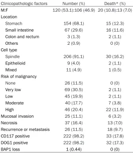
Clinicopathologic factors Number (%) Death* (%)
M:F 120 (53.1):106 (46.9) 20 (10.8):13 (7.0)
Location
Stomach 154 (68.1) 15 (12.3)
Small intestine 67 (29.6) 16 (11.6)
Colon and rectum 3 (1.3) 2 (1.1)
Others 2 (0.9) 0 (0)
Cell type
Spindle 206 (91.1) 30 (16.2)
Epithelioid 9 (4.0) 2 (1.1)
Mixed 11 (4.9) 1 (0.5)
Risk of malignancy
None 26 (11.5) 0 (0)
Very low 69 (30.5) 2 (1.1)
Low 45 (19.9) 2 (1.1)
Moderate 40 (17.7) 7 (3.8)
High 46 (20.4) 22 (11.9)
Mucosal invasion 25 (11.1) 6 (3.2)
Necrosis 37 (16.4) 13 (7.0)
Recurrence or metastasis 26 (11.5) 18 (9.7)
CD117 positive 222 (98.2) 33 (17.8)
DOG1 positive 222 (98.2) 32 (17.3)
BAP1 loss 1 (0.44) 0 (0)
*Available follow-up: 185 cases.
and arrayed in a tissue microarray (TMA) block. One core from breast carcinoma, thyroid papillary carci-noma, normal gastric mucosa, palatine tonsil, or uterine leiomyo-ma was obtained and arrayed in a TMA block, and used as control
tissues. We made total 11 TMA
blocks with 226 cases.
Immunohistochemistry
Immunohistochemistry for BAP1
was performed on an automated
Benchmark® platform (Ventana Medical Systems, Tucson, AZ,
USA). For BAP1, a mouse monoclo -nal antibody (clone C-4, Santa
Cruz Biotechnology, Dallas, TX,
USA) was used at a 1:100 dilution after onboard heat-induced epit-ope retrieval in a high-pH CC1 buf-fer (99°C, 1 h). Staining was
visual-ized using the UltraView™ univer-
sal DAB detection kit (Automa-ted BenchMark®, Ventana), which included a hydrogen peroxide
sub-strate and a
[image:2.612.91.356.85.389.2]ate the significance of the prognostic factors. Variables with significant result in the univari
-ate analysis were analyzed in the multivari-ate Cox proportional hazard regression analysis. Hazard ratios (HR) and associated 95% confi -dence intervals (CI) were calculated for each variable. All comparisons were performed using SPSS, version 23.0 (SPSS Inc., Chicago, IL, USA). A p<.05 was considered to indicate
sta-tistical significance.
Results
Clinicopathologic characteristics
One hundred twenty-six male and 120 female patients with a median age of 58.5 years (range: 22-88 years) were included in this study.
The median tumor size was 4.79 cm (range:
[image:3.612.91.526.73.236.2]1-23 cm). CD117 and DOG1 expression were detected in 222 cases (98.2%), respectively. Figure 1. BRCA1-associated protein-1 (BAP1) expression in GIST. (A) loss of BAP1 expression (B) retained BAP1
[image:3.612.91.522.311.423.2]expression (× 10).
Table 2. Univariate and multivariate analyses of clinicopathologic factors affecting the overall survival of patients with gastrointestinal stromal tumors
Variables β Univariate analysisHR (95% CI) p β Multivariate analysisHR (95% CI) p
Small intestine vs stomach .715 2.044 (0.998-4.183) .050 -.719 0.487 (0.204-1.160) .104
Colorectum vs stomach 1.874 6.517 (1.471-28.860) .014 -.079 0.924 (0.159-5.356) .924
Other location vs stomach -10,245 0.000 .978 -12.946 0.000 .979
Moderate risk vs low risk 2.330 10.281 (2.975-35.531) <0.0001 2.340 8.199 (2.308-29.123) .001
High risk vs low risk 3,043 20.974 (7.187-61.209) <0.0001 2.640 12,231 (3.692-40.525) <0.0001
Necrosis 1.549 4.705 (2.288-9.679) <0.0001 .146 1.157 (0.484-2.768) .743
Recurrence or metastasis 2.246 9.448 (4.700-18.995) <0.0001 .945 2.573 (1.163-5.692) .020
HR: hazard ratio; CI: confidence interval.
Table 3. Univariate and multivariate analyses of clinicopathologic factors affecting the disease-free survival of patients with gastrointestinal stromal tumors
Variables Univariate analysis Multivariate analysis
β HR (95% CI) p β HR (95% CI) p
Small intestine vs stomach .903 2.467 (1.218-4.997) .012 -.484 0.616 (0.269-1.414) .254
Colorectum vs stomach 1.930 6.888 (1.559-30.433) .011 -.493 0.611 (0.106-3.522) .581
Other location vs stomach -10.167 0.000 .982 -12.320 0.000 .979
Moderate risk vs low risk 2.244 9.432 (2.715-32.761) <0.0001 2.021 7.550 (2.095-27.202) .002
High risk vs low risk 3.323 27.751 (9.433-81.636) <0.0001 2.568 13.038 (3.886-43.743) <0.0001
Necrosis 1.507 4.513 (2.199-9.264) <0.0001 -.126 1.157 (0.484-2.768) .743
[image:3.612.92.523.480.593.2]SDHB negativity was observed only in two
wild-type gastric GISTs in a 56-year-old female and
15-year-old male patient. These SDHB-negative
GIST sexhibited diffuse strong positive CD117 and DOG1 staining.
Immunohistochemistry of BAP1 and its clinical significance
Of the 226 GISTs, only one case (0.44%)
exhib-ited a loss of BAP1 expression (Figure 1A). All other cases exhibited diffuse homogeneous
BAP1 positivity (Figure 1B). We next stained a
whole section from a representative tumor
block to confirm the loss of BAP1 expression,
and observed that the tumor cells were
com-pletely negative for BAP1. Statistically, we ob-served no significance associations between loss of BAP1 expression and clinicopathologic
factors. In univariate analysis, small intestinal and colorectal GISTs, GISTs with necrosis, re- currence/metastasis, or a higher risk of
malig-nancy were significantly associated with a poor OS and DFS. In a multivariate analysis, GISTs
with a higher risk of malignancy and
recur-rence/metastasis were confirmed as indepen -dent prognostic factors (Tables 2, 3).
Discussion
In a study of 226 GISTs cases, a loss of BAP1
expression was observed in only one small intestinal GIST. The indicated patient was a 33-years-old man without a family or personal
history of BAP1-associated malignancy. The tu-mor was a 9 cm × 5.5 cm-sized mass with mu-cosal ulceration, 1/50 high-powered field mito -sis rate, spindle cell histology, diffuse strong CD117 and DOG1 positivity, and KIT mutation. Irrespective of imatinib therapy, the tumor
metastasized to abdominal wall (12 months
later) and brain (90 months later), but no other malignancy was not detected for follow-up
peri-od. Recently, germline mutation in BAP1 have
been reported in causing a hereditary tumor syndrome that gives an increased risk of
can-cers [15]. Although we did not perform BAP1
germline mutation test for this case, he was
unlikely BAP1-associated hereditary syndrome because he did not have BAP1-related family history or BAP1-associated malignancy.
Studies of BAP1 expression have increased since the first report of the diagnostic utility of
this marker for mesothelial lesions. A loss of
BAP1 expression was found to be 100% spe
-cific for malignant mesothelioma and could be
used to distinguish malignant mesothelioma from benign mesothelial proliferation [9, 16]. However, studies to determine the clinical
sig-nificance of BAP1 expression in several human
tumors has yielded varied results. A
meta-anal-ysis of BAP1 expression in cancers revealed that 1) BAP1 is generally a poor prognostic
marker for cancers, except mesothelioma; 2) BAP1 mutations are associated with high-grade colorectal and renal cell carcinomas; and 3) BAP1-mutated cancers more commonly occur in women than in men [6]. In contrast,
meso-theliomas with loss of BAP1 expression showed a better outcome compared to those with BAP1
expression, and this association was notable among epithelioid cases [13]. In lung cancer,
Fan observed a 52.5% rate of BAP1 loss among
advanced cases, and this loss was associated with lymph node metastasis and poor OS [17].
However, two other studies identified a loss of BAP1 expression in only one case of primary
non-small cell lung cancer (1/101 and 1/257
cases), and suggested that BAP1 could be used
to distinguish primary lung carcinoma from ma- lignant mesothelioma [13, 18, 19]. In gastric
carcinoma, decreased BAP1 expression was
associated with a higher histologic grade, TNM stage, metastasis, and reduced OS [20]. Simi- larly, in clear cell renal cell carcinoma, a loss of
BAP1 was associated with a larger tumor size,
higher TNM stage, higher nuclear grade, metas-tasis, and reduced OS [12, 21]. In two studies of intrahepatic cholangiocarcinoma (ICC), Misu-
mi observed that a loss of BAP1, an indepen -dent prognostic factor, was associated with mass-forming small duct type ICC, reduced perineural invasion and mucin production [22]. In cholangiocarcinoma, BAP1 mutation was found to be correlate with aggressive disease and poor responses to standard therapies [23]. However, recent studies have revealed rare
losses of BAP1 in lung and other human can
-cers. A loss of BAP1 expression was observed
in only one of 306 cases of pancreatic ductal adenocarcinoma, despite the close anatomical proximity and similarities of this type of cancer (e.g., aspects of embryogenesis) with the bile
duct [24]. These findings suggest that a loss of BAP1 expression could be used to distinguish a
diagnosis of pancreatic ductal
adenocarcino-ma from ICC. Furthermore, a loss of BAP1
gynecological serous adenocarcinomas and could facilitate a differential diagnosis of abdo- minal mesothelioma [13].
A loss of BAP1 expression has shown to be 100% specific for malignancy. However, Hwang observed that losses of BAP1 were observed in
15% of sarcomatous/desmoplastic
mesotheli-omas. In contrast, no BAP1 loss was observed in sarcomatoid carcinoma [25]. These findings and our study results suggest that BAP1 loss
might not assist a differential diagnosis in a small biopsy that includes KIT (-) GIST, sarco-matoid carcinoma, and sarcosarco-matoid/desmo- sarcomatoid/desmo-plastic mesothelioma. The germline and soma- tic mutation of BAP1 is considered uncommon event in GIST, since most BAP1 mutation is highly associated with loss of protein expres- sion.
In summary, we conducted the first study of BAP1 expression status in a large cohort of GISTs. The loss of BAP1 expression was
ob-served in only one case, and it suggests that
BAP1 expression loss is a very rare event in
GIST and BAP1 would not play a major role in pathogenesis of GIST. The risk of malignancy
and recurrence/metastasis were confirmed as
independent prognostic factors for GIST. Acknowledgements
This work was supported by the 2016 Yeung- nam University Research Grant.
Disclosure of conflict of interest
None.
Address correspondence to: Dr. Mi Jin Gu, Depart- ment of Pathology, Yeungnam University College of Medicine, 170 Hyeonchung-ro, Nam-gu, Daegu
42415, South Korea. Tel: 82-53-640-6756; Fax:
82-53-622-8432; E-mail: mjgu@yu.ac.kr
References
[1] Fletcher CD, Berman JJ, Corless C, Gorstein F, Lasota J, Longley BJ, Miettinen M, O’Leary TJ, Remotti H, Rubin BP, Shmookler B, Sobin LH and Weiss SW. Diagnosis of gastrointestinal
stromal tumors: a consensus approach. Hum Pathol 2002; 33: 459-465.
[2] Hornick JL and Fletcher CD. The role of KIT in
the management of patients with gastrointes-tinal stromal tumors. Hum Pathol 2007; 38: 679-687.
[3] Ventii KH, Devi NS, Friedrich KL, Chernova TA, Tighiouart M, Van Meir EG and Wilkinson KD. BRCA1-associated protein-1 is a tumor sup
-pressor that requires deubiquitinating activity and nuclear localization. Cancer Res 2008;
68: 6953-6962.
[4] Murali R, Wilmott JS, Jakrot V, Al-Ahmadie H, Wiesner T, McCarthy SW, Thompson JF and Scolyer RA. BAP1 expression in cutaneous
melanoma: a pilot study. Pathology 2013; 45: 606-609.
[5] Wadt KA, Aoude LG, Johansson P, Solinas A,
Pritchard A, Crainic O, Andersen MT, Kiilgaard
JF, Heegaard S, Sunde L, Federspiel B, Madore J, Thompson JF, McCarthy SW, Goodwin A, Tsao H, Jonsson G, Busam K, Gupta R, Trent JM, Gerdes AM, Brown KM, Scolyer RA and Hay
-ward NK. A recurrent germline BAP1 mutation and extension of the BAP1 tumor predisposi -tion spectrum to include basal cell carcinoma. Clin Genet 2015; 88: 267-272.
[6] Luchini C, Veronese N, Yachida S, Cheng L,
Nottegar A, Stubbs B, Solmi M, Capelli P, Pea A, Barbareschi M, Fassan M, Wood LD and Scar -pa A. Different prognostic roles of tumor
sup-pressor gene BAP1 in cancer: a systematic re -view with meta-analysis. Genes Chromosomes Cancer 2016; 55: 741-749.
[7] Bott M, Brevet M, Taylor BS, Shimizu S, Ito T, Wang L, Creaney J, Lake RA, Zakowski MF, Reva B, Sander C, Delsite R, Powell S, Zhou Q,
Shen R, Olshen A, Rusch V and Ladanyi M. The
nuclear deubiquitinase BAP1 is commonly in -activated by somatic mutations and 3p21.1 losses in malignant pleural mesothelioma. Nat Genet 2011; 43: 668-672.
[8] Gossage L, Murtaza M, Slatter AF, Lichtenstein CP, Warren A, Haynes B, Marass F, Roberts I,
Shanahan SJ, Claas A, Dunham A, May AP,
Rosenfeld N, Forshew T and Eisen T. Clinical and pathological impact of VHL, PBRM1, BAP1,
SETD2, KDM6A, and JARID1c in clear cell re-nal cell carcinoma. Genes Chromosomes Can-cer 2014; 53: 38-51.
[9] Farzin M, Toon CW, Clarkson A, Sioson L, Wat -son N, Andrici J and Gill AJ. Loss of expression
of BAP1 predicts longer survival in mesothelio -ma. Pathology 2015; 47: 302-307.
[10] Yoshikawa Y, Sato A, Tsujimura T, Emi M,
Mori-naga T, Fukuoka K, Yamada S, Murakami A, Kondo N, Matsumoto S, Okumura Y, Tanaka F,
Hasegawa S, Nakano T and
Hashimoto-Tamao-ki T. Frequent inactivation of the BAP1 gene in
epithelioid-type malignant mesothelioma. Can-cer Sci 2012; 103: 868-874.
[11] Shen C, Wang Y, Wei P and Du X. BRCA1-asso
-ciated protein 1 deficiency in lung adenocarci -noma predicts poor outcome and increased
[12] Minardi D, Lucarini G, Milanese G, Di Primio R,
Montironi R and Muzzonigro G. Loss of nuclear BAP1 protein expression is a marker of poor
prognosis in patients with clear cell renal cell carcinoma. Urol Oncol 2016; 34: 338.e18. [13] Andrici J, Jung J, Sheen A, D’Urso L, Sioson L,
Pickett J, Parkhill TR, Verdonk B, Wardell KL, Singh A, Clarkson A, Watson N, Toon CW and Gill AJ. Loss of BAP1 expression is very rare in
peritoneal and gynecologic serous adenocarci-nomas and can be useful in the differential di-agnosis with abdominal mesothelioma. Hum Pathol 2016; 51: 9-15.
[14] Demetri GD, von Mehren M, Antonescu CR,
De-Matteo RP, Ganjoo KN, Maki RG, Pisters PW, Raut CP, Riedel RF, Schuetze S, Sundar HM, Trent JC and Wayne JD. NCCN task force re -port: update on the management of patients with gastrointestinal stromal tumors. J Natl Compr Canc Netw 2010; 8 Suppl 2: 4.
[15] McDonnell KJ, Gallanis GT, Heller KA, Melas M, Idos GE, Culver JO, Martin SE, Peng DH and
Gruber SB. A novel BAP1 mutation is associat -ed with melanocytic neoplasms and thyroid cancer. Cancer Genet 2016; 209: 75-81. [16] M McGregor S, McElherne J, Minor A,
Keller-Ramey J, Dunning R, Husain AN, Vigneswaran
W, Fitzpatrick C and Krausz T. BAP1 immuno -histochemistry has limited prognostic utility as
a complement of CDKN2A (p16) fluorescence in situ hybridization in malignant pleural meso -thelioma. Hum Pathol 2017; 60: 86-94. [17] Fan LH, Tang LN, Yue L, Yang Y, Gao ZL and
Shen Z. BAP1 is a good prognostic factor in ad -vanced non-small cell lung cancer. Clin Invest Med 2012; 35: E182-9.
[18] Owen D, Sheffield BS, Ionescu D and Churg A. Loss of BRCA1-associated protein 1 (BAP1) ex -pression is rare in non-small cell lung cancer. Hum Pathol 2017; 60: 82-85.
[19] Andrici J, Jung J, Sheen A, D’Urso L, Sioson L,
Pickett J, Parkhill TR, Verdonk B, Wardell KL, Singh A, Clarkson A, Watson N, Toon CW and Gill AJ. Loss of BAP1 expression is very rare in
peritoneal and gynecologic serous adenocarci-nomas and can be useful in the differential di-agnosis with abdominal mesothelioma. Hum Pathol 2016; 51: 9-15.
[20] Yan S, He F, Luo R, Wu H, Huang M, Huang C, Li Y and Zhou Z. Decreased expression of
BRCA1-associated protein 1 predicts unfavorable
sur-vival in gastric adenocarcinoma. Tumour Biol
2016; 37: 6125-6133.
[21] Joseph RW, Kapur P, Serie DJ, Parasramka M, Ho TH, Cheville JC, Frenkel E, Parker AS and Brugarolas J. Clear cell renal cell carcinoma subtypes identified by BAP1 and PBRM1 ex -pression. J Urol 2016; 195: 180-187.
[22] Misumi K, Hayashi A, Shibahara J, Arita J,
Sakamoto Y, Hasegawa K, Kokudo N and Fu -kayama M. Intrahepatic cholangiocarcinoma
frequently shows loss of BAP1 and PBRM1 ex
-pression, and demonstrates specific clinico -pathological and genetic characteristics with
BAP1 loss. Histopathology 2017; 70: 766-774.
[23] Al-Shamsi H, Anand D, Shroff RT, Jain A, Zuo M,
Conrad C, Vauthey JN and Javle MM. BRCA-as -sociated protein 1 mutant cholangiocarcino-ma: an aggressive disease subtype. J Gastroin-test Oncol 2016; 7: 556-561.
[24] Tayao M, Andrici J, Farzin M, Clarkson A, Sio
-son L, Wat-son N, Chua TC, Sztynda T, Samra JS and Gill AJ. Loss of BAP1 expression is very
rare in pancreatic ductal adenocarcinoma. PLoS One 2016; 11: e0150338.
[25] Hwang HC, Pyott S, Rodriguez S, Cindric A, Carr
A, Michelsen C, Thompson K, Tse CH, Gown AM
and Churg A. BAP1 immunohistochemistry and p16 FISH in the diagnosis of sarcomatous and