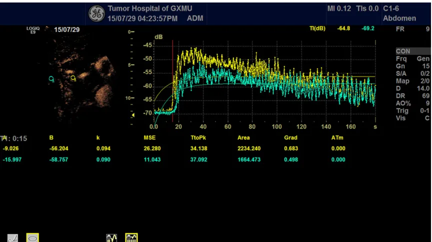
Original Article The clinical value of contrast-enhanced ultrasound and quantitative analysis parameters in the diagnosis and classification of portal vein tumor thrombus
Full text
Figure


Related documents
(PI-RADSv2): Prostate imaging reporting and data system version 2; AUC: Area under the curve; CEUS: Contrast - enhanced ultrasound; CUDI: Contrast ultrasound dispersion imaging;
Cite this article as: Gheonea et al.: Quantitative low mechanical index contrast-enhanced endoscopic ultrasound for the differential diagnosis of chronic pseudotumoral pancreatitis
Abstract: Purpose: This study aimed to determine the role of breast invasive ductal cancer (BIDC) size measured with Contrast-enhanced Ultrasound (CEUS) in the prediction of
In recent years, with the development of con- trast-enhanced ultrasound techniques, con- trast-enhanced ultrasound (CEUS)-guided lung biopsies can now clearly show the blood supply
Between different imaging techniques with contrast-enhancement, contrast- enhanced ultrasound (CEUS) and, in particular, dynamic CEUS have arisen as a promising and
According to the EFSUMB guidelines on the nonhepatic use of contrast‐enhanced ultrasound (CEUS), this method is useful to improve characterization of ductal
Figure 2 HE staining of rabbit portal vein VX2 tumor thrombus (Figure 2A), PVTT VEGF protein expression in control group, post-treatment Endostar and saline group (×400) (Figure 2B,
AIM: To analyze contrast-enhanced ultrasound (CEUS) features of histologically proven hepatic epithelioid hemangioendothelioma (HEHE) in comparison to other multilocular
