Original Article
Effects of edaravone treatment on noise-induced
hearing loss
Yi-Deng Huang1,2*, Long-Jun Wu3*, Si-Wen Xia2, Yang Zhuo2, Geng-Tian Liang3, Ning Yu1, Wei-Wei Guo1, Shi-Ming Yang1, Dong-Yi Han1
1Department of Otolaryngology-Head and Neck Surgery, Otolaryngology Institute of PLA, General Hospital of PLA,
Beijing 100853, China; 2Department of Otolaryngology, 118th Hospital of PLA, Wenzhou 325000, Zhejiang, China; 3Department of Otorhinolaryngology, The Third Hospital of Wuhan, Wuhan 430000, Hubei, China. *Equal
contribu-tors and co-first authors.
Received December 25, 2015; Accepted March 18, 2016; Epub June 15, 2016; Published June 30, 2016
Abstract: This study investigated the effects of edaravone on the cochlear hair cells of guinea pigs after noise-in-duced hearing loss and the best time for drug administration. Thirty clean-grade guinea pigs were randomly divided into three groups, and auditory brainstem responses (ABRs) were measured before, instantly after, and on the 3rd
and 7th day after noise exposure. On the 8th day after noise exposure, succinate dehydrogenase
immunohistochem-istry and scanning electron microscopy (SEM) were performed. The ABR threshold of the group that was adminis-tered edaravone immediately after the noise exposure was lower compared with that of the saline group (P<0.05). Three days later, the ABR threshold in the group was decreased more. Seven days later, it was similar to the ABR of
the normal control group, while the ABR of the saline group was significantly higher than that of the normal control
group (P<0.05). Light microscopy revealed that the cytoplasm of the outer hair cells was more intensely stained in the edaravone group compared to the saline group. The cellular morphology, immunohistochemistry, and SEM
analyses showed that the edaravone group exhibited significantly less nuclei and cilia deficits than the saline group did. The early intraperitoneal injection of edaravone had beneficial effects on the cochlear hair cells of guinea pigs
after noise exposure injury. Edaravone was effective when administered within 1 week of the noise exposure, but it was most effective when it was administered within 3 days of the noise exposure.
Keywords: Edaravone, noise-induced hearing loss, cochlea
Introduction
Noise injury to cochlear hair cells is a common cause of deafness. After noise exposure, reac-tive oxygen and reacreac-tive nitrogen contents, which reflect oxidative stress, are significantly increased in the cochlea, and the oxidative stress results in cochlear damage [1, 2]. Capaccio et al. found that oxidative stress damaged the capillaries of the inner ear tis-sues and that the cochlear ischemia and hypox-ia increased the production of free radicals, thus forming a vicious cycle that resulted in hearing loss [3].
Currently, the main antioxidants that are used clinically are edaravone, glutathione, and ascor-bic acid [4]. Edaravone is a new and very effec-tive anti-free radical scavenger that has
lipo-philic groups. Edavarone has been used in a number of neurological applications, and it exhibits significant protective effects on free radical-induced nerve cell damage [5].
sure in order to explore the most effective time for edaravone treatment on cochlear injuries and provide a theoretical foundation for the clinical treatment of noise-induced hearing loss.
Materials and methods
Animal grouping
Thirty albino guinea pigs (weighing 200-250 g, specific pathogen-free grade; Vital River Laboratory Animal Technology Co., Ltd., Beijing, China) with normal ear reflections were ran-domly divided into the following three groups: normal control group, saline group, and vone group, with 10 in each group. The edara-vone group was administered intraperitoneal injections of edaravone solution (8.0 mg/kg; Sinopharm Guorui Pharmaceutical Co., Ltd., Beijing, China) once a day within 30 min to 1 week after the noise exposure, and the saline group was injected with saline solution (5.4 mL/kg) at the same time points. The control group did not have noise exposure or intraperi-toneal injections. This study was carried out in strict accordance with the recommendations in the Guide for the Care and Use of Laboratory Animals of the National Institutes of Health. The animal use protocol has been reviewed and approved by the Institutional Animal Care and Use Committee (IACUC) of General Hospital of PLA. All of the surgeries on the albino guinea pigs were performed while the pigs were anes-thetized with 4.5% chloral hydrate (0.4 mL/100 g, intraperitoneal injection), and all efforts were made to minimize suffering.
Noise exposure
The animals in the saline and edaravone groups were caged individually. When the animals were
The normal control group was reared in a quiet environment without noise exposure.
Hearing detection
The auditory brainstem response (ABR) thresh-old was set as the observation indicator. The recording electrode was implanted under the scalp at the middle point of the connecting line of the biauricular front edges so that it contact-ed the skull. The reference electrode and grounding electrode with 1-kΩ resistances were placed in both earlobes. The stimulus was a wideband short click, with a pulse width of 0.1 ms. The scanning process was 10 ms for 1,024 superimposition times, and the sound stimulus intensity when the most stable III wave disappeared was set as the auditory reaction threshold of the ear. While anesthetized, the biauricular ABR thresholds of each group were tested and recorded four times [before expo-sure, immediately after exposure (30 min), and on the 3rd and 7th days after noise exposure].
Succinate dehydrogenase (SDH) staining
dye at 37°C in the dark for 1 h, which was fol-lowed by fixation in 4% formaldehyde solution at room temperature for 4 h. The cochlear basi-lar membrane was then isolated in 0.01 M PBS and spread in segments onto glass slides for glycerol mounting and optical microscopy examination.
Fluorescence staining of the cochlear hair cells
On the 8th day after noise exposure, 3 guinea pigs in each group were randomly selected and decapitated while they were anesthetized. The cochlea was then quickly removed, and the cochlear duct was perfused with 4% parafor-maldehyde solution and fixed overnight at 4°C. The cochlear basilar membrane was then iso-lated in 0.01 M PBS and washed with 0.01 M PBS three times for 10 min each. The mem-brane was incubated in phalloidin (P5282; Sigma-Aldrich Co. LLC, St. Louis, MO, USA) for 30 min in order to label the hair cell cilia and
epidermal plates, and the nuclear DNA fluores-cent stain 4’,6-diamidino-2-phenylindole (DAPI) was used to restain the nuclei for 10 min, which was followed by glycerin mounting and confocal microscopy examination.
SEM
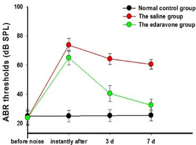
[image:3.612.90.286.70.215.2]The 2 remaining guinea pigs in each group were anesthetized, and the cochlea was removed. The cochlea was fixed overnight in a 2.5% glu-taraldehyde solution at 4°C. Subsequently, the cochlea was incubated in 10% ethylene diamine tetraacetic acid for full decalcification, and osmic acid was used for fixation. The cochlea was visualized under the microscope in order to remove the bony labyrinth, membranous laby-rinth, vestibular membrane, and covering film, and electric conduction staining and isoamyl Figure 1. ABR thresholds of the three groups before
the noise exposure, the ABR thresholds of the 3
groups had no significant difference, indicating that
the intergroup conditions of guinea pigs were of the same (P>0.05), the ABR thresholds of the Edaravone group and the saline group after the noise exposure were higher than the normal control group, with the
statistically significant difference (P<0.05), while that of the edaravone group was lower than the
sa-line group, and the difference was statistically signifi -cant (P<0.05); 3 d after the noise, the ABR threshold of the edaravone group further reduced, and 7 days later the ABR threshold recovered near to the
nor-mal, while that of the saline group was significantly
higher than the control group, with the statistically
significant difference (P<0.05). In addition, the ABR thresholds 7 of the edaravone group, measured im-mediately after the noise exposure and 7 days after
[image:3.612.323.523.73.351.2]the exposure showed the statistically significant dif -ference (P<0.05).
Tokyo, Japan), gilding (E-102 vacuum ion plat-ing apparatus), and SEM observation (Hitachi S-4800; Hitachi, Ltd.).
Statistical analysis
SPSS 13.0 (IBM Corporation, Armonk, NY, USA) was used for the statistical analysis. The fig-ures and charts were created with Sigma Plot10.0 software (Systat Software, Inc., San Jose, CA, USA) for the analysis. Tests of the homogeneity of the variance were performed first in each group. If the homogeneity of vari-ance was met, the intergroup mean values were then compared with a univariate analysis of variance (ANOVA) and t-test; if not, a non-parametric test was performed. The rates were tested with the Kruskal-Wallis rank sum test. If the difference was significant, the Nemenyi test was used to compare pairs of samples. P val-ues less than 0.05 were considered statistical-ly significant.
Results
Changes in the ABR threshold
Before noise exposure, the ABR thresholds in the guinea pigs in the 3 groups were in the nor-mal range. The ABR thresholds in the edara-vone group and saline group were examined after the last intraperitoneal injection was com-pleted, and they were increased. Three days after the noise exposure, the ABR thresholds in the edaravone group and saline group were partially recovered. After 7 days, the ABR threshold in the edaravone group was normal-ized, while the ABR in the saline group was still increased (Figure 1). These findings indicated that the first week after noise exposure was an important stage in ABR threshold recovery, and
With visualization under an ordinary optical microscope (Olympus BX51, Olympus Corpo- ration, Tokyo, Japan), the mitochondrial SDH staining area and staining intensity of the hair cells of the organ of Corti were examined in the cochlear basal turn in the 3 groups. The same parts of the organ of Corti in the cochlear basal turn were observed (Figure 2).
SEM results
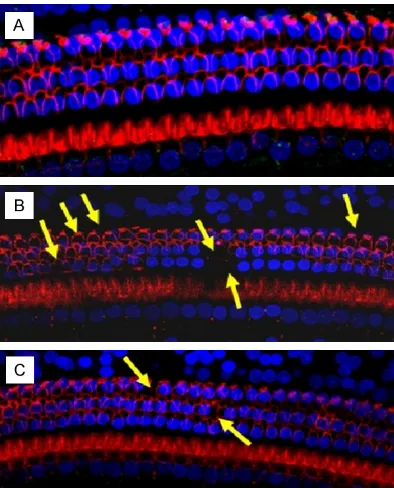
The hair cells of the acoustic papilla in the guin-ea pigs in the 3 groups were examined with SEM (Table 1). The hair cell cilia in the normal control group were arranged in an orderly pat-tern with a clear structure. After noise expo-sure, the hair cell cilia were missing, disordered, or dislodged in the edaravone group and saline group. In the saline group, the sporadic absence of lateral ciliated cells predominated. Compared to the saline control group, there were signifi-cantly less scattered and missing hair cells in the edaravone group (Figure 3).
Morphological changes in the cochlear hair cells
[image:4.612.86.528.89.153.2]signifi-cantly reduced compared to that in the saline group.
Discussion
Noise is a sound that involves erratic changes in frequency and intensity. Noise-induced hear-ing loss, which is one of the most serious occu-pational hazards worldwide [9], is thought to mainly result from mechanical and metabolic damages, such as ischemia and hypoxia, which both produce large amounts of reactive oxygen species (ROS) inside the cochlear hair cells. In addition, the overload of intracellular Ca2+ pro-motes the generation of more free oxygen radi-cals, which results in structural dysfunction of
[image:5.612.89.289.69.375.2]the cochlea and hair cells, which eventually leads to hearing loss [10]. In this study, the ABR thresholds of the guinea pigs in the 3 groups before the noise exposure, right after the noise exposure, and 3 and 7 days after the noise exposure were measured in order to determine the effects of edaravone on noise-induced hearing loss. The results showed that there was no significant difference in the ABR thresh-olds among the 3 groups, but the saline group and edaravone group exhibited increased ABR after noise exposure compared to the control group. The noise was more than 40 dB, which indicated that the noise damaged the cochlear outer hair cells of the guinea pig in only 3 days, but the ABR threshold in the edaravone group was significantly lower than that in the saline group. We therefore inferred that edaravone had protective effects on the cochlea. The ABR threshold in the edaravone group was decreased compared with that in the saline group, and it became almost normal 7 days after noise exposure.
Figure 3. SEM of cochlear organ of Corti. A. The nor-mal control group, the outer and inner hair cells were arranged in neat rows, the cilia of outer hair cells exhibited the V-shaped arrangement, while those of inner hair cells showed line-shaped arrangement; B. The saline group, the cilia of outer hair cells of organ of Corti in the cochlear basal turn exhibited
the flaky missing (shown by the yellow arrow), while
those of inner hair cells were relatively complete. C. The edaravone group, the outer hair cells were neatly arranged, exhibiting the scattered deletion of
cellu-lar cilia, while significantly reduced than the saline
[image:5.612.325.522.70.314.2]group.
The mechanisms underlying the protective effects of edaravone on the cochlea after noise exposure may be as follows. Within a short peri-od after noise exposure, a large number of free radicals appear in the mitochondria in cochlear cells. The free radicals damage the normal redox equilibrium state of the cochlea, which results in mitochondrial dysfunction and cell disintegration and death [14]. Edaravone exists in anion form in the body, and it directly clears the free radicals around the mitochondria in order to protect the cells. Edavarone transfers an electron to the free radical, thus generating an edaravone group that interrupts the lipid peroxidation chain. Edaravone reduces the oxi-dative damage of mitochondrial DNA by free radicals, protects the normal functions of the respiratory chain, and improves the mitochon-drial energy supply and antioxidant effects, thus resulting in protection of the cells. Edaravone inhibits the release of mitochondrial cytochrome C, thus blocking the JNK signaling pathway and protecting the nerve cells [15]. Specifically, edaravone blocked the shifting of BAX from the cytoplasm to the mitochondria, thereby blocking the release of cytochrome C from the mitochondria to the cytoplasm. Yoshida et al. found that edaravone downregu-lated the expression of mRNA in liver cells, upregulated the expression of BCL-2 protein, and stabilized the mRNA expression in liver cells to inhibit cellular denaturation and degen-eration [16]. Recent studies have shown that edaravone directly scavenges free radicals and significantly reduces the serum levels of inflam-matory cytokines [17-19]. These results sug-gested that edaravone ameliorates cochlear metabolic disorder, scavenges free radicals, reduces late-phase threshold shifting extents, and reduces the permanent threshold shifting in guinea pigs after noise exposure.
intracellular ROS seriously damage mitochon-dria. Thus, the SDH staining inside the hair cells will be weakened, or it will not be seen. This is consistent with Hu et al.’s results [20].
SEM further confirmed the cochlear microstruc-ture changes in the guinea pigs by showing that the noise-induced hair cell damage was mainly in the cochlear basal turn. In the normal control group, the outer and inner hair cells of the acoustic papilla were arranged in neat rows, the cilia of the outer hair cells exhibited a V-shaped arrangement, and the cilia of the inner hair cells showed a line-shaped arrange-ment. In the saline group, the cilia of the outer hair cells in the acoustic papilla of the basal turn exhibited sporadic absences, while those in edaravone group were neatly arranged with sporadically missing hair cellular cilia.
Noise-induced hearing loss involves a complex process that has multiple mechanisms and stages, and oxidative stress plays an important role in cochlear injuries. The noise directly dam-ages the cochlear structure through mechani-cal actions, and the secondary metabolic dam-age-induced oxygen free radicals destroy the cellular membrane structure. In addition, as an intracellular messenger, ROS activates signal-ing pathways and induces apoptosis, thus lead-ing to hearlead-ing loss. However, 7 days after noise exposure, the ABR threshold in the edaravone group still differed from that in the normal con-trol group. This finding indicated that there may be other factors in the noise that damage the cochlear structure.
Acknowledgements
Scien-tific Problem Oriented Project of National 973 Plan (2011cba01000), Surface Project of PLA “12th five-year” (CWSlIJ256), Project of Major Project Foundation, Nanjing Military Region (102006), Project of Major Project foundation, Nanjing Military Region (082007) and National Natural Science Foundation of China (8124- 1034, 81271081).
Disclosure of conflict of interest
None.
Address correspondence to: Shi-Ming Yang and Dong-Yi Han, Department of Otolaryngology-Head and Neck Surgery, Otolaryngology Institute of PLA, General Hospital of PLA, Beijing 100853, China. Tel: +86 10 88626138; Fax: +86 10 88626138; E-mail: shimingyangcn@126.com (SMY); dongyihanc@126. com (DYH)
References
[1] Xiong M, Wang J, Yang C and Lai H. The co-chlea magnesium content is negatively corre-lated with hearing loss induced by impulse noise. Am J Otolaryngol 2013; 34: 209-215. [2] Samson J, Wiktore-Smagur A, Politanski P,
Rajkowska E, Pawlaczyk-Luszczynska M, Duda- rewicz A, Sha SH, Schacht J and Sliwinska-Kowalska M. Noise-induced time-dependent changes in oxidative stress in the mouse co-chlea and attenuation by D-methionin. Neuro- science 2008; 152: 146-150.
[3] Capaccio P, Pignataro L, Gaini LM, Sigismund PE, Novembrino C, De Giuseppe R, Uva V, Tripodi A and Bamonti F. Unbalanced oxidative status in idiopathic sudden sensorineural hearing loss. Eur Arch Otorhinolaryngol 2012; 269: 449-453.
[4] Kamogawa E and Sueishi Y. A multiple free-radical scavenging (MULTIS) study on the anti-oxidant capacity of a neuroprotective drug, edaravone as compared with uric acid, gluta-thione, and trolox. Bioorg Med Chem Lett 2014; 24: 1376-1379.
[5] Kikuchi K, Takeshige N, Miura N, Morimoto Y, Ito T, Tancharoen S, Miyata K, Kikuchi C, Iida N, Uchikado H, Miyagi N, Shiomi N, Kuramoto T, Maruyama I, Morioka M and Kawahara KI.
Beyond free radical scavenging: Beneficial ef -fects of edaravone (Radicut) in various diseas-es (Review). Exp Ther Med 2012; 3: 3-8. [6] Gao G, Sun JJ, Gong SS, Liu Y and Jiang P.
Edaravone protects hearing from acute acous-tic trauma in guinea pigs. Zhonghua Er Bi Yan Hou Tou Jing Wai Ke Za Zhi 2009; 44: 150-153.
[7] Tanaka K, Takemoto T, Sugahara K, Okuda T, Mikuriya T, Takeno K, Hashimoto M, Shimogori H and Yamashita H. Post-exposure administra-tion of edaravone attenuates noise-induced hearing loss. Eur J Pharmacol 2005; 522: 116-121.
[8] Van Campen LE, Murphy WJ, Franks JR, Mathias PI and Toraason MA. Oxidative DNA damage is associated with intense noise expo-sure in the rat. Hear Res 2002; 164: 29-38. [9] Verbeek JH, Kateman E, Morata TC, Dreschler
WA and Mischke C. Interventions to prevent oc-cupational noise-induced hearing loss: A Cochrane systematic review. Int J Audiol 2014; 53: S84-96.
[10] Fetoni AR, De Bartolo P, Eramo SL, Rolesi R, Paciello F, Bergamini C, Fato R, Paludetti G, Petrosini L and Troiani D. Noise-Induced Hearing Loss (NIHL) as a Target of Oxidative Stress-Mediated Damage: Cochlear and Cortical Responses after an Increase in Antioxidant Defense. J Neurosci 2013; 33: 4011-4023.
[11] Takemoto T, Sugahara T, Okuda T, Shimogori H and Yamashita H. The clinical free radical scav-enger, edaravone, protects cochlear hair cells from acoustic trauma. Eur J Pharmacol 2004; 487: 113-116.
[12] Maekawa H, Matsunobu T, Tsuda H, Onozato K, Masuda Y, Tanabe T and Shiotani A. Therapeutic effect of edaravone on inner ear barotrauma in the guinea pig. Neurochem Int 2009; 54: 513-518.
[13] Asplund MS, Lidian A, Linder B, Takumida M and Anniko M. Protective effect of edaravone against tobramycin-induced ototoxicity. Acta Otolaryngol 2009; 129: 8-13.
[14] Böttger EC and Schacht J. The mitochondrion: a perpetrator of acquired hearing loss. Hear Res 2013; 303: 12-19.
[15] Fan J, Zhang N, Yin G, Zhang Z, Cheng G, Qian W, Long H and Cai W. Edaravone protects corti-cal neurons from apoptosis by inhibiting the translocation of BAX and Increasing the inter-action between 14-3-3 and p-BAD. Int J Neurosci 2012; 122: 665-674.
[16] Yoshida H, Kwon AH, Kaibori M, Tsuji K, Habara K, Yamada M, Kamiyama Y, Nishizawa M, Ito S and Okumura T. Edaravone prevents iNOS ex-pression by inhibiting its promoter transactiva-tion and mRNA stability in cytokine-stimulated hepatocytes. Nitric Oxide 2008; 18: 105-112. [17] Isahaya K, Yamada K, Yamatoku M, Sakurai K,
Takaishi S, Kato B, Hirayama T and Hasegawa Y. Effects of edaravone, a free radical