Emma J. Rose, Ciara Greene, Sinead Kelly, Derek W. Morris, Ian H. Robertson, Ciara Fahey, Sarah Jacobson, John O’Doherty, Fiona N. Newell, Jane McGrath, Arun Bodke, Hugh Garavan, Thomas Frodl, Michael Gill, Aiden P. Corvin, Gary Donohoe
PII: S1053-8119(11)01456-X
DOI: doi:10.1016/j.neuroimage.2011.12.054 Reference: YNIMG 9031
To appear in: NeuroImage
Received date: 1 September 2011 Revised date: 16 December 2011 Accepted date: 19 December 2011
Please cite this article as: Rose, Emma J., Greene, Ciara, Kelly, Sinead, Morris, Derek W., Robertson, Ian H., Fahey, Ciara, Jacobson, Sarah, O’Doherty, John, Newell, Fiona N., McGrath, Jane, Bodke, Arun, Garavan, Hugh, Frodl, Thomas, Gill, Michael, Corvin, Aiden P., Donohoe, Gary, The NOS1 variant rs6490121 is associated with variation in prefrontal function and gray matter density in healthy individuals, NeuroImage(2011), doi: 10.1016/j.neuroimage.2011.12.054
ACCEPTED MANUSCRIPT
Page 1 of 34
The NOS1 variant rs6490121 is associated with variation in prefrontal function and gray matter density in healthy individuals.
Emma J. Rose1,2, Ciara Greene2, 3 Sinead Kelly1,2, Derek W. Morris1, Ian H.Robertson2, 3, Ciara Fahey1, Sarah Jacobson2, 3, John O’Doherty2, 3, Fiona N. Newell2,3, Jane McGrath1,2, Arun Bodke1,2, Hugh Garavan2,3, Thomas Frodl1,2, Michael Gill1,2, Aiden P. Corvin1, 2, Gary
Donohoe1, 2.
1: Neuropsychiatric Genetics Research Group & Institute of Molecular Medicine, Department of Psychiatry, Trinity College Dublin, Ireland.
2: Trinity College Institute for Neuroscience, Trinity College Dublin, Ireland.
3: School of Psychology, Trinity College Dublin, Ireland.
Corresponding Author: Dr Emma Jane Rose, Department of Psychiatry, Trinity Centre for Health Sciences, St. James’ Hospital, Dublin 8, Ireland. Tel/Fax: +353-1-896-2464/3405 email: rosee@tcd.ie
ACCEPTED MANUSCRIPT
Page 2 of 34 Abstract
A common polymorphism within the nitric oxide sythanse-1 (NOS1) gene
(rs6490121), initially identified as risk variant for schizophrenia, has been associated with
variation in working memory and IQ. Here we investigated how this variation might be
mediated at the level of brain structure and function. In healthy individuals (N=157), voxel
based morphometry was used to compare gray matter (GM) volume between homozygous
and heterozygous carriers of the ‘G’ allele (i.e. the allele associated with impaired cognition
and schizophrenia risk) and homozygous carriers of the non-risk ‘A’ allele. Functional brain
imaging data were also acquired from 48 participants during performance of a spatial
working memory (SWM) task, and analysed to determine any effect of NOS1 risk status. An
a priori region-of-interest analysis identified a significant reduction in ventromedial prefrontal GM volume in ‘G’ allele carriers. Risk carriers also exhibited altered patterns of
activation in the prefrontal cortex, caudate, and superior parietal lobe, which were
characteristic of abnormal increases in activation in frontoparietal working memory networks
and a failure to disengage regions of the default mode network. These functional changes suggest a NOS1-mediated processing inefficiency that may contribute to cognitive
dysfunction in schizophrenia. While the mechanisms by which NOS1 may influence brain
structure and/or function have not yet been well delineated, these data provide further
evidence for a role of NOS1 in risk for schizophrenia via an impact upon cognitive function.
ACCEPTED MANUSCRIPT
Page 3 of 34 1. Introduction
Neuronal nitric oxide synthase (nNOS) accounts for 90% of nitric oxide (NO) in the
central nervous system, production of which is dynamically controlled during development
and in response to brain injury (Khaldi et al., 2002). NO stimulates synthesis of cGMP,
strongly influences glutamate neurotransmission (Akyol et al., 2004; Brenman and Bredt,
1997), and is involved in uptake, release and storage of other CNS neurotransmitters
including acetylcholine, dopamine, noradrenaline, and GABA (Boehning and Snyder, 2003;
Pepicelli et al., 2004). The NO synthase–1 gene (NOS1; OMIM 163731), which maps to
chromosome 12q24 and encodes for nNOS, has been associated with several psychiatric
illness phenotypes, including anxiety, depression, and schizophrenia (SZ) (Luciano et al.,
2010; O'Donovan et al., 2008; Reif et al., 2009; Reif et al., 2010), yet evidence that NOS1
can explain illness risk is mixed. For example, following four positive NOS1 candidate gene
associations in five studies (DeLisi et al., 2002; Fallin et al., 2005; Liou et al., 2003; Shinkai
et al., 2002; Tang et al., 2008), a NOS1 single-nucleotide polymorphism (SNP) rs6490121
(located in intron 10; A/G polymorphism; minor allele is G) was identified by O’Donovan et
al (2008) in a GWAS study as initially showing the strongest statistical evidence of
association (p=9.82x10⁻⁶; risk allele ‘G’). However this association was not replicated in
subsequent SZ samples either in the O’Donovan et al study or other GWAS studies
(Stefansson et al., 2009).
In contrast, NOS1 is robustly associated with cognitive variation. NOS1 mouse
knockout models have repeatedly been associated with variance in cognition (Kirchner et al.,
2004; Weitzdoerfer et al., 2004). Notably, phencyclidine hydrochloride–induced cognitive
deficits that model SZ symptoms (e.g. pre-pulse inhibition, habituation of acoustic startle,
ACCEPTED MANUSCRIPT
Page 4 of 34 prevented by interfering with NO production (Johansson et al., 1997; Johansson et al., 1998;
Klamer et al., 2001, 2004a, 2005; Klamer et al., 2004b; Palsson et al., 2007; Wass et al.,
2006). In humans, Reif et al. (2006) reported that 2 of 4 genetic markers tested at the NOS1
locus were associated with variance in performance on measures of prefrontal function (i.e.
‘Go/No-Go’ paradigm). Similarly, two more recent investigations by the same group have
found associations between NOS1 polymorphisms, cognitive performance and prefrontal
brain function in patient with SZ (Reif et al., 2011) and healthy controls (Kopf et al., 2011).
In light of these findings and our own observation that the putative risk ‘G’ allele identified
by O’Donovan and colleagues at rs6490121 was associated with poorer performance in
measures of verbal intelligence and working memory in SZ patients and healthy controls
(Donohoe et al., 2009), we conclude that NOS1’s more modest association with psychiatric
risk may reflect the moderating effects of this gene’s broader role in cognition.
With the aim of increasing our understanding of the mechanisms by which putative
SZ risk variants in NOS1 may contribute to disease risk, this study considered the role of
NOS1 rs6490121 in brain structure and function. We provide evidence that the ‘G’ allele,
previously associated with lower verbal IQ and working memory, is associated with
decreased grey matter (GM) volumes and an altered pattern of activation in complementary
prefrontal cortical regions in healthy controls.
2. Materials and methods
2.1 Participants
ACCEPTED MANUSCRIPT
Page 5 of 34 through local media advertising. Participants provided written, informed consent, in
accordance with local ethics committee guidelines. Due to data quality issues (e.g. head
motion) and matching between genotype groups, 9 individuals were excluded. The
remaining 48 individuals were all included in fMRI analysis (15 male; mean age=27.37
years; Table 1). Exclusion criteria included relevant neurological, medical or psychiatric
history, family history of psychosis (i.e. one or more first degree-relative with a confirmed
diagnosis of SZ or other psychosis), claustrophobia, pregnancy, and any other
contraindication for MRI. Psychiatric history was confirmed prior to scanning using a
semi-structured interview, in which participants responded to questions regarding whether or not
they had ever seen a medical professional for a mental health issue or if they had ever been
prescribed a psychiatric medication.
2.1.2 Structural Imaging (sMRI): Individuals were selected from the Trinity College Institute for Neuroscience imaging biobank project, which involved the opportunistic
sampling of all healthy, right-handed controls participating in MRI studies at the Institute.
Participants gave consent to the use of structural data collected under the primary study and
provided a saliva sample for genetics analysis. From this sample, 157 individuals (71 male;
mean age=27.71 years) were included based upon the quality of T1 structural data and
successful genotyping of the NOS1 (rs6490121) variant. Forty six sMRI participants were
also included in fMRI analyses.
The majority of participants (N=120) were confirmed to be Irish. Lineage information
was not available for remaining participants (N=37). However, given the relative
homogeneity of the Irish population (e.g. 92% of Dublin city population are Irish/Caucasian;
www.cso.ie/census), the vast majority of these participants were also likely to have been of
Irish lineage. Furthermore, this subgroup did not differ from known Irish participants in terms
ACCEPTED MANUSCRIPT
Page 6 of 34 >> Table 1<<
2.2 Procedure
2.2.1 Genotyping: Genetics analysis was carried out using DNA obtained from saliva samples that were collected using Oragene DNA self-collection kits (DNA Genotek). The
rs6490121 SNP was genotyped using a Taqman® SNP Genotyping Assay on a 7900HT
Sequence Detection System (Applied Biosystems). The call rate for the Taqman genotyping
was >95% and the samples were in Hardy-Weinberg Equilibrium (p>0.05). In addition a
small number of HapMap CEU DNA samples (www.hapmap.org) were genotyped for
rs6490121 for quality control purposes and were all found to be concordant with available
online HapMap data for this SNP. Genotype frequencies for fMRI and sMRI are noted in
Table 1.
2.2.2 MRI: Participants were imaged on a Philips Intera Achieva 3T MR system. Whole-brain BOLD EPI consisting of 32 non-contiguous, axial 3.5mm slices was acquired
with a 0.35mm slice gap and the following imaging parameters: TR=2000ms; TE=35ms;
FOV=224x224mm at 64x64 matrix; and flip angle=90°. The duration of functional scanning
was 220TRs/440s. Structural imaging involved the acquisition of a T1-weighted image (180
slices; duration=6mins) using a TFE gradient echo pulse sequence, with a slice thickness of
0.9mm and 230x230mm FOV.
2.2.3 SWM paradigm (Figure 1): In this block-design task participants were asked to determine whether the spatial location, relative to a white fixation cross, of a white dot (i.e.
target) and a red circle (i.e. probe) were either the same (match) or different (no match). The
target and probe were each presented for 500ms. There were three levels/conditions in the
task: A. No Delay (baseline); B. 1-dot ; and C. 3-dot. In the ‘no delay’ condition the target
ACCEPTED MANUSCRIPT
Page 7 of 34 Conversely, during the 1-dot condition (Figure 1B) the target and probe were separated by a
3sec delay. Similarly, the 3-dot condition (Figure 1C) also incorporated a 3sec inter-stimulus
interval, however in this condition the target image was comprised of 3 dots. During the
3-dot condition the probe was also a single red circle and participants were asked to judge
whether the probe was in the same position as any one of the three dots in the image that
preceded it.
There were 6 trials per block with an inter-trial interval of 2sec. Participants
completed 4 blocks of each condition (i.e. 12 blocks/72 trials total). The order of blocks was
fixed, however to account for the potentially confounding effect of block order on brain
activity across the session, there were two predetermined block orders and participants were
pseudo-randomly allocated to one or the other prior to imaging.
Participants were given left- and right-hand MRI compatible response units. They
responded with a left button press for a ‘match’ and a right button press for ‘no match’.
While there were equal numbers of ‘match’ and ‘no match’ trials, 20% trials were defined as
‘difficult’; whereby the probe was in a similar (i.e. same quadrant) but different location to
the target. The number of difficult trials was consistent across task levels and between
participants. The definition of trials as ‘difficult’ was relative to other trials, and was not
based upon participant’s perceived difficulty of these trials.
Behavioural measures of interest on the SWM task were accuracy (i.e. number of
correctly identified trials) and reaction time (RT).
ACCEPTED MANUSCRIPT
Page 8 of 34 2.3 Data Analysis
2.3.1 fMRI: Functional imaging data were analyzed using Analysis of Functional Images (AFNI; Cox, 1996). To correct for head motion, 3D EPI data for each subject were
co-registered to a base volume. The subsequent data were inspected for motion using the
censor.py application for AFNI (http://brainimaging.waisman.wisc.edu/~perlman/code/censor.py),
using the following censoring criteria: translation>0.3mm or rotation>0.3° between
consecutive TRs. TRs exceeding these parameters were recorded and were removed from
further analysis prior to deconvolution. Individuals with a censor rate >25% of TRs were
excluded from analysis (i.e. N=2). For the remaining participants (i.e. N=48) the mean
percentage of TRs censored was 3.29% (range: 0-21.82), which was equivalent to 7.25TRs
(range: 0-48). Almost 30% of participants required no censoring (i.e. 14 of 48), while 67%
had less than 25 TRs censored. The remaining subjects had 42 and 48 TRs censored each.
Visual inspection of censor plots indicated that those TRs that were censored were relatively
evenly distributed across the session and block types. Thus, we do not anticipate any
significant effect of censoring on the ability to accurately construct task regressors for those
subjects who were included in fMRI analysis. Data quality control also included the
application of an edge detection algorithm to exclude activations occurring outside the brain.
Voxel-wise multiple regression was conducted in which imaging regressors were
expressed as a delta function relative to baseline and convolved with a hemodynamic
response function. There were regressors representing the SWM conditions (1-dot and 3-dot)
and six motion parameters that were included as regressors of no interest. A voxel-wise
average amplitude change (β) equal to the percentage change from baseline was calculated
for each SWM condition. The resultant activation maps for each subject were the registered
ACCEPTED MANUSCRIPT
Page 9 of 34 Whole-brain mixed measures ANOVA were used to consider the impact of genotype
group and SWM condition (1-dot vs. 3-dot) on brain activity. In light of the relatively small
number of G homozygotes (N=7), imaging analyses focussed on the relative differences in
non-risk homozygous (i.e. AA) vs. risk carrying (i.e. AG & GG) individuals. These groups
were matched for age, gender, and years of education (Table 1).
A voxel-wise threshold correcting for multiple comparisons and controlling for
family-wise error (FWE) rate was calculated using a Monte Carlo simulation. This
deterministic sampling algorithm ascertains the frequency of significant clusters that would
occur by chance under the null hypothesis (i.e. the false positive rate). Cluster significance
was determined as equalling or exceeding a given extent threshold at pCORRECTED<0.05
following corrections for multiple comparisons at the whole brain level
Given previous findings suggesting an impact of the NOS1 genotype on mPFC and
cognitive processes that are mediated by function in prefrontal (PFC) and parietal cortices,
such as executive function, a priori region-of-interest (ROI) analyses were also carried out.
These analyses focussed on the impact of genotype on brain activity in medial PFC
(mPFC)/BA10, dorsolateral PFC (dlPFC)/BA9 & 46 and superior parietal cortex/BA7. These
regions were defined using a Talairach template in AFNI. The mean value across voxels
within each ROI was calculated for each participant/regressor and subsequently used as the
dependent variable in statistical analyses.
2.3.2 Voxel based morphometry (VBM; Ashburner and Friston, 2000): sMRI analysis was performed within SPM5 (http://www.fil.ion.ucl.ac.uk/spm) running under Matlab (v7.8;
The MathWorks) and utilising the VBM toolbox (v5.1; http://dbm.neuro.uni-hen.de/vbm).
Individual volumes were visually inspected for scanner artefacts and gross anatomical
ACCEPTED MANUSCRIPT
Page 10 of 34 WM and cerebrospinal fluid, without tissue priors and using a Hidden Markov Random Field
weighting of 0.15. Segmented images were normalised using the DARTEL toolbox
(Ashburner, 2007), in which GM and WM templates were created using standard parameters.
Jacobian scaled (‘modulated’) warped tissue classes were subsequently created for both GM
and WM for each subject. The resultant images were smoothed with an 8mm3 Gaussian kernel.
The statistical methods employed for sMRI analyses were specifically designed to
mimic those carried out for fMRI data. Mixed model ANOVA considering the impact of
NOS1 genotype (AA vs. AG/GG) on GM and WM density were conducted for both whole
brain and a priori ROI. The same regions were included as for the functional ROI analyses,
and were defined using the WFU pickatlas toolbox for SPM (Maldjian et al 2003). Although
the groups were essentially matched for age, gender, and years of education (Table 1), to
account for normal variation in brain structure, age, gender and total GM or WM volume
were included as co-variates. For these analyses, we included subject data as normalised to
native space (i.e. the DARTEL template) rather than a standard template, such as MNI.
Subjects for sMRI were sampled from a range of imaging studies that included individuals in
a range of developmental stage, including young, middle, and late adulthood, making
normalisation to a standard template inappropriate. As with fMRI analyses, significance
was defined as the voxel-wise threshold at FWE pCORRECTED<0.05 for whole-brain analyses or
for the entire volume of the mask in the case of ROI analyses. Since non-uniform
smoothness of VBM data can influence interpretation of these types of analysis (Ashburner
and Friston, 2000; Worsley et al., 1999), determination of significance included a
non-stationarity cluster extent correction, which utilized the random field theory version of cluster
inference under non-stationarity (Hayasaka et al., 2004) and was implemented using the NS
ACCEPTED MANUSCRIPT
Page 11 of 34
2.3.3 Other data: Behavioural and demographic data were analysed in SPSS (v16; SPSS Inc.).
3. Results
3. 1 SWM behavioural data
There was a significant main effect of SWM condition on participant accuracy (F(2,
90)=22.51, p<0.001). Although the average number of correct responses did not differ between the no-delay and 1-dot conditions, participants made significantly more errors on the
3-dot condition compared to the no-delay (t(46)=-5.34, p<0.001) and the 1-dot condition
(t(46)=- 6.77, p<0.001). In addition, there was a linear effect of task difficulty on reaction
time (RT; F(2,90)=18.19, p<0.001), such that as the task got more difficult RT increased.
There were no effects of NOS1 genotype on RT or accuracy, and no interactions between
genotype and SWM condition on either behavioural measure.
One potential issue here is that, due to the relatively small number of participants, our
study may have been underpowered to detect behavioural differences between the genotype
groups. Estimates of effect size (i.e. partial η2) indicated that any variability in accuracy that could be accounted for by genotype was negligible (i.e. genotype η2=0.001; genotype x SWM η2=0.005). Conversely, for RT estimates of effect size (i.e. genotype η2=0.02; genotype x SWM η2=0.04) suggest that there may be some small contribution of NOS1 genotype to variability in RT on measures of executive function. Overall, these data suggest that even at
much larger sample sizes it is unlikely that there would be any effect of genotype on accuracy
on this SWM measure, but that it might be possible to observe a small but significant effect
ACCEPTED MANUSCRIPT
Page 12 of 34 3.2 fMRI
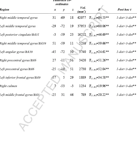
3.2.1 Main effect of SWM maintenance on brain activity (Table 2): SWM condition influenced brain activity bilaterally in two large clusters centred in the middle temporal gyrus
and which extended to occipital and parietal cortices, in the left angular gyrus/BA39,
posterior cingulate/BA31, inferior frontal gyrus/BA9 and middle frontal gyrus/BA8, in the
right culmen and the bilateral precentral gyrus/BA6. In the two primary clusters and in BA6
and BA9 this effect was result of a significantly greater increase in activation in response to
the 3-dot, vs. 1-dot, condition. Conversely, activity was lower in response to 3-dot trials in
posterior cingulate, middle frontal gyrus, and BA39. These latter regions all showed
comparatively reduced (i.e. relative to baseline) activation on 1-dot trials; an effect that was
even more pronounced for 3-dot trials. This pattern of activity is in keeping with theories of
frontoparietal activation and corresponding deactivation in regions constituting the default
mode network during executive function (Fox et al 2005; Toro et al 2008). Thus, we were
assured of the robustness of the SWM task.
>> Table 2<<
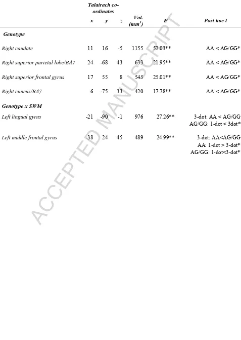
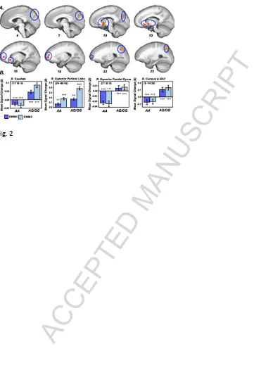
3.2.2 Main effect of NOS1 genotype (Figure 2; Table 3): Irrespective of SWM condition, NOS1 genotype impacted upon brain activity in the right caudate, superior parietal
lobe, superior frontal gyrus and cuneus/BA7 (pCORRECTED<0.05). In all significant clusters,
activation was greater in individuals who were carriers of the NOS1 risk ‘G’ allele (post hoc
t test: p<0.001).
>> Figure 2<<
ACCEPTED MANUSCRIPT
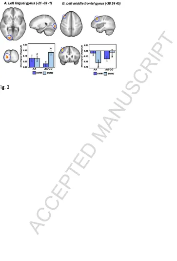
Page 13 of 34 frontal gyri. In the lingual gyrus activity in the 1-dot condition did not differ between
groups, however, activation in risk ‘G’ allele carriers was greater in response to 3-dot trials (
p<0.05). Moreover, risk carriers showed a main effect of working memory load (1-dot <
3-dot; p<0.001) that was absent in the risk homozygotes. In the middle frontal gyrus,
non-risk individuals exhibited a notable decrease in activity in response to 3-dot, but not 1-dot
trials (p<0.001), while opposite was true in risk carriers (i.e. 1-dot > 3-dot; p<0.001). In
addition the extent of deactivation in response to 3-dot trials was greater in non-risk
homozygotes vs. risk carrying individuals (p<0.05).
Post hoc one-sample t-tests confirmed that the extent of deactivation for risk carriers on 3-dot trials and non-risk individuals on 1-dot trials did not differ significantly from 0
(p>0.05).
3.2.4 A priori ROI results: Within those regions that were included in the region-of-interest analysis there was a main effect of SWM difficulty in dlPFC (i.e. BA46; 1-dot <
3-dot; p<0.05). There was also a main effect of genotype in BA10 and BA7, such that risk
carriers showed relatively greater activation compared to non-risk individuals in both regions,
irrespective of load (p<0.05).
>> Figure 3<<
>>Table 3<<
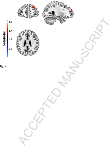
3.3 VBM
3.3.1 NOS1 genotype and GM (Figure 3): There were no regions of significantly different GM density between non-risk individuals and risk carriers that survived correction
ACCEPTED MANUSCRIPT
Page 14 of 34 The a priori ROI analysis, on the other hand, was indicative of a NOS1-mediated
influence on GM density in mPFC/ BA10, such that GM volume was relatively reduced in
risk carriers, vs. non-risk individuals, in a single cluster in the more lateral aspects of
ventromedial PFC (vmPFC; MNI co-ordinates: 19 61 26; KE=371 voxels; pCORRECTED<0.05;
Figure 4). There were no between-group differences in GM density in dlPFC or parietal
cortex.
>>Figure 4<<
3.3.2 NOS1 genotype and WM: Following correction for multiple comparisons, there were no regions of WM that were found to be sensitive to NOS1/rs6490121 genotype.
4. Discussion
In this investigation VBM and fMRI were used to determine the impact of the NOS1
variant rs6490121 on brain structure and function in healthy controls. Carriers of the SZ risk
‘G’ allele had comparatively reduced GM volume in lateral vmPFC, compared to individuals
who were homozygous for the non-risk ‘A’ allele. Similarly, risk carriers showed a pattern
of activation during performance of a SWM task which differentiated them from the non-risk
individuals. In a select range of areas in prefrontal and parietal cortices, including
BA10/vmPFC, risk carriers exhibited a load-independent increase in activity compared to
homozygous non-risk individuals. Furthermore, carriers of the risk ‘G’ allele also exhibited
load-dependent changes in activity in the lingual and middle frontal gyri, such that they failed
to disengage BA8 with increasing memory load and inappropriately increased activity in the
lingual gyrus as the task got more difficult.
ACCEPTED MANUSCRIPT
Page 15 of 34 4.1 Functional effects of NOS1
Whole brain and ROI analyses implicated NOS1/rs6490121 with load-independent
increases in activation in the mPFC/BA10. This supports prior observations showing that
activity related to cognitive tasks that involve mPFC are influenced by NOS1 genotype (Reif
et al., 2006). Functionally, the mPFC is one of the regions implicated in the so-called
‘default mode network’ (i.e. those regions that are involved in self-referential mental activity
and which show reduced activity during goal directed beahviour; Buckner et al., 2008;
Gusnard et al., 2001; Raichle et al., 2001). Similarly, of those regions where we noted
interactions between genotype and working memory load (i.e. lingual gyrus and middle
frontal gyrus/BA8), BA8 has also been shown to exhibit goal-related decreases in activation
(McKiernan et al. 2003). Risk-related variability in function also included increased activity
in BA7 and the caudate. The superior parietal region is a key component of the
frontoparietal network that supports executive function (Fox et al 2005), while the caudate is
thought to play an integral role in the cortico-striatal-cortical connections which facilitate
communication between frontal and parietal regions during working memory tasks (Joel and
Weiner 2000). Collectively, these data suggest that those who were carriers of the risk ‘G’
allele attained a level of performance equivalent to their non-risk peers via a combination of
over-activation in regions that are necessary for executive function and failing to disengage
regions that would not normally be involved in goal-directed behaviour.
It has been suggested that impaired accuracy in measures of working memory are
related to processing inefficiency in frontoparietal networks (Bassett et al. 2009), and that
cognitive deficits in SZ may reflect ‘dysconnection’ of functional brain networks that are
involved in optimal cognitive performance. Moreover, variability in SZ susceptibility genes
may contribute to dysfunctional activity during cognitive processing in SZ (Tan et al 2007)
ACCEPTED MANUSCRIPT
Page 16 of 34 influence of NOS1 on brain activity associated with SWM seen here suggests that NOS1 may
contribute to SZ risk via an impact on processing efficiency in aspects of frontoparietal and
default mode networks.
Given the paucity of human research on NOS1, it is interesting to speculate as to the
neural mechanism(s) by which the effects of the rs6490121 NOS1 variant seen here might
arise. With regards to how NOS1 may specifically impact upon brain function, there are a
number of NO attributes that may contribute to fluctuations in the BOLD signal as measured
by fMRI. For example, NO functions as a neurotransmitter (Garthwaite et al., 1988),
interacts with other neurotransmitters (Boehning and Snyder, 2003; Pepicelli et al., 2004) and
is involved in vasodilation and blood flow (Toda et al., 2009a, b). It is unlikely that
vasodilatory effects contributed to between-group differences in activity seen here, since a
global effect of NOS1/NO on blood flow (increase or decrease), rather than a region-specific
change, would be more likely, and this would produce a signal that was additive to the effect
of interest (Corfield et al., 2001). Rather, it seems more plausible that the impact of NOS1
genotype on brain activity is at the neurotransmitter level. For example, NO impacts upon
the function of other neurotransmitters such as serotonin, glutamate and dopamine, which are
functionally relevant for neuronal pathways that innervate some of the regions shown here to
exhibit an effect of NOS1 genoytpe (e.g. dopamine in the caudate and mPFC; serotonin and
glutamate in parietal cortices). NO increases GABA release (Getting et al., 1996; Ohkuma et
al., 1996), stimulates the release of noradrenaline and glutamate (Lonart et al., 1992) and
increases the release of dopamine and serotonin (Kaehler et al., 1999; Lorrain and Hull,
1993). Presuming a positive association between NO and the release of these
neurotransmitters, enhanced activity in prefrontal and parietal cortices seen here be associated
with an excess of NO in carriers of the risk ‘G’ allele and subsequent overstimulation of brain
ACCEPTED MANUSCRIPT
Page 17 of 34 4.2. Structural effects of NOS1
Our observations suggest that in addition to effects on brain activity NOS1 also
impacts upon brain structure in vmPFC. Interestingly, a recent VBM investigation of the
structural correlates of cognitive function in SZ found that reduced GM volume in mPFC,
bilaterally, was associated with dysexecutive behaviour in patients (Kawada et al 2009).
Thus, NOS1-mediated effects on GM volume may also contribute to abnormalities in
working memory function. However, the mechanisms by which NOS1-related changes in
brain structure occur may not necessarily overlap with those potentially impacting upon brain
activity. As noted, NOS1 plays an important role in neurodevelopment and the response to
brain injury via production of NO in the CNS (Khaldi et al., 2002). NO has the potential to
reduce the extent of neuronal damage via mechanisms leading to a decrease in toxicity
following reperfusion (Chiueh, 1994, 1999; Pluta et al., 2001) or reduced sensitivity to
NMDA-mediated toxicity (Khaldi et al., 2002). Conversely, excessive amounts of NO can
contribute to cell death via a variety of mechanisms (Brown, 2010) such as cellular damage
(Calabrese et al., 2007) and apoptosis (Li and Wogan, 2005). Further, excessive NO is a
common feature of neurodegenerative disorders (Calabrese et al., 2007) and although SZ is
primarily considered to be a neurodevelopmental disorder (Weinberger, 1987), neuroimaging
data in humans suggests that it is also associated with progressive neurodegeneration
(Csernansky, 2007). While the contribution of NO to SZ remains to be fully elucidated
(Bernstein et al., 2005), it is likely that downstream consequences of NOS1 variants that
compromise either the neuroprotective role of NO and/or which contribute to
ACCEPTED MANUSCRIPT
Page 18 of 34 4.3 Molecular mechanisms of NOS1
The implicated SNP (rs6490121), located in intron 10 of NOS1, has no obvious
functional impact and may reflect a proxy association with another causal variant(s). As we
noted previously (Donohoe et al., 2009), rs6490121 is not in high linkage disequilibrium
(LD; r2 >0.8) with any other common SNP at this locus based on HapMap CEU data.
Previously reported data from our lab (Donohoe et al., 2009) indicates that the risk G allele of
rs6490121 is in partial LD (D=0.70, r2=0.26, HapMap CEU) with the short alleles of a
variable number tandem repeat (VNTR) located in the promoter regions of exon 1f (known as
NOS1 Ex1f VNTR; Hall et al., 1994). This VNTR is associated with decreased
transcriptional activity of the NOS1 exon 1f promoter, alterations in the neuronal
transcriptome impacting other putative SZ susceptibility genes (RGS4 and GRIN1), and
affects both electrocortical and neuroimaging measures of PFC functions underlying
cognitive control (Reif et al., 2009; Kopf et al., 2011a; Kopf et al., 2011b). While this
suggests a possible functional mechanism of the snp reported on here, a previous study that
directly investigated the effects of this VNTR in our samples revealed no association with
any neuropsychological measure. Apart from Bunocore et al.’s (2010) allelic expression
study of another NOS1 variant (rs1047735, which is not in LD with the variants reported
here), we are unaware of NOS1 that have directly investigated the function of this variant.
Relevant to NOS1’s role in cognition, a recent study by Nicodemus et al. (2010) investigated
epistasis between SZ-implicated genes lying within the NMDA receptor pathway, including
NRGI, ERBB4, and AKT1. No evidence of epistasis was reported for the four NOS1 SNPs
studied, although these did not include rs6490121. More systematic studies NOS1’s
involvement in protein expression and regulation and adaptor proteins linked to synaptic
ACCEPTED MANUSCRIPT
Page 19 of 34 the accumulating evidence for its functional effects both here and previously (Donohoe et al,
2009; O’Donoghue et al, 2011).
4.4 Study Limitations
Inevitably, interpretation of these data is limited by a number of factors. First, due to
the relatively small percentage of homozygous ‘G’ individuals, our study was underpowered
to determine whether or not there is a dose effect of the risk ‘G’ alleles on measures of brain
structure or function. In our earlier behavioural study we observed that the deleterious
neuropsychological impact of rs6490121 was particularly noticeable in homozygous ‘G’
carriers in samples of more than 400 participants (Donohoe et al., 2009). An important issue
for future imaging studies of this variant will be determining whether the effects observed
here are particularly associated with homozygous ‘G’ carriers.
A further issue for our study, given the opportunistic manner by which the sMRI
sample was collected, was the unknown genetic background of some participants. As noted,
given the homogeneity of the Irish population, the vast majority of these cases were almost
certainly of Irish or European lineage. Further, the minor allele frequency here did not
diverge from that reported in our previous study and so is highly unlikely to have materially
influenced the significance of our results.
There are also limitations related to fMRI experimental design. Firstly, although
relatively large for a functional imaging study, our fMRI sample size was relatively small for
a genetic study. Therefore, replication in a larger sample would be advantageous. A second
issue related to experimental design is the fact that the potential influence of NOS1 in the
brain is widespread, yet we saw regionally-specific effects of genotype during functional
imaging. It is likely that this is simply a consequence of sensitivity to functional changes that
ACCEPTED MANUSCRIPT
Page 20 of 34 functional probes would reveal differences in additional or alternative regions. To clarify this
future studies should explore whether the variation in this particular NOS1 SNP also
influences other cognitive and affective processes and their neural correlates.
A third design-related factor is the lack of an effect of NOS1/rs6490121 on accuracy;
a result which is in contrast to our previous findings (Donohoe et al., 2009). Effect size
calculations indicated that this was not a power issue. These between-study differences may
be related to distinct neural mechanisms underlying the spatial working memory task used
here and the verbal task used previously. Alternatively, they may reflect the simplicity of the
experimental paradigm. For example, despite an effect of load on accuracy, performance
rates were high across task levels. This lack of overall variability in performance may have
reduced our sensitivity to NOS1 effects on accuracy. Functional imaging studies employing
spatial and verbal variants of more difficult working memory paradigms may be warranted to
further delineate the potential influence of NOS1 on working memory-related function.
It also seems pertinent to note that the effects seen here were specific to healthy
individuals, and exploration of the impact of this variant on brain function and structure in SZ
patients would be advantageous in determining the role of NOS1 in the disease phenotype.
Finally, the functional role of the brain regions highlighted in the SWM analyses are
complex and the interpretation offered here regarding the role of NOS1 on brain activity
reflects our attempt to outline the most plausible explanation. Nonetheless, there may be
other factors at play and in the absence of additional and/or replication data, caution should
be exercised in the interpretation of the precise role of NOS1 on brain activity related to
ACCEPTED MANUSCRIPT
Page 21 of 34 4.5 Conclusions
In sum, the data presented here highlight the importance of NOS1 SNP rs6490121 in
brain structure and function. The mechanisms by which this intronic SNP influences either
of these factors and whether they are causally linked remains to be established, but may result
from NO-mediated influences on neurodegeneration and neurotransmitter function. That
effects of NOS1 were seen in regions of the brain involved in executive function and default
activity in healthy individuals further supports the notion that this SNP contributes to SZ risk
via an influence on goal-directed cognitive processes. Indeed, our data suggest that the
influence of rs6490121 on SZ risk may be due to a pattern of cortical inefficiency that can be
characterized by abnormal activity in frontoparietal networks that are involved in executive
function, combined with a failure to disengage default mode regions during goal-directed
behaviour.
Acknowledgements: We would like to thank all those individuals who participated in these studies and the research associates and students who were involved in the collection of
samples for the Biobank project, particularly: Sojo Joseph, Simon Dunne, Donal Cahill,
Elizabeth Kehoe, Jason Chan, and Joanna Connolly.
Conflict of Interest: The Authors have declared that there are no conflicts of interest in relation to the subject of this study.
ACCEPTED MANUSCRIPT
Page 22 of 34 5. References
Akyol, O., Zoroglu, S.S., Armutcu, F., Sahin, S., Gurel, A., 2004. Nitric oxide as a physiopathological factor in neuropsychiatric disorders. In Vivo 18, 377-390.
Ashburner, J., 2007. A fast diffeomorphic image registration algorithm. NeuroImage. 38, 95-113.
Ashburner, J., Friston, K.J., 2000. Voxel-based morphometry - The methods. NeuroImage 11, 805-821.
Bassett, D.S., Bullmore, E.T., Meyer-Lindenberg, A., Apud, J.A., Weinberger, D.R., Coppola, R., 2009. Cognitive fitness of cost-efficient brain functional networks. Proc Natl Acad Sci U S A 106, 11747-11752.
Bernstein, H.G., Bogerts, B., Keilhoff, G., 2005. The many faces of nitric oxide in schizophrenia. A review. Schizophrenia Research 78, 69-86.
Boehning, D., Snyder, S.H., 2003. Novel neural modulators. Annu Rev Neurosci 26, 105-131.
Brenman, J.E., Bredt, D.S., 1997. Synaptic signaling by nitric oxide. Curr Opin Neurobiol 7, 374-378.
Brown, G.C., 2010. Nitric oxide and neuronal death. Nitric Oxide 23, 153-165.
Buckner, R.L., Andrews-Hanna, J.R., Schacter, D.L., 2008. The brain's default network - Anatomy, function, and relevance to disease. Year in Cognitive Neuroscience 2008, pp. 1-38. Buonocore, F., Hill, M.J., Campbell, C.D., Oladimeji, P.B., Jeffries, A.R., Troakes, C., Hortobagyi, T., Williams, B.P., Cooper, J.D., Bray, N.J., 2010. Effects of cis-regulatory variation differ across regions of the adult human brain. Hum Mol Genet 19, 4490-4496. Calabrese, V., Mancuso, C., Calvani, M., Rizzarelli, E., Butterfield, D.A., Stella, A.M.G., 2007. Nitric oxide in the central nervous system: neuroprotection versus neurotoxicity. Nature Reviews Neuroscience 8, 766-775.
Chiueh, C.C., 1994. Neurobiology of NO and OH - Basic research and clinical relevance. In: Chiueh, C.C., Gilbert, D.L., Colton, C.A. (Eds.), Neurobiology of No- and -Oh, pp. 279-281. Chiueh, C.C., 1999. Neuroprotective properties of nitric oxide. In: Trembly, B., Slikker, W. (Eds.), Neuroprotective Agents: Fourth International Conference, pp. 301-311.
Corfield, D.R., Murphy, K., Josephs, O., Adams, L., Turner, R., 2001. Does Hypercapnia-Induced Cerebral Vasodilation Modulate the Hemodynamic Response to Neural Activation? NeuroImage 13, 1207-1211.
Cox, R.W., 1996. AFNI software for analysis and visualization of functional magnetic resonance neuroimages. Computers and Biomedical Research 29 162-173.
Csernansky, J.G., 2007. Neurodegeneration in schizophrenia: Evidence from in vivo neuroimaging studies. Thescientificworldjournal 7, 135-143.
DeLisi, L.E., Shaw, S.H., Crow, T.J., Shields, G., Smith, A.B., Larach, V.W., Wellman, N., Loftus, J., Nanthakumar, B., Razi, K., Stewart, J., Comazzi, M., Vita, A., Heffner, T., Sherrington, R., 2002. A genome-wide scan for linkage to chromosomal regions in 382 sibling pairs with schizophrenia or schizoaffective disorder. Am J Psychiatry 159, 803-812. Donohoe, G., Walters, J., Morris, D.W., Quinn, E.M., Judge, R., Norton, N., Giegling, I., Hartmann, A.M., Moller, H.J., Muglia, P., Williams, H., Moskvina, V., Peel, R.,
ACCEPTED MANUSCRIPT
Page 23 of 34 Fox, M.D., Snyder, A.Z., Vincent, J.L., Corbetta, M., Van Essen, D.C., Raichle, M.E., 2005. The human brain is intrinsically organized into dynamic, anticorrelated functional networks. Proceedings of the National Academy of Sciences 102, 9673-9678.
Garthwaite, J., Charles, S.L., Chess-Williams, R., 1988. Endothelium-derived relaxing factor release on activation of NMDA receptors suggests role as intercellular messenger in the brain. Nature 336, 385-388.
Getting, S.J., Segieth, J., Ahmad, S., Biggs, C.S., Whitton, P.S., 1996. Biphasic modulation of GABA release by nitric oxide in the hippocampus of freely moving rats in vivo. Brain Res 717, 196-199.
Gusnard, D.A., Akbudak, E., Shulman, G.L., Raichle, M.E., 2001. Medial prefrontal cortex and self-referential mental activity: relation to a default mode of brain function. Proc Natl Acad Sci U S A 98, 4259-4264.
Hall, A.V., Antoniou, H., Wang, Y., Cheung, A.H., Arbus, A.M., Olson, S.L., Lu, W.C., Kau, C.L., Marsden, P.A., 1994. Structural organization of the human neuronal nitric oxide
synthase gene (NOS1). J Biol Chem 269, 33082-33090.
Hayasaka, S., Phan, K.L., Liberzon, I., Worsley, K.J., Nichols, T.E., 2004. Nonstationary cluster-size inference with random field and permutation methods. NeuroImage 22, 676-687. Joel, D., Weiner, I., 2000. The connections of the dopaminergic system with the striatum in rats and primates: an analysis with respect to the functional and compartmental organization of the striatum. Neuroscience 96, 451-474.
Johansson, C., Jackson, D.M., Svensson, L., 1997. Nitric oxide synthase inhibition blocks phencyclidine-induced behavioural effects on prepulse inhibition and locomotor activity in the rat. Psychopharmacology (Berl) 131, 167-173.
Johansson, C., Magnusson, O., Deveney, A.M., Jackson, D.M., Zhang, J.H., Engel, J.A., Svensson, L., 1998. The nitric oxide synthase inhibitor, L-NAME, blocks certain
phencyclidine-induced but not amphetamine-induced effects on behaviour and brain
biochemistry in the rat. Progress in Neuro-Psychopharmacology & Biological Psychiatry 22, 1341-1360.
Kaehler, S.T., Singewald, N., Sinner, C., Philippu, A., 1999. Nitric oxide modulates the release of serotonin in the rat hypothalamus. Brain Res 835, 346-349.
Kawada, R., Yoshizumi, M., Hirao, K., Fujiwara, H., Miyata, J., Shimizu, M., Namiki, C., Sawamoto, N., Fukuyama, H., Hayashi, T., Murai, T., 2009. Brain volume and dysexecutive behavior in schizophrenia. Progress in Neuro-Psychopharmacology and Biological Psychiatry 33, 1255-1260.
Khaldi, A., Chiueh, C.C., Bullock, M.R., Woodward, J.J., 2002. The significance of nitric oxide production in the brain after injury. Ann N Y Acad Sci 962, 53-59.
Kirchner, L., Weitzdoerfer, R., Hoeger, H., Url, A., Schmidt, P., Engelmann, M., Villar, S.R., Fountoulakis, M., Lubec, G., Lubec, B., 2004. Impaired cognitive performance in neuronal nitric oxide synthase knockout mice is associated with hippocampal protein derangements. Nitric Oxide 11, 316-330.
Klamer, D., Engel, J.A., Svensson, L., 2001. The nitric oxide synthase inhibitor, L-NAME, block phencyclidine-induced disruption of prepulse inhibition in mice. Psychopharmacology (Berl) 156, 182-186.
Klamer, D., Engel, J.A., Svensson, L., 2004a. The neuronal selective nitric oxide synthase inhibitor, Nomega-propyl-L-arginine, blocks the effects of phencyclidine on prepulse inhibition and locomotor activity in mice. Eur J Pharmacol 503, 103-107.
Klamer, D., Engel, J.A., Svensson, L., 2005. Effects of phencyclidine on acoustic startle and prepulse inhibition in neuronal nitric oxide synthase deficient mice. Eur
ACCEPTED MANUSCRIPT
Page 24 of 34 Klamer, D., Palsson, E., Revesz, A., Engel, J.A., Svensson, L., 2004b. Habituation of
acoustic startle is disrupted by psychotomimetic drugs: differential dependence on
dopaminergic and nitric oxide modulatory mechanisms. Psychopharmacology (Berl) 176, 440-450.
Kopf, J., Schecklmann, M., Hahn, T., Dieler, A.C., Herrmann, M.J., Fallgatter, A.J., Reif, A., 2011a. NOS1 ex1f-VNTR polymorphism affects prefrontal oxygenation during response inhibition tasks. Hum Brain Mapp (in press).
Kopf, J., Schecklmann, M., Hahn, T., Dresler, T., Dieler, A.C., Herrmann, M.J., Fallgatter, A.J., Reif, A., 2011. NOS1 ex1f-VNTR polymorphism influences prefrontal brain
oxygenation during a working memory task. NeuroImage 57, 1617-1623.
Kopf, J., Schecklmann, M., Hahn, T., Dresler, T., Dieler, A.C., Herrmann, M.J., Fallgatter, A.J., Reif, A., 2011b. NOS1 ex1f-VNTR polymorphism influences prefrontal brain
oxygenation during a working memory task. NeuroImage 57, 1617-1623.
Li, C.Q., Wogan, G.N., 2005. Nitric oxide as a modulator of apoptosis. Cancer Letters 226, 1-15.
Liou, Y.J., Tsai, S.J., Hong, C.J., Liao, D.L., 2003. Association analysis for the CA repeat polymorphism of the neuronal nitric oxide synthase (NOS1) gene and schizophrenia. Schizophr Res 65, 57-59.
Lonart, G., Wang, J., Johnson, K.M., 1992. Nitric oxide induces neurotransmitter release from hippocampal slices. Eur J Pharmacol 220, 271-272.
Lorrain, D.S., Hull, E.M., 1993. Nitric oxide increases dopamine and serotonin release in the medial preoptic area. Neuroreport 5, 87-89.
Luciano, M., Houlihan, L.M., Harris, S.E., Gow, A.J., Hayward, C., Starr, J.M., Deary, I.J., 2010. Association of existing and new candidate genes for anxiety, depression and
personality traits in older people. Behav Genet 40, 518-532.
Maldjian, J.A., Laurienti, P.J., Kraft, R.A., Burdette, J.H., 2003. An automated method for neuroanatomic and cytoarchitectonic atlas-based interrogation of fmri data sets. NeuroImage 19, 1233– 1239 (WFU Pickatlas, version 2.4)
McKiernan, K.A., Kaufman, J.N., Kucera-Thompson, J., Binder, J.R., 2003. A parametric manipulation of factors affecting task-induced deactivation in functional neuroimaging. J Cogn Neurosci 15, 394-408.
Nicodemus, K.K., Law, A.J., Radulescu, E., Luna, A., Kolachana, B., Vakkalanka, R., Rujescu, D., Giegling, I., Straub, R.E., McGee, K., Gold, B., Dean, M., Muglia, P., Callicott, J.H., Tan, H.Y., Weinberger, D.R., 2010. Biological validation of increased schizophrenia risk with NRG1, ERBB4, and AKT1 epistasis via functional neuroimaging in healthy controls. Arch Gen Psychiatry 67, 991-1001.
O'Donoghue, T., Morris, D.W., Fahey, C., Costa, A.D., Foxe, J.J., Hoerold, D., Tropea, D., Gill, M., Corvin, A., Donohoe, G., 2011. A NOS1 variant implicated in cognitive
performance influences evoked neural responses during a high density EEG study of early visual perception. Hum Brain Mapp (in press).
O'Donovan, M.C., Craddock, N., Norton, N., Williams, H., Peirce, T., Moskvina, V., Nikolov, I., Hamshere, M., Carroll, L., Georgieva, L., Dwyer, S., Holmans, P., Marchini, J.L., Spencer, C.C.A., Howie, B., Leung, H.T., Hartmann, A.M., Moller, H.J., Morris, D.W., Shi, Y.Y., Feng, G.Y., Hoffmann, P., Propping, P., Vasilescu, C., Maier, W., Rietschel, M., Zammit, S., Schumacher, J., Quinn, E.M., Schulze, T.G., Williams, N.M., Giegling, I., Iwata, N., Ikeda, M., Darvasi, A., Shifman, S., He, L., Duan, J., Sanders, A.R., Levinson, D.F., Gejman, P.V., Cichon, S., Nothen, M.M., Gill, M., Corvin, A., Rujescu, D., Kirov, G., Owen, M.J., Mol Genetics Of Schizophrenia, C., 2008. Identification of loci associated with
ACCEPTED MANUSCRIPT
Page 25 of 34 Ohkuma, S., Katsura, M., Chen, D.Z., Narihara, H., Kuriyama, K., 1996. Nitric oxide-evoked [3H] gamma-aminobutyric acid release is mediated by two distinct release mechanisms. Brain Res Mol Brain Res 36, 137-144.
Palsson, E., Fejgin, K., Wass, C., Engel, J.A., Svensson, L., Klamer, D., 2007. The amino acid l-lysine blocks the disruptive effect of phencyclidine on prepulse inhibition in mice. Psychopharmacology 192, 9-15.
Pepicelli, O., Raiteri, M., Fedele, E., 2004. The NOS/sGC pathway in the rat central nervous system: a microdialysis overview. Neurochem Int 45, 787-797.
Pluta, R.M., Rak, R., Wink, D.A., Woodward, J.J., Khaldi, A., Oldfield, E.H., Watson, J.C., 2001. Effects of nitric oxide on reactive oxygen species production and infarction size after brain reperfusion injury. Neurosurgery 48, 884-892.
Raichle, M.E., MacLeod, A.M., Snyder, A.Z., Powers, W.J., Gusnard, D.A., Shulman, G.L., 2001. A default mode of brain function. Proceedings of the National Academy of Sciences of the United States of America 98, 676-682.
Reif, A., Herterich, S., Strobel, A., Ehlis, A.C., Saur, D., Jacob, C.P., Wienker, T., Topner, T., Fritzen, S., Walter, U., Schmitt, A., Fallgatter, A.J., Lesch, K.P., 2006. A neuronal nitric oxide synthase (NOS-I) haplotype associated with schizophrenia modifies prefrontal cortex function. Mol Psychiatry 11, 286-300.
Reif, A., Jacob, C.P., Rujescu, D., Herterich, S., Lang, S., Gutknecht, L., Baehne, C.G., Strobel, A., Freitag, C.M., Giegling, I., Romanos, M., Hartmann, A., Rosler, M., Renner, T.J., Fallgatter, A.J., Retz, W., Ehlis, A.C., Lesch, K.P., 2009. Influence of functional variant of neuronal nitric oxide synthase on impulsive behaviors in humans. Arch Gen Psychiatry 66, 41-50.
Reif, A., Kiive, E., Kurrikoff, T., Paaver, M., Herterich, S., Konstabel, K., Tulviste, T., Lesch, K.P., Harro, J., 2010. A functional NOS1 promoter polymorphism interacts with adverse environment on functional and dysfunctional impulsivity. Psychopharmacology (Berl).
Reif, A., Schecklmann, M., Eirich, E., Jacob, C.P., Jarczok, T.A., Kittel-Schneider, S., Lesch, K.P., Fallgatter, A.J., Ehlis, A.C., 2011. A functional promoter polymorphism of neuronal nitric oxide synthase moderates prefrontal functioning in schizophrenia. Int J
Neuropsychopharmacol 14, 887-897.
Shinkai, T., Ohmori, O., Hori, H., Nakamura, J., 2002. Allelic association of the neuronal nitric oxide synthase (NOS1) gene with schizophrenia. Mol Psychiatry 7, 560-563. Stefansson, H., Ophoff, R.A., Steinberg, S., Andreassen, O.A., Cichon, S., Rujescu, D., Werge, T., Pietilainen, O.P., Mors, O., Mortensen, P.B., Sigurdsson, E., Gustafsson, O., Nyegaard, M., Tuulio-Henriksson, A., Ingason, A., Hansen, T., Suvisaari, J., Lonnqvist, J., Paunio, T., Borglum, A.D., Hartmann, A., Fink-Jensen, A., Nordentoft, M., Hougaard, D., Norgaard-Pedersen, B., Bottcher, Y., Olesen, J., Breuer, R., Moller, H.J., Giegling, I., Rasmussen, H.B., Timm, S., Mattheisen, M., Bitter, I., Rethelyi, J.M., Magnusdottir, B.B., Sigmundsson, T., Olason, P., Masson, G., Gulcher, J.R., Haraldsson, M., Fossdal, R., Thorgeirsson, T.E., Thorsteinsdottir, U., Ruggeri, M., Tosato, S., Franke, B., Strengman, E., Kiemeney, L.A., Melle, I., Djurovic, S., Abramova, L., Kaleda, V., Sanjuan, J., de Frutos, R., Bramon, E., Vassos, E., Fraser, G., Ettinger, U., Picchioni, M., Walker, N., Toulopoulou, T., Need, A.C., Ge, D., Yoon, J.L., Shianna, K.V., Freimer, N.B., Cantor, R.M., Murray, R., Kong, A., Golimbet, V., Carracedo, A., Arango, C., Costas, J., Jonsson, E.G., Terenius, L., Agartz, I., Petursson, H., Nothen, M.M., Rietschel, M., Matthews, P.M., Muglia, P., Peltonen, L., St Clair, D., Goldstein, D.B., Stefansson, K., Collier, D.A., 2009. Common variants conferring risk of schizophrenia. Nature 460, 744-747.
ACCEPTED MANUSCRIPT
Page 26 of 34 Tan, H.Y., Callicott, J.H., Weinberger, D.R., 2007. Dysfunctional and compensatory
prefrontal cortical systems, genes and the pathogenesis of schizophrenia. Cereb Cortex 17 Suppl 1, i171-181.
Tang, W., Huang, K., Tang, R., Zhou, G., Fang, C., Zhang, J., Du, L., Feng, G., He, L., Shi, Y., 2008. Evidence for association between the 5' flank of the NOS1 gene and schizophrenia in the Chinese population. Int J Neuropsychopharmacol 11, 1063-1071.
Toda, N., Ayajiki, K., Okamura, T., 2009a. Cerebral blood flow regulation by nitric oxide in neurological disorders. Can J Physiol Pharmacol 87, 581-594.
Toda, N., Ayajiki, K., Okamura, T., 2009b. Control of systemic and pulmonary blood
pressure by nitric oxide formed through neuronal nitric oxide synthase. J Hypertens 27, 1929-1940.
Toro, R., Fox, P.T., Paus, T., 2008. Functional coactivation map of the human brain. Cereb Cortex 18, 2553-2559.
Wass, C., Archer, T., Palsson, E., Fejgin, K., Klamer, D., Engel, J.A., Svensson, L., 2006. Effects of phencyclidine on spatial learning and memory: Nitric oxide-dependent
mechanisms. Behavioural Brain Research 171, 147-153.
Weinberger, D.R., 1987. Implications of normal brain development for the pathogenesis of schizophrenia. Arch Gen Psychiatry 44, 660-669.
Weitzdoerfer, R., Hoeger, H., Engidawork, E., Engelmann, M., Singewald, N., Lubec, G., Lubec, B., 2004. Neuronal nitric oxide synthase knock-out mice show impaired cognitive performance. Nitric Oxide 10, 130-140.
Worsley, K.J., Andermann, M., Koulis, T., MacDonald, D., Evans, A.C., 1999. Detecting changes in nonisotropic images. Human Brain Mapping 8, 98-101.
ACCEPTED MANUSCRIPT
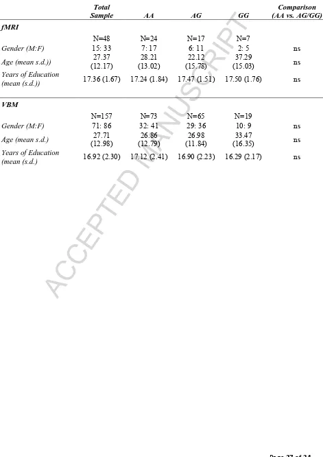
[image:28.595.70.541.120.786.2]Page 27 of 34 Tables
Table 1: Summary of participant demographics
Total
Sample AA AG GG
Comparison (AA vs. AG/GG)
fMRI
N=48 N=24 N=17 N=7
Gender (M:F) 15: 33 7: 17 6: 11 2: 5 ns
Age (mean s.d.)) (12.17) 27.37 (13.02) 28.21 (15.78) 22.12 (15.03) 37.29 ns
Years of Education
(mean (s.d.)) 17.36 (1.67) 17.24 (1.84) 17.47 (1.51) 17.50 (1.76) ns
VBM
N=157 N=73 N=65 N=19
Gender (M:F) 71: 86 32: 41 29: 36 10: 9 ns
Age (mean s.d.) (12.98) 27.71 (12.79) 26.86 (11.84) 26.98 (16.35) 33.47 ns
Years of Education
ACCEPTED MANUSCRIPT
[image:29.595.74.526.120.603.2]Page 28 of 34 Table 2: Main effect of SWM maintenance condition on brain activity. Note: all regions shown are significant at pCORRECTED<0.05 (minimum cluster extent=775 voxels); **=p<0.001.
Talairach co-ordinates
Region x y z Vol.
(mm3) F Post hoc t
Right middle temporal gyrus 31 -69 18 62077 F(1,46)=84.53** 1-dot<3-dot**
Left middle temporal gyrus -29 -72 19 37953 F(1,46)=80.06** 1-dot<3-dot**
Left posterior cingulate/BA31 -3 -59 23 16212 F(1,46)=46.69** 1-dot>3-dot**
Right middle temporal gyrus/BA39 51 -59 11 5258 F(1,46)=39.66** 1-dot>3-dot**
Left angular gyrus/BA39 -45 -72 30 3740 F(1,46)=24.61** 1-dot>3-dot**
Right precentral gyrus/BA6 27 -11 51 3420 F(1,46)=51.26** 1-dot<3-dot**
Left precentral gyrus/BA6 -25 -10 51 2730 F(1,46)=52.04** 1-dot<3-dot**
Left inferior frontal gyrus/BA9 -57 5 29 1889 F(1,46)=34.58** 1-dot<3-dot**
Right culmen 7 -53 -3 1234 F(1,46)=19.96** 1-dot>3-dot**
ACCEPTED MANUSCRIPT
[image:30.595.74.551.111.782.2]Page 29 of 34 Table 3: The effect of NOS1/rs6490121 genotype on brain activity. Note: all regions shown are significant at pCORRECTED<0.05; post hoc t-test *p<0.05; **=p<0.001.
Talairach co-ordinates
x y z Vol.
(mm3) F Post hoc t
Genotype
Right caudate 11 16 -5 1155 52.03** AA < AG/GG**
Right superior parietal lobe/BA7 24 -68 43 633 21.95** AA < AG/GG**
Right superior frontal gyrus 17 55 8 545 25.01** AA < AG/GG**
Right cuneus/BA7 6 -75 33 420 17.78** AA < AG/GG**
Genotype x SWM
Left lingual gyrus -21 -90 -1 976 27.26** 3-dot: AA < AG/GG* AG/GG: 1-dot < 3dot**
ACCEPTED MANUSCRIPT
Page 30 of 34 Figure Captions
Figure 1: Spatial working memory paradigm
Figure 2: The main effect of NOS1 (rs6490121) genotype (AA vs. AG/GG) on brain activity.
A: Regions of significant (i.e. F>12.16; pCORRECTED<0.05; minimum cluster extent=306
voxels/mm3) activity associated with NOS1 genotype - i.e. caudate (vol.=1155mm3), superior
parietal lobe (vol.=633mm3), superior frontal gyrus (vol.=545mm3) and cuneus/BA7
(vol.=420mm3). Clusters are shown on the ICBM452 T1 template from AFNI (Cox, 1996).
B: Mean signal change (β) in each of these significant clusters (co-ordinates formatted as
RAS) during 1-dot and 3-dot conditions in each participant group. Note: ***=p<0.001;
**=p<0.005.
Figure 3: Regions demonstrating a significant (i.e. F>12.16; pCORRECTED<0.05; minimum
cluster extent>306 voxels/mm3) interaction between NOS1 (rs6490121) genotype (AA vs.
AG/GG) and SWM maintenance condition (1-dot vs. 3-dot). A. Left lingual gyrus
(vol.=976mm3). B. Left middle frontal gyrus (vol.=489mm3) Note: Clusters are rendered on
the ICBM452 T1 template from AFNI (Cox, 1996). *=p<0.05.
Figure 4: Regions of gray matter exhibiting an effect of NOS1 (rs6490121) on brain volume
in healthy controls in a priori ROI analysis – mPFC/BA10; MNI co-ordinates: 19 61 26;
KE=371 voxels; cluster level pCORRECTED<0.05. Note: Clusters are rendered on the ch2better
ACCEPTED MANUSCRIPT
[image:32.595.81.444.69.612.2]Page 31 of 34
ACCEPTED MANUSCRIPT
[image:33.595.77.438.66.609.2]Page 32 of 34
ACCEPTED MANUSCRIPT
[image:34.595.76.439.74.609.2]Page 33 of 34
ACCEPTED MANUSCRIPT
Page 34 of 34