ISSN Online: 2160-8822 ISSN Print: 2160-8814
Biological Profile of HIV-Positive Patients in
Bangui, Central African Republic, in 2017
Yawo Tufa Nyasenu
1, Alain Farra
1, Brice Martial Yambiyo
2, Alexandre Manirakiza
2,
Fernand Didier Padou
1, Ferdinand Yapou
1, Pulchérie Pelembi
1, Rodolphe Mambely-Nzako
1,
Marie-Joëlle Mandeng
1, Alain Berlioz-Arthaud
1, Pierre-Alain Rubbo
1, Jean-Pierre Lombart
11Institut Pasteur of Bangui, Medical Analysis Laboratory, Bangui, Central African Republic 2Institut Pasteur of Bangui, Epidemiology Service, Bangui, Central African Republic
Abstract
Background: The biological profile of HIV-positive patients is essential for diagnosing treatment failure and the prognosis of infection. We determined the virological and immunological profiles and biological anomalies of HIV-positive people on antiretroviral therapy (ART) in Bangui, Central Afri-can Republic. Methods: We conducted an analytical, descriptive study be-tween 4 April and 30 September 2017 of all patients who had received ART for more than 12 months and who attended the Medical Analysis Laboratory of the Institut Pasteur in Bangui for a complete biological work-up, including viral load. A blood sample was taken for quantification of RNA HIV-1, CD4 lymphocytes and blood count in two tubes containing ethylenediamine te-traacetic acid, and another sample was taken in a dry tube for measurement of creatinine and transaminases. Results: The total population comprised 1748 patients, with a mean age of 38.7 years (±14.3; median, 41 years; range, 2 - 79 years); 33.3% of patients were between 40 and 49 years old. Females predo-minated (71.3%), for a sex ratio of 0.4. Immunological failure was observed in 20.2% of patients (CD4 < 200 cells/µL), and 44.5% of patients had a load of RNA HIV-1 ≥ 1000 copies/mL. The main haematological anomalies were anaemia (28.0%), leukopenia (26.7%), neutropenia (42.1%) and lymphopenia (27.2%). Blood creatinine was abnormal in 61.0% of patients, ALAT in 57.0% and ASAT in 66.9%. Conclusion: The abnormalities observed in this study concerned the haematopoietic system, the liver and the kidneys. As other or-gans and systems may be affected, periodic multidisciplinary biological and clinical follow-up is necessary for people living with HIV in order to improve their management.
How to cite this paper: Nyasenu, Y.T., Farra, A., Yambiyo, B.M., Manirakiza, A., Padou, F.D., Yapou, F., Pelembi, P., Mambely-Nzako, R., Mandeng, M.-J., Ber-lioz-Arthaud, A., Rubbo, P.-A. and Lom-bart, J.-P. (2018) Biological Profile of HIV-Positive Patients in Bangui, Central African Republic, in 2017. World Journal of AIDS, 8, 11-20.
https://doi.org/10.4236/wja.2018.81002
Received: January 13, 2018 Accepted: March 5, 2018 Published: March 8, 2018
Copyright © 2018 by authors and Scientific Research Publishing Inc. This work is licensed under the Creative Commons Attribution International License (CC BY 4.0).
http://creativecommons.org/licenses/by/4.0/ Open Access
Keywords
Anaemia, Biological, Abnormalities, HIV-1, Bangui
1. Introduction
HIV/AIDS remains a major public health problem throughout the world and particularly in sub-Saharan Africa. In 2017, UNAIDS estimated that 20.9 million people were receiving antiretroviral therapy (ART). In 2016, 6.1 million people in West and Central Africa were living with HIV, and 31,000 died of diseases asso-ciated with AIDS, whereas only 2.1 million people, 35% of those living with HIV in the region, had access to ART. Women account for 56% of people living with HIV in the region. In 2016, there were 370,000 new infections with HIV, representing a reduction of 9% since 2010, and the number of deaths from AIDS decreased by 21%. Among children in West and Central Africa, there were 60,000 new cases of HIV infection in 2016, representing a decrease of 33% since 2010 [1].
Treatment with multi-ART has prolonged survival, decreased morbidity and improved the quality of life of people living with HIV [2] [3] [4] [5]. ART, like HIV, may, however, attack various body systems and cause abnormalities in the haematopoietic system and organs such as the kidneys, the liver, the heart and muscles [6] [7] [8]. The drugs in ART may thus have undesirable, dangerous ef-fects on patients [9] [10]. Interactions between drugs given for opportunistic and other infections (tuberculosis, hepatitis B) can have dangerous consequences [11] [12] [13]. Kobangue et al. [7] reported that anaemia was the cause of death of 75.8% of children on ART who died at the Paediatric Complex in Bangui be-tween 2008 and 2013.
In the Central African Republic, there has been a decade of sociopolitical in-stability, which has resulted in population displacement and difficult access to treatment. The present study addresses the biological characteristics of patients on ART who attended the Institut Pasteur in Bangui for a free, complete biolog-ical follow-up as part of a national effort to improve their management.
2. Patients, Material and Methods
2.1. Setting and Patients
A cross-sectional descriptive study was conducted between 4 April and 30 Sep-tember 2017 at the Institut Pasteur in Bangui, a recognized centre for public health, in collaboration with the International Committee of the Red Cross and Red Crescent Societies. The institute has a medical analytical laboratory, which provides biomedical analyses for the population of the country. We included all patients with HIV/AIDS who had been on ART for at least 12 months for whom systematic biological follow-up had been conducted during the study period. The inclusion criteria were the presence of HIV-1 infection, ART for at least 1 year and a complete biological work-up, which comprised plasma viral load,
CD4 lymphocyte count, blood creatinine and transaminases and blood count. We excluded from this study: HIV-infected patients who were on treatment for less than one year, patients with incomplete biological status, and patients in-fected with HIV-2. In this study using clinical files and electronic registers, the patient’s identity was not collected in the survey file to ensure ethical clearence.
2.2. Sampling
Blood samples were taken for quantification of RNA HIV-1 and for blood counts in two tubes containing ethylene diamine tetra-acetic acid (EDTA), and another was taken in a dry tube for measurement of blood creatinine and trans-aminases.
2.3. Biological Analyses
RNA-HIV was extracted with a Biocentric kit (12.08.02-170510) on a NorDiag Ar-row (AO637R3) extractor. Biocentric kits (TR001-250IC) were used for RNA-HIV amplification by real-time PCRon an Applied Biosystems 7500 Fast System (4357362). The targeted region is on the long terminal repeats (LTRs), and the detection threshold was 300 copies/mL in a sample of 10 µL. The technique is specific for HIV-1 group M, subtypes A-H. Blood creatinine with automated Jaffe colorimetric method, aspartate and alanine transaminases (ASAT and ALAT) were measured on ABX Pentra 400 (RAB1251FR). Blood creatinine was considered normal at 6 - 13 mg/L and ASAT and ALAT at 11 - 66 UI/L and 15 - 46 UI/L, respectively. An ABX Pentra 60 (111P6010797) from HORIBA was used for blood cell counts. Cut off for anaemia was defined as haemoglobin le-vels lower than 11 g/dL in women and lower than 12 g/dL in men.
2.4. Statistical Analysis
The analysis was performed on all data from included patients (exhaustive sam-pling). Data were entered onto an Excel® sheet and analysed with Stata software. We recorded the sex and age of our study population, the viral load, presented as <1000 copies/mL (virological failure) and ≥1000 copies/mL (virological success), and CD4 cell counts, with immunological failure defined as <200 cells/mm3 and
immunological success as ≥200 cells/mm3. The World Health Organization
(WHO) guidelines recommend the use of viral load as the preferred method for monitoring treatment response over clinical and immunological approaches, and define virological and immunological failure respectively with a threshold of 1000 copies/mL and 200 cells/mm3 [14] [15]. Student’s t test, the chi-squared test
and calculation of odds ratios (ORs) with 95% confidence intervals (CIs) were used to compare viral loads and CD4 cell counts according to biological abnor-malities. The threshold for significance was set at 5%.
3. Results
A total of 1748 patients were entered into the study, with a mean age of 38.7 ±
14.4 years and a median of 41 years (range, 2 - 79); most (33.3%) were aged 40 - 49 years, and most were female (71.3%), for a sex ratio of 0.4.
3.1. Characteristics of Patients
Immunological failure was seen in 20.2% and virological failure in 44.5% of pa-tients. Anaemia was observed in 28.0% of patients (Table 1).
3.2. Biological Parameters of Patients
Patients with immunological failure had lower mean values for haemoglobin, haematocrit, mean blood count, leukocytes, polynuclear eosinophils and baso-phils and lymphocytes than those with immunological success but higher values for creatinine, ALAT and ASAT (Table 2).
[image:4.595.177.535.358.733.2]Patients with virological failure had lower mean values for erythrocytes, hae-moglobin and mean blood count but a higher mean value for ASAT. The mean haemoglobin concentration was 12.4 g/dL (10.2 - 12.5 g/dL) in patients with vi-rological success and 11.6 g/dL (11.4 - 11.7 g/dL) for those with vivi-rological fail-ure (Table 3).
Table 1. Characteristics of patients.
No. of patients 1748
Age (years)
Mean 38.7 ± 14.3
Median 41
Range 2 - 79
≤18 219 (12.5%)
19 - 29 120 (6.9%)
30 - 39 465 (26.6%)
40 - 49 582 (33.3%)
≥50 362 (20.7%)
Sex
Male 501 (26.7%)
Female 1247 (71.3%)
CD4 cell count (n = 1146)
<200/mm3 232 (20.2%)
≥200/mm3 914 (79.8%)
Viral load (copies/mL)
<1000 971 (55.6%)
≥1000 777 (44.5%)
Anaemia
Males 168 (33.5%)
Females 322 (25.8%)
Both sexes 490 (28.0%)
Table 2. Biological parameters of patients according to CD4 cell count.
Parameter CD4 < 200/mm
3 CD4 ≥ 200/mm3
Significance No. of samples Mean (95% CI) No. of samples Mean (95% CI)
Red blood cells (109/L) 232 3.77 (3.6; 3.8) 914 4.01 (3.9; 4.0) 0.001
Haemoglobin (g/dL) 232 11.08 (10.8; 11.3) 914 12.27 (12.1; 12.3) 0.001
Haematocrit 232 0.32 (0.3; 0.3) 914 0.45 (0.3; 0.5) 0.259
Mean cell volume (fL) 232 86.55 (85.1; 88.0) 914 90.59 (88.9; 92.3) 0.023
Mean cell haemoglobin content (pg) 232 29.68 (29.1; 30.2) 914 33.97 (28.5; 39.3) 0.433
Mean cell haemoglobin content (g/dL) 232 34.22 (34.0; 34.3) 914 34.22 (34.1; 34.3) 0.954
Leukocyte count (106/L) 232 4.71 (4.3; 5.0) 914 5.53 (5.3; 5.7) 0.001
Polynuclear neutrophils (106/L) 232 4.28 (1.1; 7.4) 913 2.72 (2.0; 3.3) 0.139
Polynuclear eosinophils (106/L) 232 0.22 (0.1; 0.2) 913 0.33 (0.3; 0.3) 0.003
Polynuclear basophils (106/L) 232 0.02 (0.02; 0.02) 913 0.04 (0.04; 0.05) 0.001
Lymphocytes (106/L) 231 1.35 (1.2; 1.4) 913 2.18 (2.1; 2.2) 0.001
Monocytes (106/L) 231 3.72 (1.2; 8.6) 912 1.74 (0.1; 3.3) 0.332
Platelets (106/L) 230 272.96 (256.8; 289.0) 909 285.55 (277.2; 293.8) 0.180
Blood creatinine (mg/L) 213 12.11 (9.9; 14.2) 777 9.39 (8.8; 9.9) 0.001
ALAT (UI/L) 211 34.86 (28.5; 41.1) 782 28.80 (27.2; 303.3) 0.001
ASAT (UI/L) 209 48.80 (39.3; 58.2) 782 32.53 (30.4; 34.6) 0.001
Table 3. Biological parameters of patients according to viral load.
Parameter Viral load < 1000 copies/mL Viral load ≥ 1000 copies/mL Significance
No. of samples Mean (95% CI) No. of samples Mean (95% CI)
Red blood cells (109/L) 971 4.02 (3.9; 4.0) 777 3.88 (3.8; 3.9) 0.001
Haemoglobin (g/dL) 971 12.38 (10.2; 12.5) 777 11.58 (11.4; 11.7) 0.001
Haematocrit 971 0.58 (0.3; 0.8) 777 0.48 (0/1; 0.7) 0.572
Mean cell volume (fL) 971 91.39 (89.7; 93.0) 777 87.96 (87.1; 88.7) 0.001
Mean cell haemoglobin content (pg) 971 31.76 (31.1; 32.2) 777 33.47 (27.1; 39.8) 0.554
Mean cell haemoglobin content (g/dL) 971 34.37 (34.2; 34.3) 777 34.10 (33.9; 34.2) 0.002
Leukocyte count (106/L) 971 5.27 (5.1; 5.4) 777 5.43 (5.2; 5.6) 0.199
Polynuclear neutrophils (106/L) 970 2.95 (1.9; 3.9) 777 2.48 (2.3; 2.6) 0.397
Polynuclear eosinophils (106/L) 969 0.33 (0.30; 0.36) 777 0.30 (0.27; 0.33) 0.199
Polynuclear basophils (106/L) 969 0.04 (0.03; 0.05) 777 0.04 (0.04; 0.05) 0.698
Lymphocytes (106/L) 968 2.07 (2.0; 2.1) 777 4.42 (0.2; 0.9) 0.270
Monocytes (106/L) 968 2.86 (0.8; 4.9) 776 1.77 (0.1; 3.3) 0.426
Platelets (106/L) 966 278.99 (271.1; 286.8) 773 283.53 (275.3; 291.7) 0.436
Blood creatinine (mg/L) 575 9.67 (9.0; 10.2) 564 10.02 (9.1; 10.9) 0.530
ALAT (UI/L) 584 29.22 (27.5; 30.9) 567 30.00 (27.2; 32.7) 0.631
ASAT (UI/L) 583 30.66 (29.2; 32.1) 565 39.88 (35.5; 44.2) 0.001
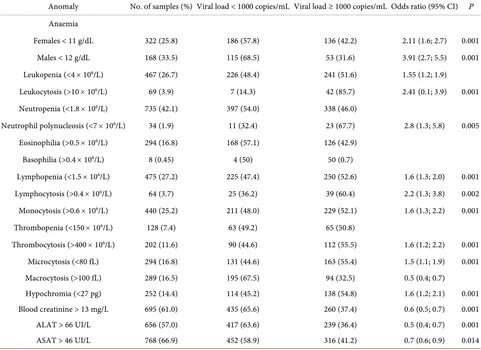
[image:5.595.71.538.426.728.2]3.3. Biological Anomalies
The biological anomalies observed are shown in Table 4 and Table 5.
Patients with immunological success were more likely to have anaemia (65.4% of females and 59.3% of males), leukopenia (66.6%), eosinophilia (85.8%), lym-phopenia (56.3%), thrombopenia (65.0%), microcytosis (69.7%), hypochromia (72.0%) and abnormal ASAT (74.8%). Of patients with a high viral load, 36.9% had anaemia, 51.6% had leukopenia, 46.0% had neutropenia, and 52.6% had lymphopenia. Blood creatinine was abnormal in 37.4% of patients with a high viral load, and abnormal ALAT and ASAT were found in 36.4% and 41.2% of these patients (Table 5).
4. Discussion
[image:6.595.61.540.366.732.2]Our finding of a sex ratio of 0.4 and a median age of 41 years are similar to those of Mouala et al. [13], who also found a sex ratio of 0.4 and a median age of 32.5 years among HIV-positive patients in Bangui, and of Loua et al. [16], who re-ported a sex ratio of 0.5 and a median age of 40 years in a study in Conakry, Guinea. Mouhari-Touré et al. [17] reported a predominance of women (68.6%)
Table 4. Biological anomalies according to CD4 cell count.
Anomaly No. of samples (%) CD4 < 200/mm3 (No. (%)) CD4 ≥ 200/mm3 (No. (%)) Odds ratio (95% CI) P
Anaemia
Females < 11 g/dL 208 (16.7) 72 (34.6) 136 (65.4) 3.07 (2.1; 4.3) 0.001
Males < 12 g/dL 113 (22.6) 46 (40.7) 67 (59.3) 4.58 (2.9; 7.1) 0.001
Leukopenia (<4 × 10⁶/L) 308 (26.9) 103 (33.4) 205 (66.6) 2.74 (2.0; 3.7) 0.001
Leukocytosis (>10 × 106/L) 49 (4.3) 7 (14.3) 42 (85.7)
Neutropenia (<1.8 × 10⁶/L) 468 (40.8) 97 (20.7) 371 (79.3)
Neutrophil polynucleosis (<7 × 10⁶/L) 30 (2.6) 8 (26.7) 22 (73.3)
Eosinophilia (>0.5 × 10⁶/L) 183 (16.0) 26 (14.2) 157 (85.8) 0.6 (0.3; 0.9 0.028
Basophilia (>0.4 × 10⁶/L) 3 (0.3) 0 3 (100)
Lymphopenia (<1.5 × 10⁶/L) 334 (29.2) 146 (43.7) 188 (56.3) 6.2 (4.5–8.5) 0.001
Lymphocytosis (>0.4 × 10⁶/L) 39 (3.4) 0 39 (100)
Monocytosis (>0.6 × 10⁶/L) 285 (24.9) 51 (17.9) 234 (82.1)
Thrombopenia (<150 × 106/L) 83 (7.3) 29 (34.9) 54 (65.0) 2.3 (1.4; 3.8) 0.001
Thrombocytosis (>400 × 106/L) 133 (11.7) 31 (23.3) 102 (76.7)
Microcytosis (<80 fL) 195 (17.0) 59 (30.3) 136 (69.7) 1.8 (1.2; 2.5) 0.001
Macrocytosis (>100 fL) 176 (15.4) 23 (13.1) 153 (86.9)
Hypochromia (<27 pg) 168 (14.7) 47 (28.0) 121 (72.0) 1,6 (1,1; 2.4) 0.007
Blood creatinine > 13 mg/L 238 (20.8) 49 (20.6) 189 (79.4)
ALAT > 66 UI/L 205 (17.9) 34 (16.7) 171 (83.4)
ASAT > 46 UI/L 301 (26.3) 76 (25.3) 225 (74.8) 1.4 (1.0; 2.0) 0.012
Table 5. Biological anomalies according to viral load.
Anomaly No. of samples (%) Viral load < 1000 copies/mL Viral load ≥ 1000 copies/mL Odds ratio (95% CI) P Anaemia
Females < 11 g/dL 322 (25.8) 186 (57.8) 136 (42.2) 2.11 (1.6; 2.7) 0.001
Males < 12 g/dL 168 (33.5) 115 (68.5) 53 (31.6) 3.91 (2.7; 5.5) 0.001
Leukopenia (<4 × 10⁶/L) 467 (26.7) 226 (48.4) 241 (51.6) 1.55 (1.2; 1.9)
Leukocytosis (>10 × 106/L) 69 (3.9) 7 (14.3) 42 (85.7) 2.41 (0.1; 3.9) 0.001
Neutropenia (<1.8 × 10⁶/L) 735 (42.1) 397 (54.0) 338 (46.0)
Neutrophil polynucleosis (<7 × 10⁶/L) 34 (1.9) 11 (32.4) 23 (67.7) 2.8 (1.3; 5.8) 0.005
Eosinophilia (>0.5 × 10⁶/L) 294 (16.8) 168 (57.1) 126 (42.9)
Basophilia (>0.4 × 10⁶/L) 8 (0.45) 4 (50) 50 (0.7)
Lymphopenia (<1.5 × 10⁶/L) 475 (27.2) 225 (47.4) 250 (52.6) 1.6 (1.3; 2.0) 0.001
Lymphocytosis (>0.4 × 10⁶/L) 64 (3.7) 25 (36.2) 39 (60.4) 2.2 (1.3; 3.8) 0.002
Monocytosis (>0.6 × 10⁶/L) 440 (25.2) 211 (48.0) 229 (52.1) 1.6 (1.3; 2.2) 0.001
Thrombopenia (<150 × 106/L) 128 (7.4) 63 (49.2) 65 (50.8)
Thrombocytosis (>400 × 106/L) 202 (11.6) 90 (44.6) 112 (55.5) 1.6 (1.2; 2.2) 0.001
Microcytosis (<80 fL) 294 (16.8) 131 (44.6) 163 (55.4) 1.5 (1.1; 1.9) 0.001
Macrocytosis (>100 fL) 289 (16.5) 195 (67.5) 94 (32.5) 0.5 (0.4; 0.7)
Hypochromia (<27 pg) 252 (14.4) 114 (45.2) 138 (54.8) 1.6 (1.2; 2.1) 0.001
Blood creatinine > 13 mg/L 695 (61.0) 435 (65.6) 260 (37.4) 0.6 (0.5; 0.7) 0.001
ALAT > 66 UI/L 656 (57.0) 417 (63.6) 239 (36.4) 0.5 (0.4; 0.7) 0.001
ASAT > 46 UI/L 768 (66.9) 452 (58.9) 316 (41.2) 0.7 (0.6; 0.9) 0.014
in a study in Togo. These findings indicate that HIV infection is a problem mainly among young, sexually active women. UNAIDS reported in 2016 that women represented 56% of all people living with HIV in West and central Africa [1].
In our study, 79.8% of patients had a CD4 count ≥ 200/mm3, and 44.5% had a
viral load of <1000 copies/mL. Lozès et al. [18] found that, after 12 months of treatment, 83.0% of patients had an undetectable viral load and 86.0% had re-covered their immunocompetence. Our immunological result is therefore simi-lar, but a high viral load persisted.
Haematological anomalies in patients on ART are the result of both immu-nodeficiency and dysregulation of the immune system. Anaemia was observed in 28.0% of patients in our study. This was the principal haematological anomaly found in other studies of people living with HIV: in 75.8% in Bangui [16], 95.2% in Zimbabwe [7], 54.5% in Conakry [17] and 13.8% in Togo [19]. We found lower mean values for haemoglobin, haematocrit, mean cell count, leukocytes, polynuclear eosinophils and basophils and lymphocytes but higher blood creati-nine, ALAT and ASAT values among patients with a low CD4 count. Immuno-logical failure may be due to complications of opportunistic bacterial, viral,
rasitic or fungal infections or to the direct effect of the virus on certain haema-topoietic progenitors, giving rise to anomalies in all cell lines [20]. Multiple ART and their secondary effects on patients’ organ systems may be responsible for renal and hepatic lesions [21].
Patients with virological failure had lower mean values for erythrocytes, hae-moglobin and mean blood count but a higher mean value for ASAT. The mean haemoglobin concentration was 12.4 g/dL for patients with a low viral load and 11.6 g/dL for those with virological failure. Nacoulma et al. [22] found a hae-moglobin level of 12.2 g/dL in a study in Burkina Faso. Such anomalies are due to the various mechanisms of HIV infection, which lead to lesions in different cells and organs. Immunological and virological failure evolves to therapeutic failure when patients are exposed to drugs that are toxic to cells.
Patients with a high CD4 cell count were more likely to have anaemia, leuko-penia, eosinophilia, lympholeuko-penia, thromboleuko-penia, microcytosis, hypochromia and abnormal ASAT. These anomalies may be due to various inflammatory mechanisms of HIV infection and secondary effects of ART and of treatments for opportunistic infections. Beuzit et al. [8] found rates of 74% for anaemia, 20% for neutropenia and 15% for thrombopenia in central Africa, and Erhabor et al. [23] found rates of 80%, 24% and 10%, respectively, in Nigeria. The high frequency of anaemia in these two studies was measured before initiation of treatment.
Creatinine was abnormal in 61.0% of our patients, and the transaminases ALAT and ASAT were abnormal in 57.0% and 66.9% of patients, respectively. These results corroborate those of Mouhari-Touré et al. in Togo, who found elevated ASAT in 55.9% of patients and elevated ALAT in 29.8%, with a mean creatinine level of 9.6 ± 5 mg/L. HIV infection, its treatment and the associated opportunistic infections are all chronic conditions, with harmful effects on stromal, haematopoietic, hepatic and renal cells and the organism’s defence me-chanisms.
Patients with a high viral load were more likely to have leukopenia, leukocyto-sis, polynuclear neutrophilia, lymphocytoleukocyto-sis, monocytoleukocyto-sis, thrombocytosis and hypochromia and less likely to have anaemia and abnormal blood creatinine, ALAT and ASAT. These biological abnormalities are due to the immunodefi-ciency induced by HIV and also to complications of treatment, opportunistic infections and side-effects of ART.
5. Conclusions
We found immunological failure in 20.2% of patients and virological failure in 44.5%. The main haematological anomalies observed were anaemia, leukopenia, neutropenia and lymphopenia. Blood creatinine was abnormal in 61% of pa-tients, and liver transaminase levels were high. Biological profiling is essential for the diagnosis of therapeutic failure and for the prognosis of HIV infection. The anomalies observed in this study mainly affected the haematopoietic system,
the liver and the kidneys. As other organs and systems may also be affected, people living with HIV should undergo periodic multidisciplinary clinical and biological follow-up in order to improve their management.
6. Strengths and Limitations
Since the recent introduction of HIV-1 viral load measurement in CAR, this study is the first to describe the complete biological profile of Central-African patients infected with HIV. The virological, immunological and haematological parameters were studied as well as liver and renal functions. Anemia was the most frequent abnormality described, as reported in other studies [7] [8] [20] [21] [22]. The limitation of the study concerns the poor access to laboratory fa-cilities in the provinces. Since the Pasteur Institute is located in Bangui, most of the patients included come from the capital city. We recommend national poli-cies together with International support will be able to implement the follow-up of HIV infected patients in the provinces.
References
[1] UNAIDS (2017) Fact sheet World AIDS Day. UNAIDS, Geneva, 1-8.
http://www.unaids.org/sites/default/files/media_asset/UNAIDS_FactSheet_en.pdf
[2] Centers for Disease Control and Prevention. (1993) Revised Classification System for HIV Infection and Expanded Surveillance Case Definition for AIDS among Adolescents and Adults. Morbidity and Mortality Weekly Report, 41, 1-19.
[3] Chêne, G., Binquet, C., Moreau, J.F., Neau, D., Pellegrin, L., Malvy, D., et al. (1998) Change in CD4+ Cell Count and the Risk of Opportunistic Infection or Death after Highly Active Antiretroviral Treatment. AIDS,12, 2313-2320.
https://doi.org/10.1097/00002030-199817000-00013
[4] Hammer, S.M., Squires, K.E., Hugues, M.D., Grimes, J.M., Demeter, L.M., Currier, J.S., et al. (1997) A Controlled Trial of Two Nucleoside Analogues plus Indinavir in Persons with Human Immunodeficiency Virus Infection and CD4 Cell Counts of 200 per Cubic Millimeter or Less. The New England Journal of Medicine, 337, 725-733. https://doi.org/10.1056/NEJM199709113371101
[5] Ba-Fall, K., Gueye, P.M., Lefevre, N., Fall, I.S., Said Ali Saindou, N., et al. (2004) Evolution of Antiretroviral Treatment of HIV/AIDS Infection at Dakar Main Hos-pital. Médecine Tropicale,64, 292-293.
[6] Montagnier, L. (1998) AIDS and HIV Infection. Flammarion, Paris.
[7] Kobangue, L., Gody, J.C., Diemer, S.C.H., Biguene-Sapoua-Boka, Y., Dibere Kamba, G.D. and Bobossy Serengbe, G. (2016) Epidemiological, Clinical, Biological and Therapeutic Aspects of Children Dying on Antiretrovirals at the Bangui Pediatric Complex, Central African Republic. Revue de CAMES Science de Santé, 4, 15-18. [8] Beuzit, Y., Bougarel, J. and Ngouonimba, J. (1992) Peripheral and Medullary
He-matologic Changes in HIV Infection in Central Africa. Médecine Tropicale, 52, 193-199.
[9] Aceti, A., Pasquazzi, C., Zechini, B., De Bac, C., Liverhaart Group. (2002) Hepato-toxicity Development during Antiretroviral Therapy Containing Protease Inhibitors in Patients with HIV. The Role of Hepatitis B and C Virus Infection. Journal of AIDS, 29, 41-48. https://doi.org/10.1097/00042560-200201010-00005
[10] Carr, A. and Cooper, D.A. (2000) Adverse Effects of Antiretroviral Therapy. Lancet, 356, 1423-1430. https://doi.org/10.1016/S0140-6736(00)02854-3
[11] Malaty, L. and Kuper, J. (1999) Drug Interactions of HIV Protease Inhibitors. Drug Safety, 20, 147-169. https://doi.org/10.2165/00002018-199920020-00005
[12] Savès, M., Vandentorren, S., Daucourt, V., Marimoutou, C., Dupon, M., Couzigou, P., et al. (1999) Severe Hepatic Cytolysis: Incidence and Risk Factors in Patients Treated by Antiretroviral Combinations. Aquitaine Cohort, France, 1996-1998.
AIDS, 13, l15-121. https://doi.org/10.1097/00002030-199912030-00002
[13] Mouala, C., Kaba-Mebri, J., Wata, J.B. and Rey, J.L. (2006) Factors Associated with Good Adherence in HIV-Infected Patients in Bangui. Cah Etudes Rech Francophones Santé, 16, 119-130.
[14] WHO (2016) Consolidated Guidelines on the Use of Antiretroviral Drugs for Treating and Preventing HIV Infection: Recommendations for a Public Health Ap-proach. 2nd Edition, WHO, Geneva.
[15] WHO (2013) Consolidated Guidelines on the Use of Antiretroviral Drugs for Treating and Preventing HIV Infection: Recommendations for a Public Health Ap-proach. WHO, Geneva.
[16] Loua, A., Dramou, C.D., Haba, N.Y., Magassouba, F.B., Lamah, M., Camara, A., Cissé, M., et al. (2011) Hematologic Profile of HIV-Infected Patients in Conakry.
Hématologie, 17, 365-369.
[17] Mouhari-Touré, A., Patassi, A., Nabroulala, K.T., Djadou, K.E., Edou, K., Nyamet-so, D., et al. (2011) Biological Profile of Adult HIV-Infected Patients at Initiation of Treatment in Togo. Medecine Et Maladies Infectieuses, 41, 229-234.
https://doi.org/10.1016/j.medmal.2010.11.007
[18] Lozès, E., Ahoussinou, C., Agassounou-Tchibozo Djikpo, M., Dahouegnon, E., Ahossouhe, N., Acoty, A. and de Souza, C. (2012) Variability of CD4 Cell Count and Viral Load in People Living with HIV on Antiretroviral Treatment: Case of Saint Jean De Dieu Hospital in Tanguieta (Benin). International Journal of Biological and Chemical Sciences, 6, 650-656.
[19] Malyangu, E., Abayomi, E.A., Adewuyi, J. and Coutts, A.M. (2000) AIDS Is Now the Commonest Clinical Condition Associated with Multilineage Blood Cytopenia in a Central Referral Hospital in Zimbabwe. Central African Journal of Medicine, 46, 59-61.https://doi.org/10.4314/cajm.v46i3.8525
[20] Bain, B.J. (1997) The Haematological Features of HIV Infection. British Journal of Haematology, 99, 1-8.https://doi.org/10.1046/j.1365-2141.1997.2943111.x
[21] Coso, D. and Gastaut, J.A. (2005) Non-Tumor Hematological Abnormalities during HIV Infection. In: Sebahoun, G., Ed., Hématologie clinique et biologique, 2nd Edi-tion, Editions Arnette, Paris, 319-322.
[22] Nacoulma, E.W.C., Some, Y., Tieno, H., Diallo, I., Zoungrana, A., Bougnounou, R., Ouédraogo, C., et al. (2007) Evolution of Hematologic Parameters during Antire-troviral Therapy in HIV-Infected Patients in Burkina Faso. Bulletin De La Societe De Pathologie Exotique, 100, 271-274.
[23] Erhabor, O., Ejele, O.A., Nwauche, C.A. and Buseri, F.I. (2005) Some Haematologi-cal Parameters in Human Immunodeficiency Virus (HIV) Infected Africans: The Nigerian Perspective. Nigerian Journal of Medicine, 14, 33-38.