University of Warwick institutional repository: http://go.warwick.ac.uk/wrap
A Thesis Submitted for the Degree of PhD at the University of Warwick
http://go.warwick.ac.uk/wrap/61761
This thesis is made available online and is protected by original copyright. Please scroll down to view the document itself.
A STUDY OF THE CELL ENVELOPE OF HALOBt\CTERiIU!M. SAL.lNAN9M.
A ~e.t. <8Ubmltt~dinpantal fulflltnont of tbQ requiremeu.ts
fo·r the award of th•• greeof DoetorQ,f PbUQ"pby of the
University of Watwiek.
PREFACE
The work described in this thesis was carried out in the
School of Molecular Sciances of the Univ-ersity of Warwick during
the perlodOctober ~966to July 1969. Except Where otherwise stated,
it is the author's original work, and has not been submitted for a degree
at any other University.
Preface Contents Abbreviations Fraction Code Abstract CHAPTER! CHAPTER II 2.1. 2.2. 2.3.
2.4.'
2.5. 2.6.2.1.
i.~.
2.9.
2.10. 2.11. 2.12. 2.13. 2.14. 2.15. 2.16. 2.17 . 2.18. 2.H~. 2.20. 2.21. 2.22. 2.23. 2.24. 2.25. 2.26. 2.27. CONTENTS INTR.OpUCnON METHODS Organisms Cultivationliarvestlng of cells
Prepatatlonof cell envelope fragment bags P~~paratton of erude membranes
Salt ...free dry weights Protein (tQtenntnatton NucleiC acid determtnatlon Cytochrome esttmanca
Amino acid analysts -hydrolysis
AmmQ acid, analyser-equipment changes Analyele of protein hydrolysate
Amine 8\lgar analYSls
Extraetton of liPids LtpLElanaly •••
Oolumn ehromatographyof lipids - DEAE cellulose CQlumn Glll.·omatograpllyof .l\ptds -Decalso
Ool\UUJ1.ch;romat;o~aphy of liplds -Alumina ThUl"l~yer c1:}romatography
Bltl~tlQn f,lf menaqubwn,eBlldcarotenGld
Q\tlfUtration
PolYM~lamlde gel disc: el~tr.ophGre81. ISQiontopoint determination
The binding of magnesium ion to the membrane-rich fraction (1\)
Endogenous acttvlty of whole ceUs
Assay of NADH Ol(ida.88
Assay of NADH-ferrteyanide oxi~reducta8e
2.28; 2.29.
2.30. 2.31.
tv. Assay pi fel'ricyanide reductase
Incubatton of cell envelope fragment bags with hywtonic media.
S~t:raQf ltptd sample$ Cllemicale
2.16.
2.16. 2.17. 2.17. CHAPTER III RESUL,TS I
3.1.
S.2.
a.3.
a.4.
3.5. 3.6. 3.7. CHAPTERtv
•• 1,.4.2.
4.3.
4.4. 4.5. 4.6. 4.7. IntroductionG:towth'Qharacteri8t~s of the'~e11culture Cheml~al composition of whole cells and the memPraJle-rlch fraction (NM)
The Upt4s ofH. sal~riqm
Va.mtiQIl$ inthe 'lipid 'composition with the growth phase
Va.PJlUOUS of the minor lipid components with
the ~wth (ilase Conclusion 3.1. 3.3.
3.3.
3.4.
3.9. 3.10. 3.10. RE~ULTS.tltntroc.tw:rlofl
Effect ef magnesium l~n on the membrane Purification of the membrane -rtch fraction Characteristics of the protein fraction RP Characteristics of the protein fraction P Characteristics of the lipoprotein fraction R Conclusion
4.t • 4.2. 4.6. 4.6. 4.7. 4.7. 4.16.
CHAPTER V RESUL1;'8 III
S.1. 5.2. 5.3. , 5.4. S.5. 5.6. 5.7. S.S.
Introduction 5 . 1 .
General solubility properties of the membrane 5. 1 . Disaggregation of the membrane illsodium dodecyl
sulphate (SOS) 5.2.
Gel ftltration of Sps -solubUlsed membrane 5.3.
Density gradient centrifugation 5 .5.
Reaggregation of the Itpoprotetn fraction R 5 .5 . Disaggregation of the membrane ill multiple
d.etergents 5 .6.
CHAPTER VI 6.1. 6.2. 6.3. 6.4. 6.5. 6.6. CHAP'l'ER VII 7.1. 7.2. 7.3. 7.4. 7.5. 7.6. 7.7. RESULTS IV Introduction
The action of urea and guanidine on R fraction Gel filtration of urea -solubilised membrane Electrophoresis of urea-solubilised membrane Fractionation of guanidine-solubUised membrane Concdusion
RESULTS V ENZYMIC STUDIES Introduction
Loeahsetten of adenosine triphosphatase Leealtsatten of NADH oxidase
Chaot:ropl~ agents
The effect of ehaetrepte agents on cell envelope
fragment bags
hlcubatlonoi cell envelope fragment bags with hypotonic media
Conclusion
CHAf'I'ER VlU DISCUSSION
ABBREVIATIONS
Abbreviations listed in Pollcy of the Joumal and Instructions to
~thor8 (Biochemical Society) are used, where relevant, undefined.
vii.
FRACTION CODE
For clarity, membll'ane fractions have been given a letter code;
and are redefined at first mentlon in the
t_.
The followtng is a general guide :CEFB
NM
R
RP
RU
RUF
P
SP
ceU en\telQpe fragment bags
erude, nuclease-treated membrane-nch fraction
P'lilrift$dmembrane, free item amino sugar andnuclootlde protein te'lea.ed with nueleotlde from NM
residual membrane frflotUm after treatment of R with ur.
prote'lnfragments released from R bytreatment With'urea
AffiTitACT
The plasma membrane of Halobactertum_saltnar1.um, strain 1, an
ex-tremely halophtltc bacterium, has been Isolated and characterised. The cell
envelope fraction (CEFB) was isolated from a eeU homogenate by differential
centrifugation. After dialysing the cell envelope fraction against distilled water
and treating with nucleaaes, a fairly pure preparation of plasma membranes (NM)
was obtained by centrifuging. The membrane -contatntng fraction was purtfted by
gel filtration on Agarose, which yielded a purified membrane fractton (R) and a
protetn-aucleottde fraction (RP). A small nwlecular weight protein fraction P
was separated frQID the pu:rtfied membrane fraction etther by gel fUtration on Sephadex in a buffer containing phosphate and fluoride tons or by ultrafiltration. Protein fractions RUP1and RUP2were separated from the purified membrane by
gel filtrattonon Agarose in the presence of 6M urea. The remaining
membrane
-containing fractton, which was eluted in the void volume of the urea-Agarose gel,
was coded RU.
The organism was studied in batch eulture; maximal growth was reached after 48 houra, after which time the cells were inthe 'stationary phase.' The
endogenoUIJresph:atory activity of the ceUs roaetc a maximum at 40 hours and dIen declined steadily, but the vtable count rematned fairly steady.
Analyses of the membrane fraction NM were made at various Iilases of
growth upto a maxtmum of 160 hours. AU the ceU liptd was fotmdto be CQn-centrared Inthe membrane traetton. The amount of lipid expressed as a per-centage of the salt-free dryweight of the cell remained constant, but both the total cell protein and the membrane protem feU durtng the period between 16 and 112 hours of growth. Also, the proportton of membrane to whole cell fell during this period. The menaqutnone and the carotenoid pigment were localised
ix.
The crude (NM) and punfied (R) membrane fractions were both affected
by magnesium ion. Inthe absence of the ion, the membrane disaggregates to small lipoprotein particles. Magnesium ions also assist inthe btnding of the amino sugar layer of the cell envelope and of the Pand RPfractions to the mem-brane.
The physico-chemica! properties of the fractions NM, R. RP, RUP and Phave beeninvestigated by a combination of amino acid analysts, gel electrophoresis. sucrose density gradient centrifugation and gel filtration on Sephadex or Agarose •
In addition, the binding of magnesium ion to the membrane and the isotonic point of the membrane rractton R have been determined.
The fraction R was found to be free from amino sugar and nucleotide. Fractions NM, R and RU contain all the cell lipid, and eytechreme, Fractions
NM and R, and probably also fraction RU, centatn the cell menaqutnoneand
carotenoid. Evidence is presented that suggests that the fraction R contatns the
NADH oxidase and adenostne triphosphatase of the electron transport system. The action of the detergent sodium oodecyl sulphate (SOS) on the membrane fraction R was to break up the membrane
moo
smaller particles. The disaggregation occurred intwo distinct steps. The dtsaggregated particles could be reaggregatedto a fraction which resembled the original membrane by removing the SOS by dialysis or gel fUtrat\on. The dlsaggregated partteles and also the reaggregated
membrane fraction were subjected to gel fUtl'atton, gel electrephorests and sucrose density gradient centtUugation, inorder to determine whether or not the lipid
and protein comPQn~ts of the membrane bad been separated.
A fraction which may be analogous to mttochoudrtal structural prote.ln
1.1.
CHAPTER 1
INTRODUCTION
The term halophtlic means salt -levtng , Halophtltc bacteria are organisms
that have an essential requirement for sodium chloride for growth. Marine bacteria are halophilic, generally requiring about 2 to 5% sodium chloride as an
.
essential component of their growth medium. Some bacteria are capable of
sur-viving in media containing higher concentrations of salt and may even require such
high concentrations. Bacteria which grow best inmedia of about 5 to20% salt are termed moderate halophUas. Bacteria which grow best at between
20%
andsaturated (about 30%) salt are termed extreme halophUes. The extreme halophUes generally have a minimum requtrement of about 10 to 15% salt for growth. Potassium
tons, and to a lesser extent, magnesium ions, can partially replace the sodium
l· ( .
~ons (Brown & Gibbons, 1955; Christian, 1956 ; Weber, 1949) but no salts have
yet been found that can completely replace the sodium chloride and yet maintain
,
growth.
The extreme halophtles have a complex. organic nutritional requirement.
They do not generally metabeltse carbohydrates, preferring proteins or amino adds as carbon sources. Chemically defined media (Dundas, Srinivasan &
Halvorson, 1963; Onishi, McCanCe & Gibbons, 1965) have included at least ten amino acids and also nucleotides and glycerol. The extreme haJophUes are usually
cultured on protein dtgests(tryptone, peptone, yeast autolysate) ceatatamg' suitable
. "
amounts of sodium chlortde , Ifcrude' alt is used as the source of sodium chloride and if the medium. is made upintap water, the relativelysma.lJ require.ments for magneetum and potassium (BrownSt Gibbons, 1955) and trace elements (Sehgal &
Gibbons, 1960) wUI normally be satisfied.
The extreme halopbt!es are wtdely distributed innature, mainly occurring
inbrines containing very high concentrations of salt. They are particularly con" spicuous intheevaperatton pans of salt wolks and ~ natural salt lakes of high
f.2.
large numbers that they impart a pink colour to the brine. Darwin had observed one such lake on his travels in1833, and bad attributed the red colouratton to
'some infusorial antmalcula ' (''Voyage of H.M.S .Beagle, "eh,IV, first published
1843). Many early reftu~ences to the red eolouranonor salt and salterns, including
a Chtnese 'referene,e of c .2500 B.C. and a quotation from PlinyI are collected in Baas Becking (1931).
The extreme haloIi1Ues are also found in saltne muds and soUs throughout the world and are capable ·ofsurviiVal in salt itself. Mined salt or reek sal~ is
usually free from the Grgani&ms butlOlal' salt, derived from the evaporation pans,
frequently contains large numbers of the extreme haloj)ilUes (Larsen, 1962).
Al-thoughstoXiage of the salt under dry condition,s 'tenda to reduce the viable count, the bactena. have been successfully rreeee-drted inthe presence of salt medium. It is possible therefore that the organisms can remain viable insalt crystals when blown about by the wind, or otherwise tranaported across land,
The presence of extremely haloJiiUic bacteria tn crude solar salt is of constderable economic importance inmany pans of the world. Salt is ve.ry often used as a preservative against microbial s po.U;ageof food and other perishable gQodB•.and the use pf contaminated salt may well defy its ownobject. Visually, the bacteria reveal themselves as pink or red patches of slime; hence the "pink"
01; "pinkeye" of salted ti6h (Harrison 8t KennQdy, 1922; Finn, 1941 ; .Shewan .. 1942)
and the "red heat" of salted hides (Lloyd, MarriQt &:Robertson, 1929; Lochhead, 1934). In severe cases the bacteria may completely penetrate the product. Such Iqicrobial spoUage ts, ~fcourse, llDdesi~able, particularly if tbe bacteria causes putrefaction aa weU as 4tscoIQurarton of the food product. However. in almost
aUcases investigated .~LFsen, 1962) the cause of the contamination has been
traced te the salt t.e)iand8polla~ may be avoided by the UNof rock salt or sterUised sea salt.
However, it was the reddenlng of salted fish that fir.t aroused interest in
the extreme hal0phUes folloWlng the dlscevery that the dlseol(i)uration was bacterial
s.s.
the extreme halophtles. Many of these are listed in Larsen (1962),a good
tntl'oductory review of balophUtiSm.
Most, if not all. of the extreme halophUes are bacteria. Other halophilic organisms which have been reported have proved to bet moderate rather than ex" treme, Thus, probably most of the species (approximately 21) isolated from 'tile Great Salt Lake (Broelc" 1969) are not optimally adapted to the high salt envlmnment 'and only a few are extreme balophUel.
The main genus of extreme halopaUes is Halebactertum, Bergey's manual (7th edition) Itst$ five epeotes, H. salinanUDl, .11.cuttruhrum, H.halobium,
H.ma.rislOOrtui and H.trapanicurn. In addition, twospecte8 of the famUy
MiCrocectaeeae, Mlerococcu. m.orrhuae and.Sarelna UtoraUsare extreme balo-phile.; these species may be vartantJ of each other. Att.empts toclasslfy the
various species ofHal()~terium by their UlUntlllslogtcal reaponsa have been made
recently by ZwUUng, Rowen it Stotzky (1969). althoUgh H. aaltnartUD.l, strain 1, was net included Ut the survey. 'l'bose strains Whlchwe,re tested ieUintQ four
mutually excluSive grGUps. A common genus ...pectfic antigen and three group-speetft.c anttgenswe:re demon.trated. The only other bacterial taxonomic group
reported as having a representatIve whioh. Is an _reme halophUe is the photo ..
synthetic bactetla '(Anderson, 1958; Ray:mond &
St.~m,
1967).Thistheei. 18prtmanly concerned wttibthe' halobactertll, tnputtcular H. saJ.inar\um, atratn 1. The halohactenaare' obligate aerohQ.. and are usually red Ol"plnk. They appear red and ,tranlparent an slide cultures if'DOn-vacuoiated.
Va.cuolated strain' are pink and; opaque but appeal" "ed and transparent under pressure. ~ balobaoterla ant normally rod sbapee, Gram-negative, aen-apore forming and are tophottichoualy fla.geUated if motUe. H.sattnarium, strain 1, is non -vacuolated and .f\ghtly metile. Cultur. aemtt a cbaraeten.ttc odour
which t. partly fitttrtbutable to bydNgen8ulph:ide.
meI-ji.4.
shaped intermediate stages, without any change intotal cell volume. One rod-shaped organism normally gives rise to an average of about twesph_res. On
further diluting th~susp$l8.ion to about 10%(w/v) sodium chloride, the indiVidual spheres suddenly lyse. It has beemshown that some iOIlS tend to protect the rod
+ +t = .
-shape, e.g, K, Mg • SO4' CHgC02 ,whilst others promote the defo.rmation
+ +t - -
-of the ::ns, e.g. N1i4,
cs ;
CNS , CI04• CC13C02. The ttansforma.tion and ly.ls proceaees dO. not appear to be enzymically mediated, although they are pH(and,temperature dePQl1dent.The observation that the rod -sphere transition occurs without an apparent
ebange in total oeU volume (Moh...Sit Larsen, 1965)sugge8ta that esmottc equUtbrium
is maintained throughQut the diluting,of the cell 8uspellsion. Cells grown in the presence of dif;fercmtconeentr'@.tion8of 8a1thave .aa iBtemal salt content which
depends an, and approaches, the ~onccmtratton Of.alt Ut the medium (Gibbons &
Baxter, 1953; Holmes, 1904; Christian, .956; Chri8.tianItIngram, 1959).
+
-
+However, ID additton tp Na and Cl • K ia a dominating eomponent of the interne!
salt and 1ts concenuattoll .a.pproachos the solubUity limit of KOl.(Christian at
+
Waltho, 1962). 'I'ho ,CQl1cel1trattondifference of Na acrose the.eoll envelope Is very approximately equal t~ that of K+but
ts
of opposite .stgn.The J.'e,pons(toftbe extreme halopbUe to '¢lumge. in the salt concentration of the medium t.related to the tnatahtUty ·ofthe ceU envelope of the organism when eXPOSed to hypotonto me.dw. (La-reea. 1961). Veryshatlar structural tranafo:J.'Dl,a" tions and lysla patte~ are observed with lsQlatedcellenvelQpe prepaXtattona. lio.wever, K+ i.alI!.BO·St; as effective as Ne.+in preservmg lntaetcell envelopes and Mg++ia much more effective inmaintaining the' integrity ofceU envelopes than it ls inmatn~:a.tnlngthe rod-shape of whole eeUs.
The isolated eDZyJUO' of halobacterta also ohtbit complex ion ....ffeota·. .It Is to Baxter & Gl.hbon.
U.s..
1956. 19S1)tbat we owe the important dleco:very thatthe enzymes are them.eelve. 'bal.,l*Ulc·, 1..,e. are adapted to the high
,intra-cellular 'alt coneontZ'atton. They mQ.t1y·e~it relatt'Y:..ly higbor even llWd.mal
;1.5.
f)f the enzymes testedexhtbtt higher tnaJCtmal aCtl'ritlEu1 in:KClthanbt NaClaolutions. "rhe salt relations ·ofa.number 'of lndtvtdtlal enzymes has been reviewed by Larsen (1967). A few rep)(nzts·eonaemtng addtttona;lenzymeaha:veappeated subsequently. Brown (1"966)has rep;urted,an NADH ,oxidase and Steven-sen & Brown (1967), an
ad..lWstne trt(:ilospha~8e f¥OmH.sa1tnanUrn, stl'atn 1. Lanyi &,Stevenson. (1969) have reported a catalase fromIi.eutlrub~ftli. ad Norberg Si HGfsten(1969) have.
reported 'eltt'tace1lulaiJi' prot eases,from H.saJ;mariUJ.n, 8tratns 1, 1M and S,. AU
.Be' enzymeB exhlbltone er other 'of the salt·re8p0nBe curves typical of halo{i1tltc Em2ytnes (see Ftg.J.1 .• . However. the ptOteaBes are reported to be more active ,biKelman,in ,NaC}, unltke the majority of 'tbeother enzymes (see above), The
salt relations of the EfuZymos are more fully dls:¢usaed inChapter Vlt.
The ,question of whem.r et flat 'the halobactena po.s~eSBa cell wall
ts
still debatable. PerlWUl¥ year. the bhlobacteda Wer., tbeughtto befllrno8t lmlque
in the microbial lW).t16m 'Pl\lHetl8ingonly astngle liimlting plasma nt~aile for
8 cell: etlveJ:o.pe.
tn
tbe elEliCtrell8lietGCQpe oilly the typiCal.three-Iayeted SttUCture :eharaetel1stte of the unit membrane (RobertS01l., 1966) was observed, although thestructure was some SO - 7S%wt_li thaa the _re u8U$l 75., - 801,. MOJ;'e'
reo.nU,y,
however t the anatomy 'ofthec.u on'Ve1opeofdlo hruoba.eterta haa been shown to be mo;recomple'lt(Lar$.QIl; 1961). With tmpl'O'Vedteehnlques ofeleetron mlCroscopy Sr&eekellt1l8 &&owen (1966. "6') andCbo~ '£loy &<Memol' (1967). working With.H'.halobtu$. md Ste$lslQld (1961.) 8l1<tSteenaland 8t Uu...~ (f%9),wotktng with
iii,sall.n:atium; snow~d that the4lnveloPfl consl_de :of:8ctt membrane overlaid by a protemace.u8 coat 15Oi; 1501 chick,. MoltG0ver" a.bllQbtum contained a much more compte •• yat.tn. o('membtanes than.H••eltnarium (S,f().ck_t~ Si Rowen, 1%7;
'StOec~_itUi &. K~\h ·t94S).
TheenvtkJPI' of tt~:8a1lnariuml
"rain
t
t bas M_further chataetet\$edbySteen.bmd (1967b).· 'the Q.ut~rprotetn coat, whteh Is responsible for the. characteristic
8urfaeepattl.J:Jlof Wholeeell.an4ll&lat.d en1'elo",a, ia, _parated frOID the
t'fhnatntng env.lope d\:lring eXPGlllH of tbe ,ceUeor .velope. to m.d'a of lErw
al_ r.I_Jed, as l'DOnlto_4by the _leoeof aminosugar eompon_te'
t~m
the,.~rat1e.
", ..
tlty ch.am_
.\l~l.yel' conre."otu:l&,1:& ••mueo1.»l'Y"
S'8Ccbadela,er I1C11~' found in~~GW~teria althoughother com'"
prill_til Ci%haract$~,(Jf'm.ucopol,.aeeh4d*
a'ppea:r
to b$absent. Mul'am. acdd baa not Been·foun'.ltb ft by Brown&. SlmJe, (tHl) in K ••!llh)Ju1_ andH.ba1oblumor by Kuabnelt. aayley •.Bortng. Kates &Gtbbons (1964) inH.~utirubrum.
No dlamUlopimelte ~td, (;!outd.be dot.ned InceD. of
!!...
b1aI1UIDdcS H.htdo~.
by BrDWIl,St Shorey(tl63, ,.d Steansland (ttU)collftftlledthe, ahlaace oftbt$ e~ompon.entin
H.sa1~um.
~...
Ku.8hnel' ItOIlt*
(t903. half. repo,i't$d the aNanft of' ... ,
-D~amtooact.
ano
*ecltOte
.$Cid _mpon_ta" hydro};yat •• of tbe ... pe:01'H..cut11'Ubnun. hGlUeft_cel!8'Of H. at ... stuveto pera~dltJl 'but
~uaatfecte4 'byD~k).ttrte (1ft Lat•• , f;N').
Hewav ••• die .. t"e efth ,·amlDe 'ugu'componep.tl baa been $11'1, w(i)11
e8tabll"d by Mjelde (1,96'.t968, 1,969). A glucosamtne ftJllduela tinted 'to a aCl¢ondunldentlttedamU\o .•• 1'(JIOt muramlc Oid) wh1ch ls 'furthU ItnnQ t.I) U
ptpttdeehain •
ca
dilseiutton of e 11enveJQptt'At:tn __ t',both H ~bal.,btum .an4 H.,cUdfUbtutngive only one 'ol"twomalOl' c.omponent. aa 'udged
bY'th
.edlmentatlonpawsm_
thelOl\1tlQul tu the anldytteal ultl'acentl'lf.up ~ tt63; Ontaht &:Kusbner,tK.6)'O At the t1_. l'tW. ganeraUy ti1bught,tlat the cUfU,.velopeao'
the
h4lo:<i bactenae •• t.ed., of the plasma memi)r4U'le, ,110the•• author. DatutaBy auriS_a the a".lIl1J1tatkmpattem. t» It.-p__ .ubunlt.of the membrane.HG\.ftVCIr, It
t.
,postUie_t .e_.1,~S
partkla. are.UlImI8d. .\lb-units .of •• 'outer
,.v_pe,
themembr.4t
adD"IIIn,
u tragment, whlch sdm.tl"oJatt'Ri, qu$c.tlV. Thu •• 819ft &N~,(t9&1) obMt\fed'a __component that a~..a 4unng t;h•• cel~
.ttho
c_rU'uge D.4:QC»1I8lUlated01\ the
De_mo'
til.e_,.
The: ma.jot .1ovAr .. __ .,
pa~.
(U.wn '. 'N.. hlty, •• 1),basan
appar_t molecuhw \VelFt
efafO,_ ..
~1'*lft4 vol1lnlf.t of 0,,,f90 ••/S.
high compa red to the average.'glebulall protetn(yang. 1961) and may,indicate, that
the particles 'a:re,Ul a more extended form than tnthe, native envelope tIl ..M NaCl.
-lnthe eleen:onmtc;roscepe, the outer protein coat appear, as a he¥ago:nal array
of subUliitS. approximatelyt30A tn diameter (Larson, 1967). An analysis of a fraction which probably corresponds, to the outer layer in H.halQbtWll bali been
presented by Marshall. Wick01lat Brown (1969).
Only under earefully c:ontrolle.d co:ndt.tlons ls a preparation of relativ~y pure plasma membrane obtained (Steensland, 1967b; Steen&land at Larsen, 1969;
Stoeekenius Lt Kunau, 1968). The preparative method involves primarily the ex-posure of the ceUenvelope to dlatUled water, and centrifuging the membrana
frag-ments fram centaminating enve1o{)Ci'and eytQpla.smtc material. With H.halobium,
(Stoeekenius at KWlau; 1968) a relatively co,mpllcated fractionation procedure must be employed to separate the various membraneous component., (t,e, plasmamem-brane, intracytoplasmiC membrane, gas vacuole, purple membrane). With
H.8allnarlum,stratn 1, the procedure is simplified by the presence of only the
plasma membrane.
Th~choteeof an appreprtate biologtcalsystem for an investigation into the
structure and function ,of the cell membrane is a difficult one to make , The choice of. the bacterial plaBtna membrane, specifically of H.saltnarium, strain 1, was
Gne'that·was based largely on the grounds of the ease of preparattonot the mem-brane fraetion. Inganel"al. the ilOlatiOBof bactel'laJ: membranes's an easle:J:'
problomthan the tao.lation of membranes
fx:om
antmal ceUs" with the possiblee~eption ief the. erythrocyte plasma membrane. However, the erythxocyte is the siteoruyof Um.ited biolo~gtcal activity. The bacterial plasma.
membrane-mesoseme. fraction centatna many bioehemlcal activtties,. inparticular the electron transport system (Salton, 1964), the (a8soelated) ion tran.port system (Kaba.eh
"Stadtman, 1966), and po.albly i8 also the site of protein synthe8i$ (Butler,
1.8.
For most bacteria. the isolation of the plasma membrane-mesosome fraction
is complicated by the presence of cell wall material external to the plasma
mem-brane. For Gram-positive organisms, the cell wall can usually be cleanly removed
by lysozyme treatment. Alternatively, the 'ready made ' L-forms may be utilised prior to lysis of the cell inhypotonic solution. Gram-negative organisms, how-ever, have a more complicated membrane structure (Murray, Steed & Elson, 1965; dePetris .• 1965, 1967) consisting of an inner (cytoplasmic) and outer membrane on either side of the cell wall lipopolysaccharide/peptidoglycan. Only recently
(Birdsell & Ccta-Rebles, 1967) has it been reported that the plasma and outer
membranes have been isolated relatively free from the wall layer in Gram-negative organisms. TIle ROTA-lysezyme treatment involved also resulted in
partial separation of the inner and outer membrane structures , With H. saltnarlum,
which contains no apparent mesosome system and which possibly eontatns only a degenerate mueecomplex layer, the plasma membrane may be separated with
comparative ease.
The other obvious candidates for a study of membrane structure are the species of My!?op!aSl!!!. Like the L-fonns of Gram-positive organisms, they
apparently contain little or no cell wall external to the plasma membrane.
MEG-nlasma have been fairly thoroughly investigated by Razt.n and his group (Raztn, Argaman & ANigan, 1963; Razin, Morowitz & Terry, 1965) but little Work has been done on the envelope structure of the halobacterta. Also, a representative
of the extremely ha1oJi'tUic bacteria was chosen because the organisms are
them-selves of great interest on account of the unusual environment inwhich they live.
As mentioned above, an interest in the halophilic nature of the cells leads directly
to an tnvestig.'ltton of envelope structure and hence also. to an investigation of the plasma membrane. Further, a speetficsalt requi'tement has been traced to
an active transport system (Stevenson, 1966) and posatb!y also to oxidative
phosphorylation (Brown. 1966). This again creates an tnterest inthe plasma membrane - the site of these biochemical activities. Ifthe membrane itself exists
1.9.
and non-polar interactions wi:~lthen become more important. Hence. the possibility
arises that the non-polar tnteracttons, which are becomtng increasingly considered
2.1.
CHAPTER II
METHODS
~.1. Organt,ms
Haloba¢tertumsalinarium (strain 1)~ Halo~tertum .saltnartum (stram 1M,
. - .
a colourless mutant ·Qf attain 1) and lial9baet,r1um~vtirubrpm were glfts ft9m PrQfessor Helge Laraen, Iastttutt for Tekntst BlokJemi, Norges Tekniske Hslgskole, Trondhetm, Norway.
2 .2:. .,Culttvatl()~, _...,
The organisms can be maintained lyephUised for long periods. Routinely they were preservedtn an actively growing state by regular transfers from a 'shake'
cutture,
For the work concerned with variations incomposition of the membrane of H.s~inartUIll..with the age alld nutritional status of the Qrganism, ('a.ge.ing ,studies'),
the ltqutd culture medium had the fellowtng composition (% w/v): 25 erude80lar
salt, 0.5 KCI, 0.5 NH4Cl, Q.S MgS04' 7H20 and 5%(v/v)yeast autolysate nutrient, pH 7 ~O, which con1J1tned20% (w/v) DUct) yc;;mliltex;traet and 10% (w/v) Oxoid tryptone.
The salts solution and nutrient were made up intap wa~er and were autoclaved
separately. When cells were needed for analysts, an enrtched culture wa~ prepared;
the normal inoculum of 5% (v/v) was increased to 20% (v/v) and the:nutrlent inthe medium was increased to 1Q%(v'lv). For both routine and enriched cultures, 50 ml.
volumes were incubated at 37° in250 ml. conical flasks in a Gallenkamp orbital tneubater at 25'0fev./mln. Two enriched Cultures, three days' old, were then
used
eotaceulaee
500 nll.volumes of enriched Culture medium. These were thenincubated at 31G in51.
round
bottomed flasks I$.etat 45° to the horizontal androtated at 200 rev./mm.
For subsequent experiments; descrtbed Inchapters IV to VU. a slightly different tu1turihg precedure walt employed. The yea'st autolysate nutttent bl the
2.2.
intermediate, 50 ml , enriched culture stage was omitted, routine shake cultures
being used to inoculate the 51. round bottomed flasks, inwhich the volume of ·(unenriched) culture medium was Increased to 11.
!.:.!:.
Harvesting of cellsThe large cultures, 560 or 1100ml. each, were harvested after 1 to 5 days' growth ('ageing studies') or at the end of the exponential phase, 66 hr. of
growth (other experiments), respeettvely. The cultures were cooled rapidly to
-10°, sedimented and washed ina Sorvall RC2-B centrifuge at 4000 g (10 min.) using
rotor_
08-3ot
GSA. depending on the volume to be harvested. Usually three cultures were handled at a dme.:2.4. Preeration of cell envelope fragment bags (CEFB)
An equal volume of glass beads (Glasperlen, 0.17 - 0.18 mm. diameter,
B. Braun, Me18tmgen) was added to a pellet of washed cells and the whole was
8U$penc,t.,d inAnalatllalts medium of composition (%w/v):25 NaC!, 0.5 KC!,
0.5 NH4Cl and 0.5 MgSO4' 7H20, containing 0.1 ~ tris. HCI, pH 8.0. The buffeted Analar salts medium lsdestgnated buffer'
a.
'The'suspension washomogenised for2
min.
at full.peed in a Braun homo~ent'ser, pre-coole-d with liquid CO2, Gla8s beads andcell debris weresedtmented at 4000 g (10 mtn.) ·tillingthe Sorvall GSArotor.
The red supernatant 'was then centrifuged at 21'.'00()g (1 hr.) to yield a reelpellet. .
We' suI'>emat!tnt wasdi8tarded and thepellet was resusp$lded inbuffer a and re-centrtfuged. first at 4000 g (pellet d!8carde'd) 'and then at 27.000 g (supernatant dlscardecl).
l_..5.
_!rtp!ratlon of cruQri:membr~es ~M). A pellet <>fwa.hed cells ('agetng studt" ') or a pellet 'ofCEFB (other expertQ1eIilts)was resuspended in buffet a (5 volume8)and was dialysed against
1250 volume. of_;distUled water (14 hr.) and then against a further 1250volumes e
of distilled water (1 hr.). AU 4lalY8es
wero
~rformed at 2 . Thesu.penfliotl2.3.
adjusted to 0.1
mM.
tris. HCl, pH 8.0 and 10m.,M,MgCI2' 30 ~g each ofdeoxy ..ribonuclease and :ril:JonuO,leasf#lmg. pretetn Were added, and the 'us pension was warmed to l'OQUl temperature and stirred gently for 1 hr. The nuclease ..treated
suspension Was then dilu.tedwith 4 volumes Qf tce-eeld dlstUled water, and the membrane-rteh fraction was aedimented by centrifuging at 150,000 g (4 hr.) in
a Beckman Spblco L ..2 wt:racentrUuge at 0° USing :rotor 50. The pellet was washed
by centrifuging twice from 2m~ MgC12. The ft.nal washed pellet waif designated {xactionNM.
2.6. Salt-free
m
WeightsMembrane preparations were dialysed exhaustively against distilled water
and dried to constant weight at 105°. Whole cell preparations were dried to
con-stant Weight at 105° and the weight of the restdue, obtained by ashing at 4000,
subtracted.
2 .7. Protein determination
Protein was estimated by the Folin -Ctecalteeu method (Lowry, Rosebrough,
Fart 8r. Randall, f9~U)using bevtne sertum albumlnas a standard. Whole cells were solubUised Withsodlum deoxycholate, pH 12. Samples estimated after
trtehloreacenc aetd precipitation gave essentially the lame results, ~xcept that
I
the Folin -Ciocalteauesttmatton gave a high blank and waser low l!!Iensttvityfor
"
samples in 6
M
urea. These preparations were therefore estimated preferentiallyby precipitattng theprotein With trichloroacetic setd and dtlsolvtng the precipitate insodtum deoxycholate, pH 12, before theeat'lmatton.
Nucleic acid and nucleotide were estimated by the orcinol reaction
(Schneider, 1957).
_2 _.8;..;.•__..~_uc.,...·.leie add det~rmina~ion
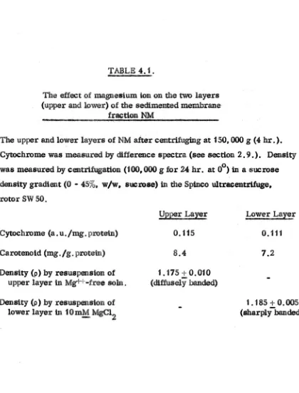
2 . 9 . C>,!ochl'ome. estb.natt.2!,
C:ytoc:hromeswet'e estimated, by difference spectra, using S()diwn dtthionite
(Bednar. 1965). For concentrated membrane samples, a Unlearn SP500 or Zeiss
PMQ II was used for the spectral analysts (wavelengths 2nmhigher on Zetss). Whole cell preparations were solubUised by diluting wtth 10 volumes of distilled
water and treating with nucleases , A Beckman OK ..2B spectrophotometer was
used for the spectral analysts.
For the analysts of minor cytochrome components, preparations from
a colourless mutant of H.salinarium, strain 1M were used. The absence of
carotenetd (ltght-absorption max. tn water 500nm) facilitated cytochrome
estimation. Membranes (NM) were prepared from cells of the mutant, omitting
the CEFB stage, and ascorbate-N,N,N',N'-tetramethyl-p"'phenylenediamine
was used as the reduCing agent. The Beekman instrument was used for the
spectral analysis.
L.!.Q.
Amino ,.!lctd analys~s .. hydrolysisThe protein or lipoprotein samples weredialysed against at least eight
changes of distilled water over a perted of 3 days at 2°. Duplicate altquots
con-taining approxtmatelyt mg. protein were ptpetted into heavy walled Pyrex
digestion tubes (18 x 120 mm. with a 100 mm. 'stem of 6 mm. inner diameter)
and were frozen in liqUid nttrogenand lYOIi1Utsed. 0.25 ml. micro-analytical grade concentrated HCI (freshly opened bottle) and 0.25 ml. 0.001
M.
norleucine in 0.01M
HCI were added and the tubes were immersed inliquid nitrogen until the contents were just frozen. The tubes were then sealed under vacuum«
50 mte rone) alloWing the contents to thaw inorder to remove traces of"
dissolved air.
The tUbes were then maintained at
11.0°
:ti°
for 22 hr. 'in a heating blockinan air oven provtded with forced air circulation .After cooling to Nom tem-perature, the tt1bes were opened and the HOI removed under vacuum.
HCI was normally removed after hydrolysts by plactng the tubes in a deSiccator over KOH in preference t-o the use ola rotary evaporator, Since
2.5.
,this p:rocedure may lead ~. decreased }'i.Ids
et
serine and threonine and the appearance of several.mallnew peaks onthe·ch3;Omatograma.lk ..wa itSuell (1961) haye suggested that the formatk>n of glutamyl 8e$e is al80 greater
d\trIn,
theevaporattoD ifv:a¢uum de81eeauon lBem}ioyed Insteadf>t r&tall'eYaporatton.
No elUomatograti1le ·evtdeDCe wu fGund fo~ tqly of these artefact. In accordance with'C;r.•stiteld, MQol'e 8& Stein (t963)who found only barely detect-aW. quantittes ·after
2f
hr. in a desiccator.When the hydrolysate WN dry (normally afteE'4 - 12 hr. in the desiccator) the fUm was di8801ved tu 5 mi .. of freshly pr~Qd pH 2.2 buffer (Moore Si:
o
,S~tn, 1954) and stored at 0 until analysed.
!:!!.
Ammo aetd ,8llQly.er.- equipmentehans:es' _
Theanal'yal8 'of protein or UpoptcOtetnbydJio"lye.ate. w.asperfotmed on a
Beckman model 1,20C amtoo acid analy.eir set up as,described. iD 'the Instruction
.Manual, (A"1M"St 1'"5)With the fGllowUlJ modifleatjou :
The AA 2' :reBUtsupplied for the analpl. of baste amino acids gave a.poor
resolution of tJtyptOpbsn and lysine. It was' thar_re replaced by the PA 35 ..e. in
whtch was origtnaUy 8upplied for: }i1y.tologtca1 fluldaanalysi8 and which gave a
_tter
reeelutloD pe~unit
of colvmn length than the AA 27 resin. bl addition, thelength of the re.ln¢olumn was increased from 5 'cm. to 1 cm.
Occuionally veltyhlgh Deck pre•• ur•• were buUt up kl the 7 cm.column due to compre8sionof the reain, which ,tended to lDcreuethmughoutthe, duration
ot
e$h .analy.". The high operating pressures ¢QuId be relieved by presoaking the :chromatography columnm
50% NaOH Qverntght., whtehpreliumably etcbed the'Column and mlntmtsed movemeat 01·there.in in the column when under prequre. To meteB.$e thesen$ltlvityof the apparatue fO.I" dltdetectlonof '.ub ..tnlcro
diameter and 10 mm. long. Windows and tubing ecnaeettens were fitted as
des-curibed by JoneS & WeiB:$.(1.965). Instead of fittiug a slide holder to the cuvette mounting plate, the adQitiQnal flow cell and the alte.mate 2. 2 mm. flow cell wel",e mounted together between brass plates aad the whole Q.S$embly was inserted in
a groove cut inthe lllQW1tlng plate. The slide assembly could be shifted
re-producibly between twQ positions governed hy end Stops. Both cells when positioned ID the Ught path were at the original optical centre of the colorimeter, so that no
modtncarteaa ·ofthe light seuree were necessary. The long path length ceU gave
an atnpllftea.ttonof peak area, compared to the 2.2 mm. cell, of 4.60. Ifthe amplification were strictly linear mthpath length, the faetor would be ".55.
!:
12. Analysis of protein JlY<:b!lysatesThe analysts of protein hydrolysates was perfotmed as described inthe Instruction Manual (A-tM-S, 1965)supplted With the Beekman model f20C amino add dnalyser used. '11leshort column analysis time was increased slightly due
to the lengdlened resmcoluBlIl (see 'Equipment changes'). A modified long column
analysis was employe" for amblQ s~aX' detenn~atten. (.see 'Amino sugar analysis').
In aQ4ition to the standard Pl"Oteinhydrolysate ·am.kloacitil (SeQkman "caJ,lbranon
mix") the fQUowtngamino aeida were observed on chromatograms :
!l
Cysteine, cystine and cYsteic acidTIl.cystine peak obtained when a hydrelysate was analysed wasnot symmetrical; racemlsat:ion of eysttae du.ring hydrolysis gave rise to meso-cystine a8 weU as the D, L"racemlc mixture. The meso-form was almost
com-plt~tely separated from the racemic mixture and was positioned 5.5 min. ahead
of the racemic mixture. Cysteine, when present, was positioned 2 min. ahead of proline hut could be easily r~osntsed by comparing the ratio of the readings obtained at 440nm.a:nd 570nm. Cysteic acid, whe:u present, was positioned
30 min. before aBpattlc acid.
2.7.
,3mta, ahead,
!>
Methionine 8u1phoxtde$.MethlonbleswItlowldea, which emerged jtlStahead ,ofQspattlc acid, 'Were
assumed to be·dQrlved. fr0InOxidatton of methtonine, and methionine values quoted include methlontne sUlphoxides.
!9.
Methionine sulphoneMethionine sulphone was pestttoned 3.5 min. ahead of threonine.
fl
NJo
-ilJGle~tneAUQ-tsoleuctae, which emerged 3. 5mtn. ahead of isoleucine was assumed to be derived from isoleucine, and 1801euc~e values quoted Include aUo -tsoleuctne ,
£} Nerleuetne
Norleuckle, uHd flS an kttemal standa17d, was, positioned 3.5 mtil. afte,r
leuCine.
g). Et;h!nolr,alnine
Ethanolamine wa. pg.ttloned 1. 5
mtn.
atter 8lIlIOOnia; two other peak,. on the ahort eolumn,one t,.9 mln. after lysine and one 4.2 min. after arginine, were not ,tdentlfted.Corrections
Correction has beenmade for expected 108se8 of serine (10%), threonine (5%), tyrostne (5%) and glutamic acid (5%) as estimated by Hirs, Moore &Stein (1_956). Corrections have been made to the value obtained for ammonia by
t) subtracting moles of ammonia equivalent to
the expe¢ted 108ses of serine and threonine (above) and
it) subtracting moles
ot
ammoniathat were ex-pected to have been absorbed by the sample from the atmosphere during the hydrolySlSand ehromatograPty (computed from a blank
2.8.
Contributions from tile by<uolysis 'oflipid pJ!'esentIn the lipoprotein samples was ex~ted to be low, ..wee the lipid is very low innitrogen (Steenslan4& Larsen,
1969). N~ertheless, ammonia values quoted aee Pl:Obably, inmost c:aaes, tQ()
high; particularly inthe case Qf samples exposed to urea.
Values for ttYIJtOi7banand cysteine (sum of cysteine, cystine and cysteic
acid) are also ~luded, but these are probably minimum values, No attemp; was
made to estimate thes~ aminQ aclds by more reliaple methods, except for tryptophan, which was estimated
tu
R fraction spectropb,otx)Juetrically by the method ofBeaven & Holiday (1952).
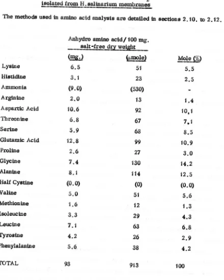
The amlnQ~td compositions of fractions are tabulated as anhyc;lto
amtno
acidl 100 mg. salt free dry weight and as mole
%,
excluding ammonia. The recovery of amino acids through the hydrolys's procedure and chrQmatograIily wasestimated by the use Qf norleucine as an internal standard (Walsh &Brown, 1962). Recovery of nodeUCine was on average 93%.
!.
131-. Amtno suer 811aly.t.Fractions for8llalyatl were concentra.ted by oxtractlilg the liptd and the
majority of the P1'Qtein by u.tng the lipid extraction procedure of HUgh&:oytt1> (1959), e.f. section 2. 14. The lower chloroform phase, containing the lipid, was diSCarded together with the layer of denatured protein which collected at the tnterfaeeof the two phae,elil QncentrifugatiQIl. The upper methanol-water (ilase
was reduced to small bulleina rotary evaperatar at, 400 and exceas salt tUtered
, o'
off. Aliquot8 were then hVclJ:nlys~ for 4 hr. at 105 In 4 ~ Hel inaealed tubes undl;tr vacuum. After cooli1lg, tlle tubes were Q~ed and the Hel evaporated under
,
-reduced pressure. Aliquots of the residue were dissolved in pH 2.2 buffet
_ 0
(Moore &: Stein, 1954) and stored at 0 until analysed.
Samples Were then p~ceesed as tor a normal amino acid analysis. Amlno sugar (as giue8samme) could be detected on the ahQrt column, between the 'acids
2.9.
gluoosamtne and galactosamtne, and alse muramic acid, diamlnoptmellc actd
and netleuelns to be resolved from the standard protein hyd:tolysateamtno acids.
ne pH 3.28 to 4 ..28 buffer c:hange was sett at 137m.m. and dle.hut-down timer
at 250 min.
1.:
14.; ExtrapJ~on of Up~dsTotalllpt.ds were extracted from preparations by the method of Bligh &
Dyer (1959, except that the fUtratlon step Was replaced by centrtfug1ngin glass bottles. The 80rvall GSA l'ot01' was used inthe RC2-B centrifuge; spins were for 10 nnn. at 2000 g. After centrifuging. a buff.sttcky layer of denatured
protein and nucletc actd collected at the interface of the methanol and chloroform layers .This was re ..extracted by repeating the ortgtnal extraction. Combined chloroform layers were then reduced to ,small volume by d1st1l1atton and taken 1;0'
dryness under nttregen, The residue was re -extracted Wi·thdrychloroform to constant weight,· The extraetton procedure ad aU subsequent manipulations were eartiedG\lttnllubdued ltgbl!. Llptd88mples were stored at ..200 under vacuum
inthe presence of a self"lndleattng desslcant (sUiea gel). Non-saponifiable lipid waf extracted. by the method of Kat;es, Palameta,
JOG.
Kushner & Gibbons (1966),2,15, LtEl~analy.es
Total liptds were estimated gravtmettteaUy. Ltptd phosphorus was
estimated by the method of Rhee81 Dugan :(1967) usingsulphurlc setd -
1C1fo
(w/v) pel"ohlol'ie acid (1 : 1v/v) as digesting reageBt and t-amlno-2-naphthol ...4"sul{ilonlcactd as reducing agent. Similar results were obtabled by the method ·of MeClare
(private communication) ustng
7rm
(w/v) perchlortc actd aa, digest1ng reagent and ascorbic acid a.reduCIn, agent. Vicinal gly¢Qls were 'determined by pertodate oxidation (KamavsIcy & Brumm. f95.5) feUowed by a fotmaldehyde 4etetmtnat1oDwith chromotroptc acid (MacFadyen. 1945),
-The genell'al method employed for the analytical fractionation of the
com-pIe. Itptd extJ."actobtained from H, sa1tnarlum was ElcoJ,t\bLnati&n of DEAE "Cellulose
~-. .
~ol\UlUl~hr(;)lnatQpaphy and th1D-layer chrolXmtQgraphy (Rouser. Gallt, Lieber.
2.10.
to separate majol1lipid olsaaes .• thts method being used
m
preference to methods'of separatt<>n based on the st!lubtltty properties of lipid classes, as recommended
by Lees (1957). The precipitation of phospholipids from chloroform solution by
acetone (Faur4, Marechal tit Troestler, 1964) had,
m
any case, not been successful. 2.1~~olumn ehromatosraphy of itptds .. DEAE.-ceUulose"Themethod used was essentially that of Rouser, Kritchevsky, Galli &
Hellet(1965). DEAE"'CeUulose (5g. Whattnan DE-50) was pre-washedwtth three
cycles of 1!!! HCI and 0.1
!::!
KOH, washed withwatel" (3 vol.). acetic acid (3 vol.) and methanol (3 vol.) and then air dried and deSiccated over KOH. The resin wasconverted to the aeetate form bystandtng ttovemtght in. acetic aetd. The resin was' packed in acoiumnof,1 cm. inner diameter" and washed wtth methanol (4 vol.) and chloroform (4 vol. )'. The mixture to be arutlysed· (100 mg.) was applied to the top of the column as a chloJ;ofotm solution. NOD'.-.poIarltptds were eluted from the
column with chloroform. polar ltptda with methanol "'chloroform (1 :-2, v/v) and
chloroform-meth.anol-28% (w/v) aqueous ammonia. (8:4 :1, by vot.}, After use, the resin was regenerate(} by washing with methanol and chloroform.
2. 17. C~lu1lUlchromat08!aJ!lyo~ ltptdS.... De¢a1so
Deeal80 (5 g. Permutltt) was ·packedina column of 1 em. diametet, fitted
. .0
with a tap lubricatedWith charcoal, as a slurry in light petroleum (40 -60). The mtxtureto be analysed, the non-polar Upids from the DEAE-cellulose fractionation
(see sectloD. 2.16). 1Q mg., was appl1edrQthe tOp olthe column as a suspension in
Itght petroleum, after pre ..washtngthe column with light petroleum. Non·polar
lipidS wereelut.d wlth ltghtpettoleum (100
w. ).
A fraction eontatmng menq .. quinone waBeluted With 2% (v/v) of diethyl ether'ln light petroleum (15 mI.) and finally most of the remat,ntng lipid was eluted With methylal (dtmethoxymethane).!.:.
18.. Column chromatoEa~y ·oflipids .. Alumina.) ..
Acid-washed alumina (5 g. Woelm. Grade II) was packed
m
a column of1.6 cm. dlameter as a slurry
m
light petroleum (40 - 60°). The lipid fraction toI
2.1f .
inltgh* petroleum tQUowed by tner(l!llsmgeQlle$lltt.art.ons (v/v. 2%. 4%t 6%. 8%, etc:.),,)f diethyl 'eth .. ,tn
lilht
petroleum.2.19, "m,~-laY~ll'ehl;9m.~~oS!ael
2S0~thlck plates. 20 em. X 20 em .• of sUtea gel H(Merek) were prepared and seated as in the tITLe path method'· of Blank. Schmidt &: Prtvett(1964) to
lmptove resolution. Plates were activated by drying at 11Cl' for 1hI'. Afte~ the appltcatiol!lof the' lipid samples" plates were dev$;tOpfbd Invartoss solvent systems
in¢onv.tlonal tad. lined With fUter paper. For die analysts of tGtai lipids or of phospholipids,. 'eotvent system A was used. whleh was eh1orototm·lllethatml-watet (65 t25
:4, by
vol.). For the analyst. of neutral lipid traction,$" elther solvent system B"dt,ethyl ether-light petroleum (40 - 60o)-acedc actd ,(30: 110 :1, by vol. Jor a01v$ltsymemC. me$anol-ehloroform (1 :4. vIv) were used.Lt,ples we,re
vt,suaJ;tsoo
by eh.arrmg with SO%('v/v) 'H2SO.€ur byspraying With litbadan1tne60 (Ma:rtnetti, Brbland St Koohen.195'1). Pho.sP1ollplds were detected Wildt the Hane$ ..lshe~()od $pray (Halles'1$her\v00d.
f,949).Cts-.glyools w~re detected by the periQdate-Sehlff J'eutloll (SastrY& Kates, 19(4). 2 .20.Jt8tlnl~t'O~~ ~{Me~iUitl.one and, «~f~teno14
... _. ."' .. . _ it, 1$. . _,' _. - ( , •. _-r, _ ~__, .. ,_,i - y
MoaquUtoile was estlmaled d\rectly ftom the fraction ,of non"wla.r lipid 'see section 2, 16. )ehtted from the Decal_column In 2%dtethyl ether
1U11tl4t
" ;,.
pettoleum (8" "~otl,2.11.) from ~e u.V. abS()~on at.2491un. Ine141ob~e
".
.
ii.
..
.'
-S:$lll(lOJil uatng ·.$Jl1
t\~.
val~e ·of2,4 (Bishop" ,_~ & Km" 1962). A gravt ...melrie ~.t1mat\on o~ ¢lis fJ1aatlon bl4tQa,te4that.
it
wa.90 ... 100% lllm'laquUlGDC'(tJ$t.tmat:ftds~¢ttopbQtQm~t~ioaUy) •
Cs;J;$tCilllCi>tdaW.l.tfe(elftraeted from. p"~at\()nl ftu!$~de4 In fS% ,(Vi/V)
NaC! 8~1\lt1onWith 2 Y0l. a~_()ru~, a.n.dt~@ete\fr~ 1)0 ~the;r. An E
!;.
vaiu4),of2500 at .9. nm. was U$e'd (Ltaaen J(tlt$@n, 1962)., $p!K'.t.opbot~IX.l~td~dat$. W4iJJ;:e
obtamed on ~ Zet$s PMQIp~t.t0pbo·t()m.et. ~~2
t.
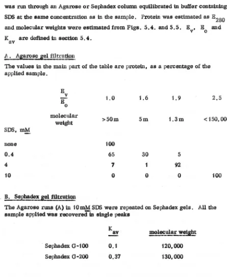
,Gel fttt~atktn2.12.
was used. For molecular weight estimations and for various fractionation
precedures u8tngdete,l'gent or 111'$4, .mallel' diametel'Golumns were used;
50 - 100 em. ~ 1.6 em, U1n~:rdiameter. tUliUgBlo-Gel A-150m or A-SOm, or Sephadex 0-200. 0-100 Qr 0-25 (Phannacla) as gel filtration re'in.
InmoSt expe.rim<m.t" a baste buffer, Qeslgnated buffer ~, of O. 1 M
. .
--NaCl, 0.05
M.
trts. HC1. pH St0, and 0.02% 'Qdium azide (as a bactertostat), was used. Insome e"peI1men.ts 1 - 10~MgC12' 0.4 -10~80dium dodec:yl sulphate (SDS) 01' 6 M urea were added to the-'
baste buffel'..
Columns Were run under reversed flow, as this miniml$ed eolUDll'l,padctn8
and resultant high back pressures. Buffer Was pumped upward$ through the column
by means of a POristaltic pump (LK3 4917,A)and the sample was applied by
temporartly removing the tubing cenneettenIrom the buffer reservoir and placing it inthe sample tube,
ay
this preeedare, ~twa.s rendered unneeessal"Y to stop the flow of liquid; through the column, which tended to cause disturbances intheUvicord .. The small bubble. introduced were removed by the use of a bubble trap.
Effluent fl'aeUona were monitored on an LKS Uvicord (type .710A) and collected
on a fraetion coUeeto~ (LKB .3404B) o~rateQ on a ttmed auto change.
Samples applied were approximately 20 mg. protein per cm. cross section
of resin and flow rate& Were apprextmately 18ml./hr. except for the columns run
inbuffer Containing
6
...
Murea, when the flow rates were' 10ml./hI'
,Blue dextran ·:ZOOO(Pharmacta), aldolase. hexokinase, haemoglobin and ribon1.ielease A (Sigma.)and digitonin (Calbtochem) were used as molecular weight standards, on columns
equ1l1brated With buffer f6l, All molecular weights obtained are calculated on the assumption that the calibrations with columns equUtbrated wtth Huffer
a
are appropriate when buffet'S containing urea or detergent were used. There la someevidence that this ~8um.ptlon may not be valtd; Rosenburg &: Guidotti. (1967) have reported that the pore siZe of some gel filtratton resins are altered inthe presence of unbuffered SOS. No attempt was made to eonitrm this or otherwise. for it was
eXpeCted that the calibration proteins would also be susceptible to, for example,
2.13.
quoted are 'apparent, 'J In the (lase of samplee UJ. dete.x:gen.t,the apparent molecular
weights may be adjuSted to a£c'oun.tfor the,'(knqwn) baunElSDS. 2.22. Poly!crllamide
s!l.
disc electrQ@oreslsAll the electroti1o:teti~ expertments were run byMr. F. Bellingham (With
the exception of some abortive experiments With eetlutese acetate membranes) but
are included in this meets since they provldeva:luable supplementary data. R.e1evant
detatls of the methods usedare as follows :
Samples, 200lJ.g. of protein, were layered onto
a.
small-pore gel (7%)over-laidwtth a large-pore spacer gel (3.5% acrylamlde). The buffer used was O.03~ sodium borate, pH 8.0, and the lower electrode was the anode. After developtng,
the gels were fixed intric.hloroaeetic acid and statned for protem (Co0massie blue), lipld(Otl red 0) or nucleic acid (methylene blue).
Some of tl:u:,'polyacrylamide gel disc eleetrophoresis analyttcal runs were scaled up and run on a p-repa;rati¥e electrophoresis apparatus. The electrophoresis
column was; elut.ed near the base Qf the gtl by passing a stream ef bL!1ferf3- without NaCl, through a thin section cut thrQugh the gel, whUst the run was in progress.
Fraettoll.w.:te ooH.:ted and monitored as fo'rtbe:gel fUuatton e.:petimenta.
2.23. lsotC!!licpoint ~termtnadon
Purified membrane-rich fraction (R) in0.1 M NaCI, O.05M trts,
nci,
-
-pH 8.O. WI;tS coneentratedby ultrafiltration (Arnicon'Dtaflo, fUter UM 2) to 10 •
20 mg. protein/ml.The sample was then desalted by passage through a column
of mixed ion exchange resins as described by Nom!and Tanford (1967).The solution of the desalted lipoprotein obtained from the ton-exchange column had a
pH which vaned With its protem concentration, as measured by tts absorption at 280nm. A plot of the pH of the desalted Itpeprotetn solutton again. the reciprocal
of its protein eo~~en~1"atton'WasItaear Over th.$ reglon E280nm c: 0.2 to 2.0 and a
reproducible value could beobtained for the tsotcmlc point of the lipoprotein by extrapolation of this plot to infinite protein c&neentranon. t..Jnustially low values
!.24. 'The btndipj' of ~auestum ton to the membrane-rich fraction (Rl
R. ftutioo, 20 mg. protein, was dialysed exhaustively against 1 ~ EDTA pH 7.0 and then d.elontsed. water. Magnestum' binding was determined by the
equtltbrtum dlalVSi$ method. The llpopr0tein was placed in a washed dialysis
bag, vol. 30ml ,; and Unmersed in .Ionised water, vol. 170ml ,; to which
known qUahttties
of magnestum,
a$ MgC12, wereadded.
Both the internal andthe external solutions were stirred for 24hi.". at 2°. After equilibration,
magnesium inthe external medium was detelimlned by atomic abserptlon spectrophotome·try. It WaS assumed that the llpoproiem was isotonic and that thenet ehuge on thelipoprotein WAS negl1glble.(Stetnhardt &: Beyeh0k,f'964).
Bound magnesium was ealculated by the lllethod of Seatchard, Scheinberg 8£ Arm8ttong (19$0) aa8utnllig no ChlOride iOll was !»md. Blank mealiurementCi,
Without magne'I,UIn ehlortdt and Without lipoprotetn,wereailO made.
~. 25. Endogenous act,1;vt!'Xot whole eeUs
Cell. were l'UU!'V$8tedas described above and washed several time.s With butfer (X until free of gtowth medium. For each wash, a ceU pellet was
re-suspended with ~etne eare inbuffet ex; the peUet flrllt being "softened' by
expo.uro to buffer torSO 'min. and then resu$I*ided in 50 volumes of buffer
Usmg a pipette With a wide o.rtflce (1 mm.},
c~n$
Were maintained at 0° at alltbtlee except dunng oent~g, when theywere maintained at _100• For the
tnvemgs;tion of the .tf~t of the age of the cUlture on the endogenous activity of the eeU8, cultures
were
grown sueh that they could all be assayed on the sameday.
A eonventiebal Clark type of oxygen electrode was used to measure
endogenou8oxygen uptake polarograliltcal1y. TIte eathede was maintained et
...;-O.6vand the generated ou.rrent Measured with a Kent mark 3 (0 -1 mv) or
ServoSCril:Mt(0 ~2 mY) reeoJfder. The endogenous actiVity o·feells, .suspended
'tn bUffer etWQ aesayed inbuffet ex at Soo. Due al1~WattCeewas made tor the
ltmtted .olUblllty ofQX':ygenincon.eentrat~d "alt solUtions, en estimate of'the
$obibtltty of oxygen
in".3M
NaCI "tng iUttrp$lattd ftomthe
reind,s fi)f-2.15.
Smith (1928).
2.26. Assay of NADH oxidase
_ _. "_. ,1. L
The NADH oxidase activity of whole cells and of envelope fractions was
measured in two ways. Insome experiments oxygen uptake was measured, as for the endogenous activity of whole cells. with an oxygen electrode. In some experiments. NADH oxidation was followed spectrephotometrteally. A Zeiss
M4Qll monochromator was linked to a Gilford 2000 recording spectrophotometer e
and the cuvette assembly was maintained at 30 . NADH oxidation was followed at
+
340nm (NAOH. ,
=
6220. Horeeker & Kortnberg, 1948; NAD •• = 20, Stegel, Montgomery& Bock, 1959). Approximately 1 mg. protein of enzyme was usedper assay, Activities were measured at the optimal NADH concenttatlon(O.04mM)
-for the NADH oxidase of an envelope fraction (CEFB) obtained by Brown (1966).
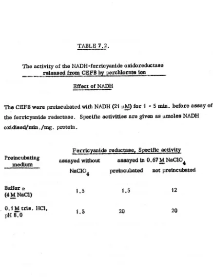
2. 27~./:..'"Y of
~4DH
"ferricyanide oxidoreductase ("diaphorase.:1.All. enzyme that catalysed the transfer of electrons from NADH to
tem"
Gyantde (as the PQtasS,iWUsalt) was assayed spectll'QphQ.tometrically on the -Gilford-ZeiSS assembly aIJwas used for the NADH oxida$. ~s8ay (secUon 2,26.). NADH oxidation was followed at 840nm, and ferricyanlde reduction at 420nm. (ferrtcyanide
and fer(~yanide ·extinetion coefficients obtained from Cohen & Plane, 1957). The
assays were performed at substrate concentrations of 21IJ.MNADH and 50lJ,M
-
-. '" o. .. .
K3Fe(CN)6 inbuffer·(l at 30 . Due allowance was made for residual NADH OXidase activity and for the, non -enzymic reduction of ferrlcyanlde by NADH.
whtch were both 'small compared with the diapbo1'8lfe activity. As the reaction was stotehiomet;tic (1 mole of NADH reduced 2 mole K3F~ (CN)6); the rea.c;tionwas
normally llftOn\tored by NADHO;l{tdation only.
Inorder to enhanee the observed actlvity (sometimea attrtbuted ,toa "diati1orase"-typeEmZ~), pteparattons Were" in some caeee, treated wtth chaotrepie agents, (DaVis ~Hateft, unpublished communtcatiGll). As wUl be Been
~ow (sectton 7 .4.) c~troptc agents. espeetally thi~yanate (SeN-) 811.4 pe.tehlorate (CIO~),
~9ie~'e
an NADH-ierzWytUli. ~JQ;do~e<luctase("ferrtcyantdeo~doreduet~") of aetivlty up toQJl$.hundred.
time. the
cltaphorue aethtty. Inthese experiments, buffer at was replaced by 0,1 M tris.
ncr,
pH 8.0, 100 mM Mg++.
-2.16.
2.28. _ Assay of ferrtcyaulde reductase activity
Tht. activity is defined as the NADH-ferneyantde oXidOreductaSe actiVity released by ehaotropte agents from prepa.ratlonsof CEFB. The effectiveness of
various chaotr()pie agents tn releaSing thiS a.otlvtty was investigated. The trtal assays were as for the diaphorase a$.ay, but chaotropte agent. were included in
~e assl;lYmedilJm at concentrations upto 4
M,.
Unless otherwise stated inthe results, theenzyme
waa added, to w.easeaymed1wn
last, hliJtin $(i)Dlecaae. the enz·yme was pre-tnc~~ed with MADH Orwith .theqbaotrep1cag.t.As a result of tb.eee trtalasaaYI, ·the ~esult8. of which are prCt.8eDtedin seetl0n
1.5.,
a standard aSlaytor
thefe:utC:~l~
re4\Jeca,e was devised J;n which the 'chaotrGPic. ag_t was perchlorate at a concentration of 3.3M_.
Although thiocyanate wasmoreefftclent, mole
for mole. than perehlQfate inreleasing the ferneyantde redu4_e aettvtty, perchlorate was initially used in preference. because itcould be obtained in a htgher state of pUrity. Insome later experiments, when the potMttally _plosive nature of pel'Chiet'ate was realised, thlc>cyanate
was used in preference. Inaddition, when the stimulatory effect of imidazole
had been observed, the tria buffer in the assay medium was replaced by tmtda.z()le at the same n»lar1ty
and
pH.2.29. Incubation of cell enve.lope fragment baas
with
bmtl)ule
m~a .Concent:rate<!sus.peneions of CEFB(1€)0 mg. protein/mi.) inbuffer ~ were exposed to hypotoniC media at 0·. The diluting buffer consisted of 0.1 M tria.
-HCI, pH 8.0 containing up to 100
In!:!
MgC12 and up
to 33m!:!,
spenntne;N,
N'-bts(3-amtnopropyl) 1,4-butanedtamine. Normally the dUut10n was 1 :50 (v/v).t,e. the salt was dtluted to 86 mM,
-
but in&Gme experlments higher dUutlonswere obtained by ¢entrUugtng (1 SO,
000
g .for2 .. 4 hr , in the Sptneo w.tracentrlfuge,rotor 50) and re,uspendtng several times tnthe diluting buffer. 'The dUuted prepara-tton~,or the final pellet and the various washings, were as.ayed for the cruwtropicaUy released ferrieyantde reductase al described in eeot\on 2.28 .
2.17.
urea in O.1
M..
ms. Hel, pH 8.0, aad again assayed for ferricyanide reductase.In some experiments the effect ofsubstltuting O.1
M.
imidazole buffer forthe trts buffer in the above incubation was studied. For these experiments the
revised thiocyanate-Imidazole assay was used for ~e ferrtcyanide reductase activity
(see
section
2.2'.).2 . 30. .Spect:ra ofl1P!q samP;les
I.r. speetraof lipid samples were taken onthin fUms deposited from chloroform or light petroleum (40 - 6fl") sotution onto NaCl plates. S~mples were
desiccated for 1hr. bef(i):respectra were taken.Thespectrophooometers used were the Perkin -Elmer 131 (Unear wavelengt'hscale) and the Unlearn SP-200 (linear wavenumber seale),
U.v. spectra wesreta.ken
m
cyelohexane, ethanol or other 8ultalllesolvent on the Unlearn 81'-800.2. 31 •..CbeIJ.)lcal.1
Unles. otherwise, ,w.ted,all chemicals were obtaine4 from Hopkin and WU11ams or Brittah Drug »0\1808 Ltd.; biochemicals were obtained from the
3.1.
CHAPrER ttl
RESULTS I
VARIATION tN THE COMPOSITION OF THE PURIFIED
M!MBaANE PRAe1'TON
<t!M}.
yvrlTH 'THE ~S OP GROWTH3.t. Int~t;ton
It is welt kno:wnthat variations
tntte.U
,cl;)lnpG'8itt&ba.tld the' aettvity of'cell enzymes octrull"~the gtowth e~le0f baete:rta (Netdhatdt, t963; Glase:r, 1%6), Van~tf.Gns iatlyoccur dtmtng the logarithmic phase (fgrGWtbj)
at) In A~rO'baeter se:!'OF!e!,s (bean &; HlnS'belwood~f959) ot during the statwnazy phase ('agemg')as tn &i$cherichta celi (CUft0rtJtM6) aBa/or in the trBDSiti&n ~eEm the
torari'thmic
aa4 stationary' {ilate8, aetil'f!aemol!!Uttspara:Jnflu~ae
(Wlllte~f962) er inBao8ltE$.sUttlts (Coleman, f96.7).There i;also evidence
-th&t'(ave!'tt,e) eellst!Ze
may
changedltritig thegrowth and agemg'Of'a culture (Dean '- Mm8~lwood, t.'959; Salle, 1tt').
It\Vas of mte~tt0$ee whether these overall change. durtng gxowth and ageh1g might be t.,fi,"ted
tn
¢hanges at the:mentbrane level. At the time' that thl:. tnve'$dgauonwall
begttn, no data were available :eoneemtng changes inthegto •• eoUlpesttttiib~f membranes eertve. ftom Gram -negative GJtganl,sm8.
Change••
t,n ml$b~tI~vel.. and
.0mpO$ltttiBduiing th~tttase ef
growth had,'_\t~.r. betn noted!ctr a nmnber ef Otam';"p0$ldveergatlleIJ .. f:tom wbleh ptGt!()plua! '(~lu.... tomes) eoa1clbe·readU~7'obtatn.e:d(aattGnlFreer, 1965
Ut MteJl'GcGec~.ll.detkqcu.altdSateUm !ut~a: ShOttimtUt'. Kolb. JJalc:ay, CQ)l~v.t
at
'TCi)'!tJUli.8" 1963 lD!,!tt!!C!»t¢u •.f.~tl.U.). ''ot
Gram"9uve Qrganisms onlySOWlJW'hat bldb:ect eVidence was3.2.
these changes were primarily a response to changes inoxygen tension as all three
organisms are facultative anaerobes (Wimpenny, 1969).
With this background the aim of the present tavesnganon was to determine
the variations, if any, inmembrane composition during the growth cycle of !!..:.!_aUnarium. The cells were grown under conditions where the lag (ilase was
ofm'inlmal duration (use o,f ammonium chloride,. 0.5%, w/v, inthe growth medium, Onishi, McCance &: Gibbons, 1965; use ofa high inoculum, Holmes, D\mdas &
Halvorson, 1965), and cultures were harvested and examined at intervals
through-out the logarithmic pha$e and from early to middle stationary phase (Fig. 3.1.).
The gross composition of the membrane (preparation NM) was studied, both protein to lipid ratios and membrane to whole ceU dry weight ratios, and the
composition of the membrane lipid was invest1,g:atedfurther, as a function of cell age. In the absence of a previous analy8is of membrane or whole celllipi.ds of
~.8alinartum, a 'fingerprinting' technique was used to monitor the levels of the
major 1ipidcomponents, sbnUar to that used by Daw80n (1966) for the analysts
of amillG acid pools durtng growth b1 Can:dldautUte. Nevertheless. With the aid of partial analyses of whole eeU ltplda of ether speetes of Halobacterlum(H.cutirubrum; Sehgal. Kates &. Gibbons, 1962; Kates, Sastry &. Yengoyan, 1963; Kushner,
Bayley, Boring, Kates
at
Oibbons., 1964.0;Kates. Yengoyan &. Sastry. 1965; FaUX'&, Marlehal &. Tt'Qestler. 1963. 1964) pal't1ai dlar_tertaatlO11 of the membranelipids of H. sa1lnarlwn Wa$ ~hleved. It should be added that a later report (Kates,
Palameta, Joo, Kushner &Gibbo.ns, 1966) had suggested that the cellllpids of a 8t_raln of
.Ii.
aa1btarlwn were 81mUar te those,of H.c:utiwrum, and also that it bad been reporteCllthat the eell envelepe, and thel"ei0-re possibly the, membrane.of H.cuttrubrum (SmttiUes, Gibbons &. Bayley, 1955) and of H.halobtum. (Brown, 19(5) consisted of lipoprotein.
The balte a.ppzoaeh used for the aaalyt1cal fract;ienation of thecomplu
ltptd mbtt\:ll'e was a combi,natton ef DEAB-q<.d1u1osecolumn ehroma~()gJ"aJibYWith
th.tn"la~r ChlOmatop:4.pJly (l\ow.er, GaUl. Lieber, maak It Privett. 1964) although 80me .ubtltdtary column work was done for the fracttonation of non -polar lipids
o
...,.,
1_... :
,m_._
1aIt-"_
.,..
nil.11.,..
/1 •....
;
~
.,~
...It..
t,mi...
o
1
so
7-
(J) (J) H H ::l ::l ...., 2 .-<--
...., ::l .-< H ::l U (J) U ...., 40 ClJ' .-<El .-< El
• .-< <, <,
H
bJJ
0 ...
... I
0 [fJ 0
U rJJ
... C':.l
(J)
El x
iJ.l [fJ
... ... .-<
.-< .-<
ID II
30 (J)
hi U U
;:l ..,_,
(J)
....,
0
.-< ...-<
::l ...., ..Q
~ ..c: CIl
,...
.~ ... ;> 01 II >-. ~ 0 ...., ... >-. Z '0 :.5 H "Cl HII 20 .j....>
:J
-
r<E--< ·H(J) ;3
0 c,..., I U ...., (J) .-< CIl ... VJ ..Q CIl • .-< > 10 o
16 32 48 64 80 96 112
3.3.
3. :2. Growth cha.racter~stlcs of the cell culture
A growth curve for the cell culture is giv~ inFig. 3. 1. A plot of turbidity or of weight yield of harvested caUs against age of culture (which are
supertmposable) both indicate that there is no observable lag phase. The
turbidity of the culture was measured inan Eel eolorimeter (filter 608. Sml.
o e
cells) at 30 using a blank ·Qfbuffer Of. Corrections were made for non-Itnarity
1\
of the scale above about :)units. The end of the logartthmtc phase of growth Is
at about 48 hr. and the begbming of the stationary phase at alx;)ut64 hr.
ThreughOllt the whole of the growth curve (upto 160 hr. growth) the cells remain as slender l'QQs. The appearance of (dub-shaped or s}ilerica! forms was assumed to be indicatiVe of anunhealtby cul.ture. and such cultures were discarded.
No change inceU siZe coUld be detected at any stage. although estimation of cell volumes by conventional light microscopy is subject to considerable error
(Patnter & Marl', 1965). If anything. the cells were slightly smaller inolder culturea, and there was possibly a higher pror;»rtloD of very long cells invery young cultures (between 0 and 16 hr.).
The viable count is alee given in Fig. 3. 1. The viable eeuat was estimated by the method of MUesand Misra (Cruickshank, 1965). Serial dilutions ·of the
cultures inbuffer·at were drop-eounted on agar plates having the same compos.tttoB as the culture medium (see section 2.2.) wlththeaddition of 1% (wIv) ·Oxold agar CM3.
The endogenous aeth1ty of washed cells (see Sect;iOB2.25.) reaches a
maximum at the end of the exponential phase and then declines rapidly(Fig.3.2.).
3.3. Chemlcaleowpo8ttion of wholeceUs
!!!.~e me~~e~rieh
ix-action &M)Salt "'free dfy
wetgbls.
protein and tetalllpid were estimated in s8.Q\p1esofwhole eells or in preparat.lons of NM (section 2.5.) as described tn'Methods'
"..
...
,..,..
~...
.,.
.
El
<,
<lJ
~
'-'
0.
;:::l
<"l
o
[fJ
<lJ
... o
El
S
o~ __ ~~
~~
-+~
~~
~~
v...
_t~..,.
.,...
et ..
et
··.ua•
• p~;
.. lipid;
50
rJl
... ...
(])
o
(])
...
, 0 ~
'-H
40
0
+-'
..c:
co
'Q)
;3:
>.
H "Cl
(]) 30 (])
H 4-<
I
+-'
...
Cil
rJl
tf2
20
..
O~ __ ~ ~~ L- ~ ~
60
- 10
, V ,..., ...
.,.
.,_
.
10 70 f=l 0 ......., C) c1j H 60 4-< OJ f=l c1j , H .0 E Q) E 50 4-< 0 ...., .c eo ... Q) ~ 40 :>. H "0 OJ Q) H '+-< I ,-' 30 ~ c1j [fJ 4-< 0 eo'" 20