REVIEW ARTICLE
Principles and Limitations of Computational
Algorithms in Clinical Diffusion Tensor MR
Tractography
H.-W. Chung M.-C. Chou C.-Y. Chen
SUMMARY:There have been numerous reports documenting the graphic reconstruction of 3D white matter architecture in the human brain by means of diffusion tensor MR tractography. Different from other reviews addressing the physics and clinical applications of DTI, this article reviews the compu-tational principles of tractography algorithms appearing in the literature. The simplest voxel-based method and 2 widely used subvoxel approaches are illustrated first, together with brief notes on parameter selection and the restrictions arising from the distinct attributes of tract estimations. Subsequently, some advanced techniques attempting to offer improvement in various aspects are briefly introduced, including the increasingly popular research tracking tool using HARDI. The article explains the inherent technical limitations in most of the algorithms reported to date and concludes by providing a reference guideline for formulating routine applications of this important tool to clinical neuroradiology in an objective and reproducible manner.
ABBREVIATIONS CC⫽corpus callosum; DTI⫽diffusion tensor imaging; FA⫽fractional anisotropy; HARDI⫽high angular resolution diffusion imaging
A
t the beginning of the 21st century, the radiologic com-munity witnessed the tremendous technical progress to-ward noninvasive investigations of the white matter architec-ture in the central nervous system. Thanks to the advancement of DTI plus the ever-growing computer technology of the so-called tractography methods, a comprehensive visualization of the white matter fiber bundles and their relationships to tumors (Fig 1) can be readily displayed without the need for any surgery.1Needless to say, the potential implications inpresurgical planning are huge, particularly if the related tech-nology can be executed objectively and automatically on a daily basis, with the reliability approaching a level that is highly acceptable in routine practice.
This article attempts to provide a pedagogic overview of the aspects of diffusion MR tractography that clinical neuroradi-ologists may find helpful for routine use. Because an overview of data acquisition via DTI and potential clinical applications has been provided in several review articles,1-3only the
com-putational means to perform the tractography will be intro-duced here. In addition, only selective methods will be men-tioned, because detailed comprehensive explanations about the massive amount of newly proposed algorithms are neither necessary nor insightful. At the same time, the ultimate restric-tions originating from the computation methods will be
ad-dressed. With the basic concepts understood, extensions to more sophisticated algorithms would ideally be easily compre-hensible. Finally, we conclude with a suggestive guideline for optimal use of diffusion MR tractography in clinical neurora-diology, emphasizing particularly the preparatory work that ought to be done before routine application. The mathematics in this entire article, though inevitable, will be kept at a mini-mum to ensure readability.
Diffusion Tensor Ellipsoid
Because the primary focus of this article is on the principles and limitations of tractographic computations, review of physical principles related to DTI by using echo-planar imag-ing will be omitted. Interested readers are referred to the series of 2 review articles by Mukherjee et al2,3for a comprehensive
overview of the physical principles and scanning optimization for DTI. At this point, it suffices to simply remember that the From the Department of Electrical Engineering (H.-W.C.), National Taiwan University,
Taipei, Taiwan, Republic of China; Department of Radiology (H.-W.C., C.-Y.C.), Tri-Service General Hospital, Taipei, Taiwan; and Department of Medical Imaging and Radiological Sciences (M.-C.C.), Kaohsiung Medical University, Kaohsiung, Taiwan, Republic of China.
H.-W.C. and C.-Y.C. received support in part from the National Science Council under grants NSC 98-2221-E-002-095-MY3 and NSC 97-2314-B-016-028-MY3. M.-C.C. was supported by the human resource grant sponsored by the Ministry of Education to National Sun Yat-Sen University.
Please address correspondence to Cheng-Yu Chen, MD, Department of Radiology, Tri-Service General Hospital and National Defense Medical Center, 325 Cheng-Kung Rd, NeiHu, Taipei, Taiwan, Republic of China; e-mail: [email protected]
Indicates open access to non-subscribers at www.ajnr.org
DOI 10.3174/ajnr.A2041
Fig 1.Colored white matter tracts rendered for a patient with a falx meningioma (yellow) on the right hemisphere near the location of the precentral primary motor cortex, superimposed on a coronal T2-weighted image. Blue is used for the corticospinal tracts, for which the right-sided fiber bundles are displaced by the presence of the meningioma. Note that though differential diagnosis for meningioma is relatively straightforward using conventional T1- and T2-weighted MR imaging, hence making diffusion MR tractography unnecessary, the example shown here at least demonstrates the validity of the displaced tracts reconstructed from DTI data.
REVIEW
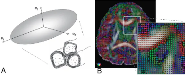
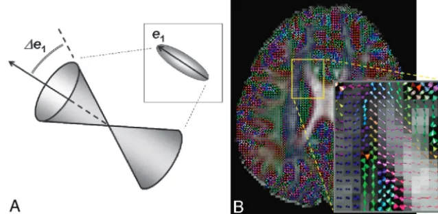
[image:1.594.339.494.292.434.2]diffusion tensor ellipsoid reflects the tendency of diffusion along various directions and that this information can be ob-tained from the original diffusion-weighted images on a voxel-by-voxel basis (Fig 2). Therefore, the direction of the longest axis of the ellipsoid, mathematically derived as the principal eigenvector of the 3⫻3 tensor matrix, corresponds to the major orientation of the axonal fiber bundle in the image voxel. The FA, a quantitative index expressed as a numeric value between 0 and 1, reflects the directionality (or equiva-lently, how elongated the ellipsoid is) of the tissue in the image voxel. Note that for most of the current tractography algo-rithms reported to date, the orientation and FA of the diffu-sion tensor constitute the sole information used to reconstruct the 3D white matter fiber tracts. Naturally, as will become clear in the following sections, the ultimate limitation of tract esti-mations essentially originates from the inadequacy of the in-formation provided by tensor orientation and FA.
Basic Principles of Tractography
Voxel Connection Based on Major Directions
One of the simplest methods for performing tractography is to connect the neighboring image voxels that are thought to be-long to the same white matter fiber tract, starting from 1 pre-assigned voxel called the “seed” voxel. This can be achieved by examining the directional consistency of 2 relationships: one between the principal eigenvectors of the 2 neighboring voxels under examination for connectivity and the other between the fiber direction and the vector connecting the 2 voxels (Fig 3A).4 The angle between 2 vectorsa andb can be easily calculated with the following inner product formula:
cos ⫽ a䡠b |a||b|.
When both vectors are normalized to unit length (ie, length⫽ 1), the denominator vanishes and the examination for connec-tivity becomes computationally very economic. After examin-ing the angular relationships between 1 certain voxel and all its neighboring voxels (totally 3⫻3⫻3 3D voxels), one forms connections by choosing those with angles smaller than a pre-specified threshold. The process then proceeds to the next voxel to extend the fiber tracts until certain stop criteria are reached, followed by volume- or surface-rendering for 3D ste-reo visualization of the tracts. Two stop criteria most fre-quently used in current tractography algorithms are the turn-ing angle and the FA value. Namely, for an acute turnturn-ing white
matter tract like the Meyer loop, the connection of 2 neigh-boring voxels would unlikely be formed at such a large turning angle. On the other hand, when the FA value of a voxel is less than a prespecified threshold, tract tracing should stop be-cause the voxel under examination is likely to contain mostly gray matter or the CSF.
The definition of the voxel neighborhood for the examina-tion of connectivity can be extended from the simplest case of 3⫻3⫻3 to, for instance, 5⫻5⫻5. This extension would allow a “jump” of the reconstructed tract in case an underlying voxel contains an erroneously estimated fiber direction due to fiber crossing or noise contamination, at the expense of com-putational complexity. The optimal choice of the voxel neigh-borhood definition is certainly a function of the spatial reso-lution and the width of the fiber tracts under consideration.
Several issues are noted here regarding the so-called dis-crete tractography algorithm illustrated above, with similar considerations largely held true for many (though not all) other tractography algorithms.5First, image acquisition with an isotropic voxel dimension is preferable for fiber tractogra-phy, though not absolutely necessary.6 For imaging data
whose section thickness substantially exceeds the in-plane voxel width, the examination of the relative orientation of the vector connecting the 2 neighboring voxels, for example, would need separate calculations along the section direction. Second, determination of the voxel connection based only on the principal eigenvector of the diffusion tensor essentially means that the information regarding the second and the third eigenvectors is entirely excluded (except partially taken in the FA value used as one of the stop criteria). Possible usage of the entire tensor information in tract tracing will be explained in a later section. Third, the examination criteria of directional consistency for connecting voxels is in favor of reconstructing white matter tracts that do not exhibit sharp turning angles. Tracts known to show prominent directional turning, such as the Meyer loop, thus represent difficult cases for computa-tional tractography algorithms.
Placement of the seed voxels is another factor that could influence the tractography results. To identify white matter tracts passing through certain regions of interest, one can pre-assign seed voxels in, for example, the internal capsule to start the tract tracing. Alternatively, the seed voxels can be placed globally within the entire brain region, such that all possible tracts within the range of computational considerations are detected at the expense of huge loads to the computations (hence called the “brute force” approach).7 Subsequently,
Fig 2.A, Schematic drawing showing the relationship be-tween the diffusion tensor ellipsoid and the underlying white matter fiber bundle. The direction of the longest axis of the ellipsoid, mathematically called the principal eigenvector of the diffusion tensor (e1), corresponds to the major fiber
[image:2.594.55.374.44.174.2]tracts passing through the designated regions of interest are then picked up by filtering out those undesired ones.
Fiber Connection in the Subvoxel Coordinate System Although conceptually straightforward, the simplified meth-ods mentioned in the previous section are rarely used in prac-tice for one obvious reason: The white matter tracts obtained therein are restricted to the original spatial resolution of the image acquisition. This is especially important because the signal-intensity-demanding echo-planar diffusion imaging sequence provides voxel widths of approximately 2 mm for the usual 128⫻128 acquisition matrix.3Limited spatial resolu-tion not only leads to loss of the smaller fiber bundles but also causes voxel-quantization errors in the tract orientation.2
Therefore, most of the tractography algorithms proposed in the literature actually attempt to trace fiber tracts in a subvoxel manner.
One of the first and rather popular methods uses subvoxel fiber tracking with the principle shown in Fig 3B.8,9Starting
with seed points placed at certain locations within a voxel, the tracts simply follow the principal eigenvector of the diffusion tensor ellipsoid until reaching the voxel boundary, after which the tracts enter a neighboring voxel to continue the tracking process. Stop criteria using the turning angle and FA value thresholds, just as in discrete tractography, are used to deter-mine the termination of fiber tracking. Note that different seed points, even if placed in the same starting voxel, likely lead to distinct tracking results (Fig 3B). This approach, therefore, allows identification of numerous thinner fiber bundles ex-ceeding the spatial resolution of the original images. During the tracking process, the computer software needs to record all
the location coordinates of the tracking steps for every single tract, instead of simply highlighting certain voxels as in the discrete tractography approach. Consequently, memory re-quirements are much more demanding in these so-called con-tinuous tractography algorithms.8
The white matter tracts can be made even smoother by using smaller steps during the tracking process. This can be achieved by assigning a predetermined step size (eg, one-tenth of the voxel width) and performing continual alterations of the tract direction according to the expected fiber orientation at each subvoxel location (Fig 3C). The fiber orientation at each subvoxel location, in turn, would need to be estimated by using the tensor field originally acquired at a much coarser spatial resolution. Distance-weighted interpolation is fre-quently used for this purpose: Namely, the fiber orientation should be closer to that of the principal eigenvector of the voxel when the voxel is close to the subvoxel location under consideration and it should be affected by other nearby prin-cipal eigenvectors in a decreasing manner as those voxels move farther away.8One simple interpolation example may look like the following equation:
ec⫽bea⫹aeb a⫹b ,
whereec,ea, andebare the vectors representing the fiber direc-tion to be estimated and the principal eigenvectors of the 2 diffusion tensors used for the interpolation calculation, anda andbare the distances from the location to the centers of the 2 nearest voxels, respectively. A more sophisticated interpola-tion algorithm to minimize angular error accumulainterpola-tion can
[image:3.594.53.389.43.361.2]certainly be used.10The equation above can also be extended
to⬎2 voxels for a maximal inclusion of all the major fiber information available in the neighborhood.
When stepping through a curve in the tensor field, the ra-dius of curvature of the identified tract is strongly dependent on the step size (Fig 3C), one of the many tunable parameters in most of the current tractography algorithms. Obviously, a smaller step size allows the tractography program to follow more closely the curvature of the tracts, at the expense of in-creased computational loads. In addition, just as explained previously for discrete tractography, placement of the seed points to start the tracking process also has critical determin-ing effects in subvoxel continuous tractography. Because there are virtually infinite possibilities in choosing the number, lo-cations, and separation of the seed points, the resulting white matter tracts derived from the calculations may exhibit a somewhat different appearance, even if the same algorithms are used with identical choice of the region of interest to place the seed points. Regions showing branching or even diverging fibers are examples most likely demonstrating discrepancies in tractography results due to varieties in seed point assignments (Fig 3B).9,10
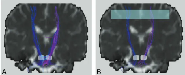
One method to increase the tract consistency is to select multiple regions of interest to retain the specific tracts that pass through all these regions. For instance, the white matter tracts within the corticospinal tracts responsible for motor function of the lower limbs can be isolated by encircling the medial precentral gyrus, the posterior limb of the internal cap-sule, and the cerebral peduncle in the midbrain. In this man-ner, unrelated tracts are filtered out, and the chance for false inclusion of spurious tracts is reduced. This technique is sometimes called the tract editing or virtual parcellation9,11,12 and is very useful in removing false-positive tracts (Fig 4). Certainly, the selection of regions of interest is based on prior knowledge of the neural transmission pathway. Application suitability in pathologic situations should, therefore, warrant careful attention.
One should keep in mind that tract editing is not only un-able to reduce the false-negative rate but may also reduce the true-positive rate in tractography. Together with the depen-dency of tract curvature on step size as mentioned before, possible inability to follow a curve accurately could lead to a loss of tracts (ie, decreased sensitivity and underestimation of tract extent). The phenomenon is especially prominent for peripheral fibers that stray out of the main tract and later get filtered out by, for example, inappropriate tract editing. For neurosurgeons, this could be an important issue.
Adjustment of Tract Orientation by Using the Entire Tensor Information
Tract connection based solely on the principal eigenvectors of the diffusion tensors (ie, the longest axis of the ellipsoid) in the voxels ignores information contained in the second and third eigenvectors (ie, the short axes of the ellipsoid). In cases of disk-shaped ellipsoids whose 2 longer axes are roughly equal, as in the case of 2 intersecting fiber bundles roughly perpen-dicular to each other, the direction determination of the prin-cipal eigenvector is prone to prominent errors in the presence of noise. One method uses the diffusion tensor matrix multi-plication to adjust for fiber direction.13For a preassigned step size, the fiber orientation incoming vector,vin, encountering a
voxel with diffusion tensorDis adjusted to produce the out-going vector,vout, for the next tracking step, by using the
fol-lowing formula:
vout⫽D⫻vin.
The physical meaning of the matrix multiplication can be viewed as a decomposition of the incoming vector into the coordinate system formed by the 3 eigenvectors of the diffu-sion tensor ellipsoid, with the 3 components weighted by the corresponding eigenvalues:
vout⫽D⫻共␣1䡠e1⫹␣2䡠e2⫹␣3䡠e3兲
⫽␣11䡠e1⫹␣22䡠e2⫹␣33䡠e3.
Consequently, after matrix multiplication, the tracking direc-tion is altered somewhat toward the direcdirec-tion of the vector with the largest eigenvalue. When the 2 larger eigen-values are close in magnitude, the amount of deflection toward the 2 directions is nearly equal. In the special case of fiber-crossing that yields a nearly isotropic diffusion tensor (ie, FA⬃0), the equal weighting on the 3 orthogonal direc-tions simply causes little change in the tracking direction. One may even use adaptive stepping to alter the step size according to local tensor characteristics for performance optimization.14
Certainly, deflection of the fiber tracking direction by using the entire diffusion tensor information can be extended to more sophisticated functions rather than the simple matrix multiplication, because over-reliance on the entire diffusion tensor is perhaps as unnecessary as reliance solely on the prin-cipal eigenvector for tractography. After all, anisotropy per se is only an index instead of the absolute measure of the direc-tionality of the voxel under examination. Fiber bundles known to be highly unidirectional in the corpus callosum of healthy subjects, for instance, can exhibit regionally
[image:4.594.53.374.44.175.2]dent FA values that may vary from 0.35 to 0.65 due to differ-ence in the microarchitecture.15,16As a consequence,
non-linear operations for tensor-based tractography, such as a reinforcement of alignment in the presence of a highly uni-directional tensor and a direct passing of the tracking vector when encountering an isotropic tensor, can be performed by examining the magnitudes of a series of shape-describing pa-rameters of the diffusion tensor (called the linear, planar, and spheric coefficients) to select the best suitable operations for the voxel under examination.17Interpolation of the tensor at
subvoxel locations can also be used for refinements.10,17
More Advanced Tractography Algorithms
The number of tractography algorithms available for diffusion MR imaging keeps increasing at an incredible speed. Com-pared with those illustrated in the previous sections that use relatively straightforward computational approaches, sophis-ticated algorithms proposed at later times all claimed superi-ority in various aspects under certain conditions. While it is unnecessary to explain the complicated principles used in all the methods, it would be worthwhile to give a brief introduc-tion to some of them to realize the potential advantages brought about by these means.
Fast-Marching Algorithms
“Fast-marching” refers to the computational process where the passage from 1 vector to another is done in a way similar to the propagation of a water wave from the seed point.5,18The
speed of the “wavefront” toward all directions is determined by the colinearity of the principal eigenvectors in the neigh-boring voxels, following which the direction of fiber tracking in a region-growing manner is assigned as the one with max-imum propagation speed.19In more advanced fast-marching algorithms, the propagation speed can also be modified on the basis of the magnitude of the eigenvalues rather than on the directions of eigenvectors alone.20Other variations capable of
resolving crossing fibers have also been proposed.21
Fiber Tracking via the Fluid Mechanics Analogy
The analogy of fiber tracking to the stream of viscous fluid flow inspired the use of computational fluid mechanics in tractography. The information from the diffusion tensor is included by a modeling process by using the Navier-Stokes equation, an equation well-known in fluid mechanics gov-erning a wide range of physical phenomena, to simulate an artificial fluid flow through the entire imaging volume.22
Sub-sequently, the most likely connection paths between pre-assigned points in the imaging volume are estimated on the basis of certain metrics (the gradient of vector flow) derived from the fluid velocity vector field. Because the diffusion ten-sor anisotropy is inherently incorporated into the local “vis-cosity” of the fluid, there is no need for a white matter mask as used in many tractography algorithms.22
Global Optimization Approaches
Tracking of the white matter fibers by honestly following the principal eigenvectors of the diffusion tensors is prone to error accumulation when the estimation of direction is uncertain in the presence of noise.10Consequently, as the distance from the
seed point becomes larger, fiber tracking accuracy would
de-crease. Global optimization approaches were proposed to remedy this problem.23-25In the simplest sense, the principles
are analogous to curve fitting as opposed to connecting all points in a scatter plot. Often some modeling is performed, first for a predetermined geometric smoothness of the fiber tracts; then the shortest or the most suitable path is searched so that a general agreement with the principal eigenvectors can be reached.23To facilitate the fitting calculation, one must define
some “energy” and “penalty” and evaluate them iteratively to reach the balance between tract smoothness and tensor con-sistency.24Needless to say, a huge number of tractography
algorithms belong to this broad family.26 In general, better
immunity to cumulative effects of noise can be expected.25
Tractography Incorporating A Priori Anatomic Knowledge
The fiber tracking algorithms mentioned in the previous sections are based solely on directional consistency or tract smoothness. In other words, judgment of fiber connection is computational rather than anatomic. Incorporation of prior anatomic information can be done via a multiple-step ap-proach, where the application of the tractography computa-tion is preceded by a global searching procedure to find the anatomic connections used to guide the subsequent fiber tracking process.27With anatomic guidance, improvements in tracking accuracy are reported, especially in the region of com-plex fiber crossing. On the other hand, because normal anat-omy is not to be extrapolated to pathologic situations, some studies even used anatomic templates constructed from a spe-cific group of patients to assist tractography.28
Probabilistic Tractography
Algorithms that construct a 3D view of the white matter con-nection pathways are sometimes referred to as “deterministic tractography.” Rather than giving direct visualization of the fiber bundles, probabilistic tractography informs the users about the estimated probability distribution of how likely 2 or more certain regions are interconnected (ie, confidence map-ping). The typical method to estimate the probability of fiber connection is to perform repeated image acquisitions from the same subject and, subsequently, use 1 specific algorithm on these datasets to obtain several tractograms.29The probability
that certain pathways exist in this subject is then found by counting the number of successful trackings, divided by the total number of datasets. As a result, the final display for prob-abilistic tractography provides the users with only a quantita-tive guidance, for which the users have to determine by them-selves how trustworthy the results are. More on this issue will be mentioned in a later section.
Bootstrap Probabilistic Tractography
The need for multiple DTI datasets to derive the fiber tract passing probability as stated in the previous section is certainly accompanied by an inevitable lengthening of the image-acqui-sition time. It is, therefore, not an attractive approach in the busy clinical environment.30Alternatively, if multiple
ap-proach).31 Moreover, when ⬎6 directions are used for the diffusion-sensitizing gradients with a single signal-intensity average, it is possible to estimate the diffusion tensor fitting uncertainties, from which the probability of fiber tracts pass-ing through certain regions can also be computed.32,33In any case, the computations needed for probabilistic tractography are generally much more demanding than deterministic trac-tography algorithms.
Fiber Tracking Based on HARDI
In the presence of intravoxel fiber crossing, the inadequacy of the diffusion tensor model to fully describe multiple fiber bun-dles has led to the development of data-acquisition ap-proaches called HARDI.34,35With sufficient data sampled at
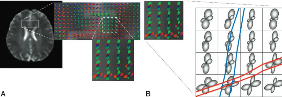
various diffusion-encoding directions uniformly covering the so-calledq-space, the directionality of multiple fiber tracts can be depicted by using a complex 3D shape called the orientation distribution function (Fig 5A). Just as in the case of a diffusion tensor ellipsoid, for neuroradiologists it suffices to remember that the orientation distribution function simply reflects the tendency of diffusion along various directions. This informa-tion can again be obtained from the original diffusion-weighted images on a voxel-by-voxel basis, albeit necessitating a much larger amount (sometimes⬎100 diffusion-encoding directions) of image data than DTI.
The directions showing the locally fastest diffusion in an orientation distribution function correspond to directions lacking substantial diffusion barriers (myelin sheath, cell membranes, organelles, proteins, etc) for the water molecules. Hence, as in the case of the longest axis of a diffusion tensor ellipsoid, directions of these local maxima are likely consistent with those of the underlying fiber bundles. In the case shown in Fig 5A, 2 major white matter tracts seem to cross each other in the voxels of interest. With all possible fiber directions in-cluded, tractography by using algorithms mentioned in the previous sections can be performed in a more comprehensive manner. Briefly speaking, all connection possibilities are con-sidered together, and the highest directional consistency with neighboring voxels is selected to form the most probable con-nection (Fig 5B). The major difference from DTI tractography
lies in the fact that multiple choices rather than a single choice are now available. Because each single voxel now contains multiple fiber bundles with different directions, angular con-sistency is relatively easier to achieve, rendering generally more fibers to be tracked.
With the presence of many fiber tracts in a large portion of the image voxels, it is not difficult to imagine that the increase in tract connection flexibility has resulted in a massive number of various tractography algorithms.36-41Almost all algorithms demonstrate advantages over other methods in certain, albeit perhaps limited, aspects. Even though the HARDI acquisition is rather lengthy and may not be suitable for clinical practice, trying to shorten the total scanning time without much com-promise in data quality has been attempted with satisfactory experience reported.42,43
Bayesian Probabilistic HARDI Tractography
Sophisticated algorithms dedicated to a combination of essen-tially all the major advantages in algorithms mentioned above also exist. One example is the Bayesian approach for probabi-listic HARDI tractography, which uses a partial volume model containing single or multiple fiber bundles with isotropic dif-fusion from free water for every single voxel.44,45With more
than 50 diffusion directions encoded in a 20-minute acquisi-tion, derivation of the uncertainty in the principal fiber direc-tion can be done without the need for multiple acquisidirec-tions or bootstrapping.26 Following voxelwise modeling, one
calcu-lates the global connection of the tracts on a whole-brain basis under the condition of voxel-by-voxel principal fiber direc-tion and uncertainty obtained from the diffusion imaging data (hence the name Bayesian to stand for conditional probabili-ty).45Better accuracy is obtained with more diffusion
encod-ing directions acquired. The result is displayed as a function of connection probability with the seed points estimated by the algorithm (Fig 6). As stated before, for probabilistic tractogra-phy in general, the users have to determine by themselves the probabilistic threshold at which the final graphic results should be shown, such that a good balance can be reached between increased false-positives versus lost tracts.
One important characteristic of the Bayesian algorithm is
[image:6.594.59.531.43.206.2]that the construction of the tracts is based on an integration of the fiber direction uncertainty into the global optimization approach. When only a single fiber is modeled in a voxel, the presence of intravoxel fiber crossing would simply lead to a relatively high uncertainty for the principal fiber direction, rather than an erroneous orientation.44Tract reconstruction
based on conditional probability thus still allows a connection if the nearby neighboring voxels exhibit largely consistent fiber directions. As a consequence, stop criteria using a turn-ing angle or FA value play a relatively minor role, with the accompanying advantages of avoiding some of the tract loss from the settings of stop criteria.36
The advancement of HARDI tractography is currently an ac-tive field of research and development, as can be evidenced by the increasing number of investigations in the literature. No matter whether one prefers the use of model-free algorithms34,35,38or the
model-based approaches,26,44,45 it is anticipated that the
im-proved differentiation of crossing fibers in HARDI tractography will find unique neuroradiology applications that are hardly achievable by simple diffusion tensor tractography. The poten-tially critical role of HARDI tractography can, therefore, be ex-pected for clinical routines in the near future, particularly if the heavy computational loads could be solved with continuous im-provements in computer technology.
Limitations of Diffusion Tractography
In the authors’ strictly personal opinions, the most important limitations of tractography in clinical neuroradiology lie in the nature of computational estimation based almost entirely on fiber directions in the presence of noise. There are actually 3 key words embedded in the previous statement: computa-tional estimation, fiber direction, and noise, whose effects are obviously mutually integrated. Although each single factor could theoretically be dealt with by using proper methods specifically designed to reduce tracking errors, it is difficult to isolate the combinational influences independently. There-fore, even if the number of tractography algorithms stated above is overwhelmingly large, the fact that they are all using directional consistency as the major constraint should give one an idea of their ultimate limitations.
First, all computations are simply estimations. For any estima-tion method, the false-positive and false-negative rates determine the extent to which the method can be clinically useful. Unfortu-nately, diffusion tractography for the human brain is a case in which the results are hard to validate in vivo; hence, false-positive and false-negative rates are both unavailable. Under such
circum-stances, comparison with physical or mathematic phantoms27
and verification by using animal experiments,46both having
lim-itations to be addressed below, are probably the best one can do to test effectiveness. For the human brain in vivo, investigations on the sensitivity of the tracking results to the choice of freely adjust-able parameters alone has shown that many tractography algo-rithms are not simple robust turn-key systems. For instance, a recent study examining the corticospinal tract concluded that the placement of seed points significantly influences the tracking re-sults.47This outcome is not surprising considering what we have
illustrated in Fig 3B. Other reports found that the correlation of tractography results with pathologic status depends strongly on the threshold setting of FA.48,49Moreover, in our own experience,
running the same tractography algorithm with all identical pa-rameter settings on 3 different personal computers could result in quite distinct outputs (ie, computer-dependent!), not to mention the expected confusing discrepancies in tracking results when us-ing different software algorithms.50
The example of seed point dependency illustrated above exhibits direct difficulty in the reproducibility of tractography results. Prescribing section orientation at a slightly oblique angle may lead to rather different placement of the seed points in terms of the anatomic location, even if the region of interest is chosen carefully, particularly because the seed points are likely placed in a subvoxel coordinate. As a consequence, even if only 1 tractography algorithm is used with fixed settings of all variables, the tracking results are not free from computa-tional imprecision. Visualization of the specific white matter pathway does not give accurate information in terms of the size or volume of the fiber bundle, either.51
Second, fiber tracking based totally on directional consis-tency computation risks bias in favor of connecting fibers with similar orientations. As mentioned before, highly curved fi-bers such as the Meyer loop may be easily missed. In addition, incidental consistency in fiber directions leads to identifica-tion of spurious tracts with no actual anatomic existence.52
This issue is further complicated by the presence of noise be-cause of the different levels of influence on direction deriva-tion. Estimation of the principal direction of a diffusion tensor exhibits high accuracy when the diffusion tensors exhibit high FA values and are less prone to noise influences. In contrast, for diffusion tensors whose first eigenvalue is not much larger than the second eigenvalue, the angular deviation from the true value could become rather large. Therefore, fiber tracking reproducibility was reported to be somewhat worse in the op-tic radiation and pyramidal tracts than in the callosal fibers.53
[image:7.594.134.452.42.141.2]Likewise, the existence of a large number of studies demon-strating successful tracking of major fiber bundles such as the corpus callosum should not be regarded as the validity of tracking results of the increasingly challenging tracts such as the language pathways.54,55
[image:8.594.133.452.45.201.2]Investigators having noticed the importance of estimation errors in fiber direction have proposed means to graphically depict the directional uncertainty on a voxel-by-voxel basis (Fig 7).45,56Models applicable to identifying the limitations of
tractography algorithms are also available.57While likely
use-ful in checking the reliability of fiber tracking results, the com-plexity of these tools is perhaps no less than the tractography algorithms themselves, hence substantially lowering the inten-tion of usage for the general clinicians. It would be ideal if the uncertainty assessment could be integrated with the usage of fiber tracking algorithms as part of the training program, translating imperfect tractography into a useful clinical tool,58
which certainly entails much effort.
The third factor imposing limitation to the tractography algorithms reported to date lies in the difficulty of obtaining systematic verification for the human brain in vivo. Valida-tion studies, though large in number, are mostly focused on limited aspects only and hence should not be extrapolated to the general situations unless careful attention is paid. For instance, documents demonstrating successful tractography results compared with manganese-enhanced T1-weighted im-aging on animals actually validated the identification of only 1 or some specific tracts,46instead of multiple white matter pathways as would be done with general computational esti-mations. The information obtained from diffusion MR trac-tography and manganese-enhanced T1-weighted imaging should, therefore, be treated as complementary.59In addi-tion, because clinical diffusion imaging with limited scanning time hardly reaches the data quality achievable in animal scans, the same algorithm applied to the human brain is an-ticipated to show somewhat inferior performance. As another example, most reports on HARDI tractography mainly fo-cused on the capability of resolving fiber bundles with crossing configurations (ie, the unique capability of HARDI compared
with DTI).38-40Few have taken into account the white
mat-ter tracts exhibiting high curvature.44,60Naturally, validation
studies showing the resolving of fiber crossing on phantoms only demonstrate the limitation of the diffusion tensor mod-el,61rather than the real success of HARDI tractography.
All issues mentioned above indicate a clear rule in the clinical use of diffusion MR tractography: Caution is always highly warranted when performing sophisticated algorithms. Unfortunately, articles showing positive results tend to out-number those with negative ones in the literature, giving the general audience the strong (and actually correct at least par-tially) impression that the noninvasive diffusion MR tractog-raphy is clinically effective and useful. Letters about negative results,55being extremely limited in journal publication space,
often do not detail possible sources of errors in a clearly repro-ducible manner. As a consequence, even if many pilot studies demonstrated encouraging hope,62,63one really needs to go
through the tedious procedure of a reproducibility study for an assessment of the precision. Not only are comprehensive investigations examining the clinical effectiveness of diffusion MR tractography in surgery strongly encouraged64-66 but
comparative studies of the postmortem human brain speci-mens using a large cohort are also highly desirable.67
With continuous technical advancement in computational algorithms or via careful use of the current tools by experi-enced operators, precision and accuracy of diffusion MR trac-tography are expected to increase to a clinically satisfactory level. In such an ideal situation, one should still keep in mind that other ultimate limitations exist. Differentiation of affer-ent and efferaffer-ent fibers is curraffer-ently impossible with diffusion MR tractography.1,2,5 Ambiguity between fiber kissing and
fiber crossing is another example that is hard to solve even with the HARDI data acquisition.10Misregistrations arising
from geometric distortions of the original images acquired by using the echo-planar technique have to be corrected. Fur-thermore, directional consistency between neighboring voxels (and hence showing successful tract connection) does not guarantee fiber integrity, even if directional consistency and fiber integrity are generally believed to be positively correlated.
Nonetheless, we would like to provide some basic guidelines in the following section, to help the clinical neuroradiologists reach the level of these ultimate limitations.
Guidelines toward Applications in Clinical Neuroradiology
Always Control the Original Image Quality
Obtaining the original images with sufficient quality is al-ways the first step in convincing people of the validity of any processing or analysis subsequently applied to the imaging data. In diffusion MR tractography, it should be clear that the accuracy of the fiber directions is positively associated with the signal–to-noise ratio (Fig 8), which can be in-creased usually at the expense of scanning time or spatial resolution. Optimization of the routine protocol in a busy clinical environment is, therefore, an important prerequi-site toward successful tractography.2,3In addition, for
cer-tain tractography algorithms, there may be special require-ments for the format of the original images, such as separated storage of individual acquisitions before per-forming signal-intensity averages68 or the preference for
multiple diffusion-encoding directions over multiple sig-nal-intensity averages.2Before pooling the large amount of
original data for the generation of beautiful 3D tracto-grams, it is better to make sure that the image-acquisition protocol has been adjusted in accordance with the require-ments posed by the tractography algorithm.
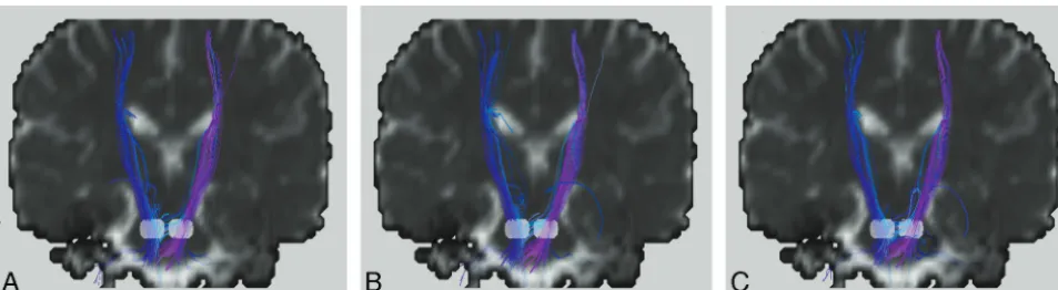
Choose the Algorithm You Understand
Sophisticated computational tractography algorithms, though supposedly performing better in some aspects, are not neces-sarily suitable for the needs in routine practice. Differences among various algorithms can be subtle (Fig 9). For example, one method may be superior to another only when the signal– to-noise ratio is above a certain level, and hence the compari-son results obtained at 3T might show a reversed trend at 1.5T. Because the characteristics of algorithms vary to a great extent, choosing the one with details understood by the operators is likely the dominant factor in successful usage. When a com-parison of different tractography algorithms must be made, it would be ultimately important to compare the visualization of fiber bundles with the same tracking criteria and seed points and on a standard subject or a group of subjects who are well coregistered in the same coordinate system. Under these cir-cumstances, the performance of tractography algorithms would be minimally data- and region-of-interest-dependent, and any newly proposed algorithms could thus be tested and compared objectively.
When the choice of tractography algorithm must be based on other criteria (eg, immediate software availability, data processing time, and so forth), an equivalent alternative is to understand the algorithm one chooses. In addition to spend-ing time studyspend-ing the computational principles and playspend-ing around with freely adjustable parameter settings, trying an uncertainty test on major and minor tracts is strongly
encour-Fig 8.A somewhat exaggerated comparison of the signal–to-noise ratio effects on diffusion MR tractography. Fiber bundles connected to the corpus callosum (mostly red) and the corona radiata (mostly blue) are shown in the colored tracts superimposed on a sagittal image (with the subject facing the right-handed side). The figures inA,B, andCare processed on original MR images acquired with 2, 8, and 20 signal averages, respectively. Note inA, compared withBandC, the accumulated errors in fiber orientation particularly in the inferior part of the descending tracts and the frontal portion of the callosal tracts, as well as the presence of spurious tracts.
[image:9.594.63.532.45.160.2] [image:9.594.56.533.217.348.2]aged56,57,69to have an idea of the algorithm’s limitations so
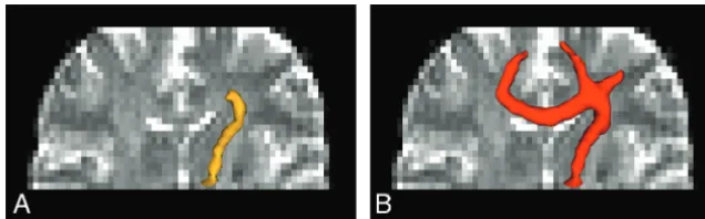
that one knows where to start or stop the interpretations of results. Another rough way to evaluate uncertainty is to check the left-right symmetry of reconstructed tracts when selecting a central region of interest (eg, the splenium of the corpus callosum) on images obtained from healthy subjects,70
pro-vided that the fiber bundles do not play functional roles with known lateralization (Fig 10).71
Perform Reproducibility Tests on Healthy Subjects A reproducibility study using highly cooperative healthy sub-jects is a very important step for tractography quality control. Performing it is relatively straightforward, though admittedly rather tedious. Healthy subjects are recommended because any missing of well-known white matter tracts is unlikely to be attributed to anatomic anomaly, hence likely reflecting algo-rithm limitations. Through an examination of the tractogra-phy results from repeated processing, one should then identify possible sources of error and attempt a remedy. For instance, an objective algorithm with parameters all set to fixed values should allow interoperator consistency as high as intraopera-tor consistency, leaving the choice of regions of interest the only degree of freedom. Comparison of within-scan versus between-scan agreements is also essential in figuring out the direction of improvement in terms of either the computations or the acquisitions. In rare instances but having been encoun-tered in the authors’ experience, the same algorithm analyzing the same dataset on different computers may create discrepant results. Hence computer dependence is also among the many factors that may need to be repeatedly tested by using healthy subjects.
Bear Limitations in Mind When Applied to Clinical Practice With all the effort spent in the preparatory work as suggested above, clinical applications on a routine basis should be largely ready. Even so, one should have a conservative attitude, keep-ing in mind all the technical and ultimate limitations of diffu-sion MR tractography. A change of the operator responsible for processing tractography or an alteration of image quality
due to upgrading of the MR imaging scanner could both affect the results in a time-varying manner. Another subtle but very practical and somewhat paradoxic challenge to the neuroradi-ology administrator is the ever-advancing computer technol-ogy: As the calculation becomes faster and the memory space keeps increasing, the desire to obtain tractograms at a higher spatial resolution in less time will, for sure, pose strong de-mands on the change of the predetermined protocol. Main-taining the pre-established quality while being continuously updated in the technical progress, at the same time noticing the current (but time-varying) limitations, is certainly no less complicated than the difficult and time-consuming radiologic diagnosis of rare diseases.
Conclusions
The clinical implications and potentials of diffusion tensor MR tractography in the noninvasive investigation of white matter tracts will continue to show promise. From a visualiza-tion of pathologic alteravisualiza-tions of the fiber bundles to presurgi-cal evaluation of brain masses, tractography provides neuro-radiologists with perhaps an elusive hope that white matter morphology can now be depicted with details exceeding the original acquisition resolution. Nevertheless, after reviewing this article, readers should agree that putting diffusion MR tractography in clinical practice with sufficient accuracy is no easy task either. With the discovery of new tracts, reasonable speculation on the technical inadequacy of the tractography algorithms remains the first healthy attitude.10Even when
ob-taining fancy tractography results highly consistent with ex-pectations, careful conservative attention in the interpretation is mandatory.
References
1. Nucifora PG, Verma R, Lee SK, et al.Diffusion-tensor MR imaging and tractography: exploring brain microstructure and connectivity.Radiology
2007;245:367– 84
2. Mukherjee P, Berman JI, Chung SW, et al.Diffusion tensor MR imaging and fiber tractography: theoretic underpinnings.AJNR Am J Neuroradiol2008; 29:632– 41
3. Mukherjee P, Chung SW, Berman JI, et al.Diffusion tensor MR imaging and fiber tractography: technical considerations.AJNR Am J Neuroradiol2008; 29:843–52
4. Terajima K, Nakada T.EZ-tracing: a new ready-to-use algorithm for magnetic resonance tractography.J Neurosci Methods2002;116:147–55
5. Mori S, van Zijl PC.Fiber tracking: principles and strategies—a technical review.NMR Biomed2002;15:468 – 80
6. Oouchi H, Yamada K, Sakai K, et al.Diffusion anisotropy measurement of brain white matter is affected by voxel size: underestimation occurs in areas with crossing fibers.AJNR Am J Neuroradiol2007;28:1102– 06
7. Huang H, Zhang J, van Zijl PC, et al.Analysis of noise effects on DTI-based tractography using the brute-force and multi-ROI approach.Magn Reson Med
2004;52:559 – 65
8. Mori S, Crain BJ, Chacko VP, et al.Three-dimensional tracking of axonal projections in the brain by magnetic resonance imaging.Ann Neurol1999; 45:265– 69
9. Conturo T, Lori N, Cull T, et al.Tracking neuronal fiber pathways in the living human brain.Proc Natl Acad Sci U S A1999;96:10422–27
10. Basser P, Pajevic S, Pierpaoli C, et al.In vivo fiber tractography using DT-MRI data.Magn Reson Med2000;44:625–32
11. Catani M, Howard RJ, Pajevic S, et al.Virtual in vivo interactive dissection of white matter fasciculi in the human brain.Neuroimage2002;17:77–94 12. Huang H, Zhang J, Jiang H, et al.DTI tractography based parcellation of
white matter: application to the mid-sagittal morphology of corpus callosum.
Neuroimage2005;26:195–205
13. Lazar M, Weinstein DM, Tsuruda JS, et al.White matter tractography using diffusion tensor deflection.Hum Brain Mapp2003;18:306 –21
[image:10.594.90.245.39.218.2]14. Chou MC, Wu ML, Chen CY, et al.Tensor deflection (TEND) tractography with adaptive subvoxel stepping.J Magn Reson Imaging2006;24:451–58 15. Hasan KM, Gupta RK, Santos RM, et al.Diffusion tensor fractional anisotropy
of the normal-appearing seven segments of the corpus callosum in healthy adults and relapsing-remitting multiple sclerosis patients.J Magn Reson Imaging2005;21:735– 43
16. Hofer S, Frahm J.Topography of the human corpus callosum revisited: comprehensive fiber tractography using diffusion tensor magnetic resonance imaging.Neuroimage2006;32:989 –94
17. Westin C, Maier S, Mamata H, et al.Processing and visualization for diffusion tensor MRI.Med Image Anal2002;6:93–108
18. Parker GJ, Stephan KE, Barker GJ, et al.Initial demonstration of in vivo tracing of axonal projections in the macaque brain and comparison with the human brain using diffusion tensor imaging and fast marching tractography. Neuro-image2002;15:797– 809
19. Jackowski M, Kao CY, Qiu M, et al.White matter tractography by anisotropic wavefront evolution and diffusion tensor imaging.Med Image Anal2005;9: 427– 40
20. Ciccarelli O, Toosy AT, Parker GJ, et al.Diffusion tractography based group mapping of major white-matter pathways in the human brain.Neuroimage
2003;19:1545–55
21. Staempfli P, Jaermann T, Crelier GR, et al.Resolving fiber crossing using ad-vanced fast marching tractography based on diffusion tensor imaging.
Neuroimage2006;30:110 –20
22. Hageman NS, Toga AW, Narr KL, et al.A diffusion tensor imaging tractogra-phy algorithm based on Navier-Stokes fluid mechanics.IEEE Trans Med Imaging2009;28:348 – 60
23. Lifshits S, Tamir A, Assaf Y.Combinatorial fiber-tracking of the human brain.
Neuroimage2009;48:532– 40
24. Wu X, Xu Q, Xu L, et al.Genetic white matter fiber tractography with global optimization.J Neurosci Methods2009;184:375–79
25. Friman O, Farneba¨ck G, Westin CF.A Bayesian approach for stochastic white matter tractography.IEEE Trans Med Imaging2006;25:965–78
26. Jbabdi S, Woolrich MW, Andersson JL, et al.A Bayesian framework for global tractography.Neuroimage2007;37:116 –29
27. Cheng P, Magnotta VA, Wu D, et al.Evaluation of the GTRACT diffusion tensor tractography algorithm: a validation and reliability study.Neuroimage
2006;31:1075– 85
28. Hagler DJ Jr, Ahmadi ME, Kuperman J, et al.Automated white-matter tractog-raphy using a probabilistic diffusion tensor atlas: application to temporal lobe epilepsy.Hum Brain Mapp2009;30:1535– 47
29. Morris DM, Embleton KV, Parker GJ.Probabilistic fibre tracking: differenti-ation of connections from chance events.Neuroimage2008;42:1329 –39. Epub 2008 Jun 20
30. Chung S, Lu Y, Henry RG.Comparison of bootstrap approaches for estima-tion of uncertainties of DTI parameters.Neuroimage2006;33:531– 41 31. Lazar M, Alexander AL.Bootstrap white matter tractography (BOOT-TRAC).
Neuroimage2005;24:524 –32
32. Jones DK.Tractography gone wild: probabilistic fibre tracking using the wild bootstrap with diffusion tensor MRI.IEEE Trans Med Imaging 2008;27: 1268 –74
33. Berman JI, Chung S, Mukherjee P, et al.Probabilistic streamline q-ball trac-tography using the residual bootstrap.Neuroimage2008;39:215–22 34. Wedeen VJ, Wang RP, Schmahmann JD, et al.Diffusion spectrum magnetic
resonance imaging (DSI) tractography of crossing fibers.Neuroimage2008; 41:1267–77
35. Tuch DS.Q-ball imaging.Magn Reson Med2004;52:1358 –72
36. Behrens TE, Johansen-Berg H, Jbabdi S, et al.Probabilistic diffusion tractog-raphy with multiple fibre orientations: what can we gain?Neuroimage
2007;34:144 –55
37. Jian B, Vemuri BC.A unified computational framework for deconvolution to reconstruct multiple fibers from diffusion weighted MRI.IEEE Trans Med Imaging2007;26:1464 –71
38. Wedeen VJ, Hagmann P, Tseng WY, et al.Mapping complex tissue architecture with diffusion spectrum magnetic resonance imaging.Magn Reson Med
2005;54:1377– 86
39. Chao YP, Cho KH, Yeh CH, et al.Probabilistic topography of human corpus callosum using cytoarchitectural parcellation and high angular resolution diffusion imaging tractography.Hum Brain Mapp2009;30:3172– 87 40. Kreher BW, Schneider JF, Mader I, et al.Multitensor approach for analysis and
tracking of complex fiber configurations.Magn Reson Med2005;54:1216 –25 41. Chiang MC, Barysheva M, Shattuck DW, et al.Genetics of brain fiber
architec-ture and intellectual performance.J Neurosci2009;29:2212–24
42. Kuo LW, Chen JH, Wedeen VJ, et al.Optimization of diffusion spectrum im-aging and q-ball imim-aging on clinical MRI system.Neuroimage2008;41:7–18. Epub 2008 Feb 26
43. Yamada K, Sakai K, Hoogenraad FG, et al.Multitensor tractography enables better depiction of motor pathways: initial clinical experience using diffu-sion-weighted MR imaging with standard b-value.AJNR Am J Neuroradiol
2007;28:1668 –73
44. Behrens TE, Johansen-Berg H, Woolrich MW, et al.Non-invasive mapping of connections between human thalamus and cortex using diffusion imaging.
Nat Neurosci2003;6:750 –57
45. Behrens TE, Woolrich MW, Jenkinson M, et al.Characterization and
propaga-tion of uncertainty in diffusion-weighted MR imaging.Magn Reson Med
2003;50:1077– 88
46. Lin CP, Tseng WY, Cheng HC, et al.Validation of diffusion tensor magnetic resonance axonal fiber imaging with registered manganese-enhanced optic tracts.Neuroimage2001;14:1035– 47
47. Hattingen E, Rathert J, Jurcoane A, et al.A standardised evaluation of pre-surgical imaging of the corticospinal tract: where to place the seed ROI.
Neurosurg Rev2009;32:445–56. Epub 2009 May 13
48. Taoka T, Morikawa M, Akashi T, et al.Fractional anisotropy: threshold de-pendence in tract-based diffusion tensor analysis— evaluation of the unci-nate fasciculus in Alzheimer disease.AJNR Am J Neuroradiol2009;30:1700 – 03 49. Kunimatsu A, Aoki S, Masutani Y, et al.The optimal trackability threshold of fractional anisotropy for diffusion tensor tractography of the corticospinal tract.Magn Reson Med Sci2004;3:11–17
50. Bu¨rgel U, Ma¨dler B, Honey CR, et al.Fiber tracking with distinct software tools results in a clear diversity in anatomical fiber tract portrayal.Cen Eur Neuro-surg2009;70:27–35
51. Kinoshita M, Yamada K, Hashimoto N, et al.Fiber-tracking does not accu-rately estimate size of fiber bundle in pathological condition: initial neuro-surgical experience using neuronavigation and subcortical white matter stimulation.Neuroimage2005;25:424 –29
52. Yamada M, Momoshima S, Masutani Y, et al.Diffusion-tensor neuronal fiber tractography and manganese-enhanced MR imaging of primate visual pathway in the common marmoset: preliminary results.Radiology2008;249: 855– 64
53. Ciccarelli O, Parker GJ, Toosy AT, et al.From diffusion tractography to quan-titative white matter tract measures: a reproducibility study.Neuroimage
2003;18:348 –59
54. Saur D, Kreher BW, Schnell S, et al.Ventral and dorsal pathways for language.
Proc Natl Acad Sci U S A2008;105:18035– 40
55. Yamada K.Diffusion tensor tractography should be used with caution.Proc Natl Acad Sci U S A2009;106:E14
56. Jeong HK, Anderson AW.Characterizing fiber directional uncertainty in dif-fusion tensor MRI.Magn Reson Med2008;60:1408 –21
57. Dyrby TB, Søgaard LV, Parker GJ, et al.Validation of in vitro probabilistic tractography.Neuroimage2007;37:1267–77
58. Pujol S, Kikinis R, Gollub R.Lowering the barriers inherent in translating advances in neuroimage analysis to clinical research applications.Acad Radiol
2008;15:114 –18
59. Thuen M, Olsen O, Berry M, et al.Combination of Mn(2ⴙ)-enhanced and diffusion tensor MR imaging gives complementary information about injury and regeneration in the adult rat optic nerve.J Magn Reson Imaging2009; 29:39 –51
60. Descoteaux M, Deriche R, Kno¨sche TR, et al.Deterministic and probabilistic tractography based on complex fibre orientation distributions.IEEE Trans Med Imaging2009;28:269 – 86
61. Poupon C, Rieul B, Kezele I, et al.New diffusion phantoms dedicated to the study and validation of high-angular-resolution diffusion imaging (HARDI) models.Magn Reson Med2008;60:1276 – 83
62. Coenen VA, Krings T, Mayfrank L, et al.Three-dimensional visualization of the pyramidal tract in a neuronavigation system during brain tumor surgery: first experiences and technical note.Neurosurgery2001;49:86 –92
63. Gulati S, Berntsen EM, Solheim O, et al.Surgical resection of high-grade glio-mas in eloquent regions guided by blood oxygenation level dependent func-tional magnetic resonance imaging, diffusion tensor tractography, and intra-operative navigated 3D ultrasound.Minim Invasive Neurosurg2009;52:17–24 64. Romano A, D’Andrea G, Minniti G, et al.Pre-surgical planning and MR-trac-tography utility in brain tumour resection.Eur Radiol2009 Jun 16. [Epub ahead of print]
65. Mikuni N, Okada T, Enatsu R, et al.Clinical significance of preoperative fibre-tracking to preserve the affected pyramidal tracts during resection of brain tumours in patients with preoperative motor weakness.J Neurol Neurosurg Psychiatry2007;78:716 –21. Epub 2007 Mar 1
66. Okada T, Miki Y, Kikuta K, et al.Diffusion tensor fiber tractography for arte-riovenous malformations: quantitative analyses to evaluate the corticospinal tract and optic radiation.AJNR Am J Neuroradiol2007;28:1107–13 67. Kier EL, Staib LH, Davis LM, et al.Anatomic dissection tractography: a new
method for precise MR localization of white matter tracts.AJNR Am J Neuro-radiol2004;25:670 –76
68. Smith SM, Johansen-Berg H, Jenkinson M, et al.Acquisition and voxelwise analysis of multi-subject diffusion data with tract-based spatial statistics.
Nat Protoc2007;2:499 –503
69. Jones DK, Pierpaoli C.Confidence mapping in diffusion tensor magnetic res-onance imaging tractography using a bootstrap approach.Magn Reson Med
2005;53:1143– 49
70. Wakana S, Caprihan A, Panzenboeck MM, et al.Reproducibility of quantita-tive tractography methods applied to cerebral white matter.Neuroimage
2007;36:630 – 44