ORIGINAL RESEARCH
ADULT BRAIN
Cerebral Perfusion Pressure is Maintained in Acute
Intracerebral Hemorrhage: A CT Perfusion Study
A.S. Tamm, R. McCourt, B. Gould, M. Kate, J.C. Kosior, T. Jeerakathil, L.C. Gioia, D. Dowlatshahi, XM.D. Hill, S.B. Coutts, XA.M. Demchuk, B.H. Buck, D.J. Emery, A. Shuaib, and K.S. Butcher; on behalf of the ICH ADAPT Investigators
ABSTRACT
BACKGROUND AND PURPOSE: Although blood pressure reduction has been postulated to result in a fall in cerebral perfusion pressure in patients with intracerebral hemorrhage, the latter is rarely measured. We assessed regional cerebral perfusion pressure in patients with intracerebral hemorrhage by using CT perfusion source data.
MATERIALS AND METHODS: Patients with acute primary intracerebral hemorrhage were randomized to target systolic blood pressures of⬍150 mm Hg (n⫽37) or⬍180 mm Hg (n⫽36). Regional maps of cerebral blood flow, cerebral perfusion pressure, and cerebrovascular resistance were generated by using CT perfusion source data, obtained 2 hours after randomization.
RESULTS:Perihematoma cerebral blood flow (38.7⫾11.9 mL/100 g/min) was reduced relative to contralateral regions (44.1⫾11.1 mL/100 g/min,P⫽.001), but cerebral perfusion pressure was not (14.4⫾4.6 minutes⫺1versus 14.3⫾4.8 minutes⫺1,P⫽.93). Perihematoma cerebrovascular resistance (0.34⫾0.11 g/mL) was higher than that in the contralateral region (0.30⫾0.10 g/mL,P⬍.001). Ipsilateral and contralateral cerebral perfusion pressure in the external (15.0⫾4.6 versus 15.6⫾5.3 minutes⫺1,P⫽.15) and internal (15.0⫾4.8 versus 15.0⫾ 4.8 minutes⫺1,P⫽.90) borderzone regions were all similar. Borderzone cerebral perfusion pressure was similar to mean global cerebral perfusion pressure (14.7⫾4.7 minutes⫺1,Pⱖ.29). Perihematoma cerebral perfusion pressure did not differ between blood pressure treatment groups (13.9⫾5.5 minutes⫺1versus 14.8⫾3.4 minutes⫺1,P⫽.38) or vary with mean arterial pressure (r⫽ ⫺0.08, [⫺0.10, 0.05]).
CONCLUSIONS: Perihematoma cerebral perfusion pressure is maintained despite increased cerebrovascular resistance and reduced cerebral blood flow. Aggressive antihypertensive therapy does not affect perihematoma or borderzone cerebral perfusion pressure. Maintenance of cerebral perfusion pressure provides physiologic support for the safety of blood pressure reduction in intracerebral hemorrhage.
ABBREVIATIONS:BP⫽blood pressure; BZ⫽borderzone; CPP⫽cerebral perfusion pressure; CVR⫽cerebrovascular resistance; ICH⫽intracerebral hemorrhage; ICH ADAPT⫽Intracerebral Hemorrhage Acutely Decreasing Arterial Pressure Trial
P
atients with intracerebral hemorrhage (ICH) most often pres-ent with elevated blood pressure (BP), but acute treatmpres-ent remains controversial.1,2Despite the results of recent randomizedcontrolled trials of BP management demonstrating no excess of adverse clinical events,3,4many physicians are reluctant to
aggres-sively use antihypertensive agents in the acute phase of ICH. This relucence is primarily based on persisting theoretic concerns that there is a zone of tissue at risk for ischemic injury surrounding the acute hematoma.5In addition, more recent MR imaging studies
have suggested that subacute ischemic injury occurs in areas re-mote from the hematoma, including borderzone (BZ, also known as watershed) regions.6-11The etiology of these ischemic injuries
has been postulated to be hemodynamic compromise secondary to BP reduction.10Studies of CBF in the perihematoma region
indicate that this region is relatively hypoperfused, but not
se-Received February 23, 2015; accepted after revision July 14.
From the Division of Neurology (R.M., B.G., M.K., J.C.K., T.J., L.C.G., B.H.B., A.S., K.S.B.) and Department of Diagnostic Imaging (A.S.T., D.J.E.), University of Alberta, Edmon-ton, Alberta, Canada; Division of Neurology (D.D.), University of Ottawa, Ottawa, Ontario, Canada; and Department of Clinical Neurosciences (M.D.H., S.B.C., A.M.D.), University of Calgary, Calgary, Alberta, Canada.
This work was supported by a grant-in-aid from Alberta Innovates Health Solu-tions (G513000128) and the Heart and Stroke Foundation of Canada (G220170180). K.S.B holds a Canada Research Chair in Cerebrovascular Disease, a Heart and Stroke Foundation of Alberta Professorship in Stroke Medicine, and a New Investi-gator Award from Alberta Innovates Health Solutions. M.D.H. has a Heart and Stroke Foundation of Alberta Professorship in Stroke Medicine. A.M.D. has a Chair in Stroke Medicine (Heart and Stroke Foundation of Alberta). S.B.C. has an Alberta Innovates Health Solutions New Investigator award. B.G. and R.M. were supported by Alberta Innovates Health Solutions studentships.
Preliminary data previously presented at: International Stroke Congress, February 9 –11, 2011; Los Angeles, California.
Please address correspondence to Ken Butcher, MD, 2E3 WMC Health Sciences Centre, University of Alberta, 8440 112th St, Edmonton, AB, Canada T6G 2B7; e-mail: ken.butcher@ualberta.ca
Indicates open access to non-subscribers at www.ajnr.org
verely enough to result in ischemia.12-14Previous PET studies
have demonstrated that the perihematoma region is, in fact, hy-pometabolic, likely secondary to the primary brain injury, and that the oxygen extraction fraction is not elevated, indicating the absence of misery perfusion.12,15Nonetheless, it is possible that
reduction of BP will result in a fall in cerebral perfusion pressure (CPP), subsequently precipitating ischemia.16In the
Intracere-bral Hemorrhage Acutely Decreasing Arterial Pressure Trial (ICH ADAPT), we demonstrated that acute BP reduction is not associ-ated with a significant fall in CBF.17It has been demonstrated,
however, that CPP is more sensitive than CBF or CBV to changes in blood pressure.18The relationship between CPP and BP
reduc-tion in patients with intracerebral hemorrhage is unknown. Global CPP is normally calculated as the difference between the mean arterial pressure and intracranial pressure, which re-quires insertion of an intraventricular manometer. Monitoring of intracranial pressure and CPP is generally reserved for patients with a decreased level of consciousness and/or obstructive hydro-cephalus requiring ventricular drainage. In these cases, current consensus guidelines recommend that BP be titrated to ensure that CPP is between 50 and 70 mm Hg.19,20In addition, global
CPP may not reflect local variations in intracranial pressure due to the mass effect of a hematoma, particularly in small hemato-mas.21Measurements of regional CPP might inform clinical BP
management decisions. With PET, it has been demonstrated that CPP can be calculated as a ratio of CBF to CBV.18We adapted this
technique by using CTP source data from ICH ADAPT to assess local CPP in acute ICH. We tested the hypothesis that aggressive antihypertensive therapy reduces CPP in the perihematoma and borderzone regions.
MATERIALS AND METHODS
PatientsThe ICH ADAPT protocol (clinicaltrials.gov, NCT00963976) has been published previously.4Briefly, patients 18 years of age or
older presenting with acute primary (spontaneous) ICH diag-nosed on noncontrast CT within 24 hours of symptom onset were prospectively enrolled. Exclusion criteria included evidence of secondary ICH (ie, related to underlying tumors, arteriovenous malformations, or drug use), planned surgical resection, contra-indications to BP reduction or an indication for urgent reduction, or inability to undergo CTP imaging. A standardized antihyper-tensive treatment protocol was applied with the sequential use of labetalol, hydralazine, and IV enalapril. Informed consent was obtained from each patient or an authorized representative, and the human ethics committees at each site approved the study protocol.
Imaging Protocol
Two hours after randomization, all patients underwent a standard repeat noncontrast CT scan of the brain. The scan consisted of 5-mm sections (120 KV[peak], 300 mA per section) through the entire brain (18 –20 sections with a 512⫻512 matrix). Due to differences in CT scanner capabilities between sites, a variable 38-to 80-mm-thick section was selected 38-to assess perfusion (CTP) centered over the section where the hematoma had the greatest diameter on noncontrast CT. Intravenous iodinated contrast (40
mL) was administered at 4 –7 mL/s via an 18-ga angiocatheter in an antecubital vein. CTP images were acquired every 1 second for 50 seconds (80 KV[p], 200 mA per image), and all sections were 5-mm-thick. All patients had a repeat NCCT scan at 24⫾3 hours.
Image Processing and Analysis
Raw contrast-enhanced CT images were imported into the Perf-Scape analysis package (2.0 CT Edition; Olea Medical, La Ciotat, France) software. An arterial input function was manually se-lected over the contralateral anterior cerebral artery, while the venous output function was obtained over the confluence of the sinuses. Perfusion maps were derived from the tissue time-atten-uation curve on the basis of the change in x-ray attentime-atten-uation, which is linearly related to iodinated contrast concentration on a per-voxel basis with time. Errors introduced by delay and disper-sion of the contrast bolus before arrival in the cerebral circulation were corrected for by using a block-circulant deconvolution algo-rithm.22Quantitative perfusion indices, including CBF and CBV,
were calculated on a voxelwise basis and used to generate color-coded maps.
All perfusion maps were transferred to the Analyze 11.0 soft-ware package (AnalyzeDirect, Overland Park, Kansas).23Maps of
CPP and cerebrovascular resistance (CVR) were generated by us-ing a voxelwise calculation of CBF/CBV and 1/CBV, respectively (Fig 1), as previously described.18The perimeter of the hematoma
was outlined on the precontrast arrival CT source image by using a semiautomated intensity Hounsfield unit threshold technique, as previously described.24Internal and external BZ and 7-mm
perihematoma ROIs were manually outlined (Fig 1). In cases in which the hematoma itself involved a BZ, the latter was not out-lined. All voxels containing blood vessels were removed from the ROI by using an intensity-threshold function. On the basis of previous studies, voxels with CBF of⬎100 mL/100 g/min or CBV of⬎8 mL/100 g were assumed to contain vessels and removed from the ROI.25-27Mean perfusion indices were measured in all
ROIs, contralateral homologous regions, and the entire hemi-spheres (excluding the hematoma) ipsilateral and contralateral to the hematoma.
Statistical Analysis
Statistical analysis was performed by using SPSS Statistics 21.0 2008 (IBM, Armonk, New York). Differences in perfusion pa-rameters were assessed with pairedttests. Linear regression was used to assess the relationship between the perfusion pa-rameters and blood pressure. Differences in perfusion param-eters between treatment groups were assessed with indepen-dent-samplesttests.
RESULTS
Patient Characteristics and Outcomes
Seventy-five patients were randomized in ICH ADAPT.17Two
imag-ing was 9.8 hours (interquartile range, 6.0 –19.2 hours), and from the acute diagnostic CT to the CTP study, it was 4.8 hours (inter-quartile range, 3.4 –14.5 hours). The delay between the diagnostic CT and randomization was variable with a median of 2.3 hours (interquartile range, 1.0 –11.6 hours). Four patients in each treat-ment arm had antithrombotic-associated ICH (either antiplatelet or anticoagulant;Table 1).
The mean systolic and diastolic BP at the time of the CTP scan was 150⫾20 and 77⫾15 mm Hg, respectively. The number of patients receiving each of the 3 antihypertensive therapies along with the mean dose is recorded inTable 1. The median acute Glasgow Coma Scale and NIHSS scores were 15 (range, 4 –15; interquartile range, 13–15) and 10 (range, 1–35; interquartile range, 6 –17), respectively.
The mean intraparenchymal hematoma volume at the time of CTP imaging was 23.9⫾28.3 mL. Follow-up imaging was per-formed at a median of 21.8 hours (interquartile range, 21–23.7 hours) later. Mean hematoma volume at that time was 25.5⫾ 27.3 mL. Eleven patients (15%) had large-volume ICHs, with he-matoma volumes of⬎40 mL. Hematoma expansion of⬎6 mL was seen in 16 patients (8 in each treatment group,P⫽.86). Thirty-seven patients were randomized to a target BP of⬍150 mm Hg, and 36, to a target BP of⬍180 mm Hg. No significant differences in patient characteristics were seen between the treat-ment groups (Table 1).
There were no differences in any clinical outcome events be-tween the 2 groups (Table 1). Mortality was 19% in the 150-mm Hg treatment group and 11% in the 180-mm Hg treatment group
(P ⫽ .52). Functional disability as measured by the modified Rankin Scale score was comparable between the 150-mm Hg (me-dian, 3 mm Hg; interquartile range, 1.5–5.5 mm Hg) and 180-mm Hg (median, 4 mm Hg; interquartile range, 2–5 mm Hg) treat-ment groups (P⫽.43). No patient in the trial had an ischemic lesion on 24-hour follow-up CT.
Cerebral Blood Flow and Volume
Mean perihematoma CBF in all 73 patients (38.7⫾11.9 mL/100 g/min;Fig 2) was significantly lower than that in contralateral homologous regions (44.1⫾11.1 mL/100 g/min,P⬍.001). Mean ipsilateral hemispheric CBF (42.1 ⫾10.5 mL/100 g/min) was lower than that in the contralateral hemisphere (43.4⫾10.5 mL/ 100 g/min,P⬍.001).
There was a reduction in perihematoma CBV (3.65⫾0.70 mL/100 g;Fig 2) compared with the contralateral regions (4.21⫾ 1.53 mL/100 g,P ⫽ .001). Mean ipsilateral hemispheric CBV (3.83⫾0.63 mL/100 g) was lower than that in the contralateral hemisphere (3.88⫾0.64 mL/100 g,P⫽.033).
Cerebral Perfusion Pressure and Cerebrovascular Resistance
Perihematoma CPP (14.4⫾4.6 minutes⫺1) was similar to that in
contralateral homologous regions (14.3⫾4.8 minutes⫺1,P⫽
.93;Fig 2). Ipsilateral hemispheric CPP (14.6⫾4.6 minutes⫺1)
was also comparable with that in the contralateral hemispheric CPP (14.8⫾4.9 minutes⫺1,P⫽.28). There were no differences
in CPP within the ipsilateral and contralateral external (15.0⫾4.6
[image:3.594.54.537.46.342.2]and 15.6⫾5.3 minutes⫺1, respectively;P⫽.15) or ipsilateral and
contralateral internal (15.0⫾4.8 and 15.0⫾4.8 minutes⫺1,
re-spectively;P⫽.90) BZ regions. Similarly, there were no signifi-cant differences when the CPP in the above ipsilateral and con-tralateral external and internal BZ regions was compared with the mean bilateral hemispheric CPP (14.7⫾4.7 minutes⫺1,Pⱖ.29; Table 2). On linear regression analysis, CPP was not related to intraparenchymal hematoma volume ( ⫽ ⫺0.001 [⫺0.002, 0.001]).
Mean perihematoma CVR (0.34 ⫾0.11 g/mL) was slightly higher than that in contralateral homologous regions (0.30⫾0.10 g/mL,P⬍.001;Fig 2). Ipsilateral hemispheric CVR was also ele-vated (0.31 ⫾ 0.08) relative to the contralateral hemisphere (0.30⫾0.09 g/mL,P⫽.04). There were no hemispheric differ-ences in CVR within the external (0.34⫾0.12 versus 0.35⫾0.12 g/mL,P⫽.53) or internal (0.41⫾0.15 versus 0.39⫾0.14 g/mL,
P⫽.17) BZ regions.
Effect of Blood Pressure Treatment on Cerebral Perfusion Pressure and Cerebrovascular Resistance
At the time of CTP imaging, systolic BP was significantly lower in the⬍150-mm Hg target group (140⫾19 mm Hg) than that in the⬍180-mm Hg target group (162⫾12 mm Hg,P⬍.001). Mean CPP and CVR in the perihematoma and most BZ regions was similar between BP treatment groups (Table 2). Mean CPP in the ipsilateral internal BZ (13.5 ⫾4.6 minutes⫺1) in the
⬍150-mm Hg group was lower than that in the ⬍180-mm Hg group (16.5⫾4.7 minutes⫺1,P⫽.02;Table 2). Mean CVR in the contralateral in-ternal BZ (0.32 ⫾0.11 g/mL) in the
⬍150-mm Hg group was lower than that in the ⬍180-mm Hg group (0.38⫾0.12 g/mL,P⫽.04;Table 2).
There was no relationship between perihematoma CPP and BP in all pa-tients at the time of the CTP scan (sys-tolic BP,⫽ ⫺0.04 [⫺0.07, 0.05]; dia-stolic BP,⫽ ⫺0.10 [⫺0.11, 0.04]); or mean arterial pressure,  ⫽ ⫺0.08 [⫺0.10, 0.05];Fig 3). Similarly, perihe-matoma CVR was unrelated to BP at the time of the CTP scan (systolic BP,⫽
⫺0.10 [⫺0.002, 0.001]; diastolic BP,
⫽ ⫺0.10 [⫺0.002, 0.001]); or mean arterial pressure (⫽ ⫺0.11 [⫺0.003, 0.001],Fig 3).
DISCUSSION
This assessment of local cerebral perfu-sion pressure and cerebrovascular resis-tance in patients with acute primary in-tracerebral hemorrhage indicates that CPP is maintained in all potentially hemodynamically vulnerable regions. There is also a slight increase in CVR within the perihematoma region. Nei-ther CPP nor CVR appear to be affected by acute BP reduction.
Cerebral Perfusion Pressure Assessment
Changes in CPP are relevant to the management of BP in patients with acute ICH, though measurement of CPP in patients with ICH is challenging. In this study, we adapted the method origi-nally described by Schumann et al (1998)18to estimate regional
CPP on the basis of positron-emission tomography– derived measurements of CBF. In a primate model of cerebral ischemia, these authors demonstrated that CPP is strongly correlated with mean arterial pressure, the major physiologic determinant of CPP in patients with vascular insults. In fact, CPP (or the CBF/CBV ratio) was shown to be more sensitive to slight variations in mean arterial pressure compared with individual measurements of CBF or CBV.18These data indicated that blood pressure reduction did
not have a subtle effect on CPP, which may not have been evident on the more conventional measures of CBF and CBV alone at this single point in time. Although PET and CTP differ with respect to tracer kinetics (diffusible versus nondiffusible), the physiologic data are comparable both qualitatively and quantitatively,28
[image:4.594.55.374.56.373.2]per-mitting application of the method described by Schumann et al to both perfusion imaging modalities. Additionally, intermittent CTP studies in severe traumatic head injuries have been shown to provide additional information with respect to preservation of autoregulation compared with continuous CPP measurements.29 Table 1: Baseline characteristics and outcomes of randomized patientsa
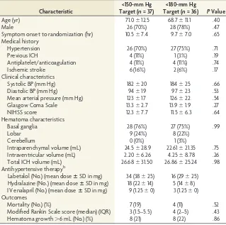
Characteristic
<150-mm Hg Target (n= 37)
<180-mm Hg
Target (n= 36) PValue
Age (yr) 71.0⫾12.5 68.7⫾11.1 .40
Male 26 (70%) 28 (78%) .47
Symptom onset to randomization (hr) 10.5⫾7.4 9.7⫾7.0 .65 Medical history
Hypertension 26 (70%) 27 (75%) .71
Previous ICH 4 (11%) 1 (3%) .19
Antiplatelet/anticoagulation 4 (11%) 4 (11%) .74
Ischemic stroke 6 (16%) 2 (6%) .17
Clinical characteristics
Systolic BP (mm Hg) 182⫾20 184⫾25 .66
Diastolic BP (mm Hg) 94⫾19 97⫾23 .53
Mean arterial pressure (mm Hg) 123⫾17 126⫾22 .54
Glasgow Coma Scale 13.3⫾2.7 13.9⫾1.9 .27
NIHSS score 12.3⫾7.7 11.5⫾6.3 .64
Hematoma characteristics
Basal ganglia 28 (76%) 27 (75%) .99
Lobar 9 (24%) 8 (22%)
Cerebellum 0 (0%) 1 (3%)
Intraparenchymal volume (mL) 24.5⫾28.9 22.61⫾21.35 .75 Intraventricular volume (mL) 2.20⫾6.26 4.25⫾8.78 .26
Total ICH volume (mL) 26.68⫾31.50 26.86⫾25.24 .98
Antihypertensive therapyb
Labetalol (No.) (mean doseⴞSD in mg) 34 (38⫾25) 16 (29⫾25) Hydralazine (No.) (mean doseⴞSD in mg) 18 (22⫾14) 5 (14⫾8) IV enalapril (No.) (mean doseⴞSD in mg) 9 (1.25⫾0) 3 (1.25⫾0) Outcomes
Mortality (No.) (%) 7 (19) 4 (11) .52
Modified Rankin Scale score (median) (IQR) 3 (1.5–5.5) 4 (2–5) .43
Hematoma growth⬎6 mL (No.) (%) 8 (21) 8 (22) .86
Note:—IQR indicates interquartile range. a
Data are means unless otherwise specified. b
Thus, CPP measured by noninvasive imaging techniques may be of particular interest in patients with traumatic brain injuries.
Perihematoma Perfusion Pressure
Our results and those of a PET study completed in patients with acute ICH12confirm that CBF and CBV are modestly reduced in the
peri-hematoma region. Although estimates of CPP and CVR were not included in the PET studies, these parameters could, in fact, be de-rived from source data in the same manner as that described by Schumman et al (1998).18The elevated CVR within the
perihema-toma region is presumably secondary to compression of the micro-vasculature by the space-occupying effect of the hematoma and edema. Maintenance of CPP suggests that this increase in CVR is not responsible for the reduction in CBF, however. Both direct (PET) and indirect (CTP) measurements of oxygen metabolism suggest that CBF reduction is related to decreased metabolic demand within the perihematoma region.12,15,30
Blood Pressure and Perihematoma Perfusion Pressure
We have previously demonstrated that acute BP reduction does not result in reduced perihematoma CBF in patients with acute ICH.15,17,31In the present study, we found no relationship
be-tween BP treatment target or actual BP and perihematoma CPP. The maintenance of CPP despite marked reductions in BP in many of our patients suggests some preservation of autoregula-tion in patients with acute ICH. This is consistent with serial mea-surements of CBF pre- and post-BP reduction by using both PET and CTP.15,31
Borderzone Cerebral Perfusion Pressure
Our CPP measurements may be relevant to the results of dif-fusion-weighted MR imaging studies indicating that subacute ischemic injury occurs in areas remote from the hematoma, including BZ regions, in up to one-third of patients with acute ICH.6-11The retrospective association shown between BP
re-duction in the setting of chronic hypertensive vasculopathy and these ischemic lesions raises the possibility that they are a consequence of hemodynamic compromise. Given that CPP has been shown to be more sensitive to slight variations in mean arterial pressure than individual measurements of CBF or CBV,18we postulated that subtle perfusion changes in the
BZ regions may only become apparent by using this analysis. External and internal BZ CPP was not different from that in surrounding hemispheric tissue and did not vary between the ipsilateral and contralateral hemispheres. The modest reduc-tion in the absolute CPP in the ipsilateral internal BZ in the
⬍150-mm Hg treatment group is of dubious clinical signifi-cance, particularly given the lack of difference in relative CPP. Furthermore, the slight decrease in ipsilateral CPP in our more aggressive BP target group is an unlikely mechanism for DWI lesion formation, which has been reported with equal fre-quency in both the ipsilateral and contralateral hemispheres.10
[image:5.594.81.501.43.337.2]Nonetheless, the possibility that very localized hemodynamic changes, including variations in CPP, are related to the devel-opment of these small DWI lesions cannot be excluded on the basis of our results.
Clinical Implications
Our findings are consistent with those in previous studies of per-fusion in patients with acute ICH, suggesting that acute BP reduc-tion is safe. These data are also consistent with the lack of indica-tors of clinical harm in randomized trials of BP reduction after ICH.17,33,34
Although concerns persist that BZ re-gions may be hemodynamically vulner-able to rapid BP reduction, we can find no objective evidence to support this hy-pothesis. We are currently testing the hypothesis that BP reduction is associ-ated with the development of MR imag-ing ischemic lesions as part of an ongo-ing randomized trial (NCT02281838).
Limitations
This study has a number of limitations. Regional CPP was measured indirectly, but noninvasively, by using the ratio of CBF to CBV maps. Global CPP has been correlated with the CBF/CBV ratio,18
but regional evaluation of CPP by using CTP has not been confirmed with direct measurements of intracranial pressure. Invasive intracranial pressure measure-ments were not included in this study; consequently, our noninvasive, indirect measurements of CPP cannot be vali-dated. Furthermore, CTP studies are single-time-point assessments in a po-tentially dynamic process. This study cannot exclude the possibility that CPP does, in fact, drop transiently after BP
[image:6.594.59.463.113.695.2]FIG 3. Plots of cerebral perfusion pressure and cerebrovascular resistance against mean arterial pressure. Theopen circlesrefer to the⬍150-mm Hg treatment group, while thefilled squaresrepresent the⬍180-mm Hg treatment group. There was no relationship between mean arterial pressure and CPP (⫽ ⫺0.08 [⫺0.10, 0.05]) or CVR (⫽ ⫺0.11 [⫺0.003, 0.001]).
Table 2: Effect of BP reduction on perfusion parameters
Perfusion Parameters and Region
Treatment Group
PValue <150 mm Hg (n= 37) <180 mm Hg (n= 36)
Absolute ipsilateral CPP
Perihematoma 13.9⫾5.5 14.8⫾3.4 .38
Hemisphere 14.0⫾5.3 15.2⫾3.9 .27
External BZ 14.1⫾4.6 15.8⫾4.6 (n⫽35) .11
Internal BZ 13.5⫾4.6 (n⫽27) 16.5⫾4.7 (n⫽29) .02a Absolute contralateral CPP
Perihematoma 13.9⫾5.5 14.8⫾4.0 .43
Hemisphere 14.3⫾5.8 15.2⫾3.8 .45
External BZ 15.1⫾6.5 16.0⫾3.7 (n⫽35) .46
Internal BZ 15.0⫾7.1 (n⫽27) 16.0⫾4.6 (n⫽29) .48
Absolute ipsilateral CVR
Perihematoma 0.32⫾0.10 0.36⫾0.11 .11
Hemisphere 0.29⫾0.08 0.32⫾0.08 .14
External BZ 0.31⫾0.11 0.37⫾0.12 (n⫽35) .05a
Internal BZ 0.37⫾0.13 (n⫽27) 0.44⫾0.16 (n⫽29) .08 Absolute contralateral CVR
Perihematoma 0.28⫾0.09 0.33⫾0.11 .56
Hemisphere 0.28⫾0.08 0.32⫾0.09 .09
External BZ 0.32⫾0.11 0.38⫾0.12 (n⫽35) .04a
Internal BZ 0.36⫾0.12 (n⫽27) 0.40⫾0.14 (n⫽29) .16 Relative CPP
Perihematoma 1.02⫾0.16 1.02⫾0.15 .96
Hemisphere 0.99⫾0.09 1.00⫾0.10 .75
External BZ 0.99⫾0.19 1.00⫾0.26 (n⫽35) .76
Internal BZ 1.01⫾0.17 (n⫽27) 1.01⫾0.16 (n⫽29) .99 Relative CVR
Perihematoma 1.16⫾0.23 1.11⫾0.17 .36
Hemisphere 1.04⫾0.10 1.02⫾0.08 .32
External BZ 1.01⫾0.18 1.01⫾0.22 (n⫽35) .91
[image:6.594.51.529.124.696.2]reduction either before or after the CTP study. Finally, these data are from a relatively small sample of patients with ICH with pre-dominately small-to-moderate hematoma volumes. In our study, only 15% of patients had ICH volumes of⬎40 mL. This is con-sistent with other ICH trials.33,35Patients with large-volume ICH
are less likely to be enrolled in trials because they are treated either conservatively or alternatively with surgical resection from the onset; consequently, these patients make up the minority in this clinical trial. All patients in our study were stable enough and lacked contraindications to CTP scanning 2 hours after random-ization, which certainly introduced a selection bias. Nonetheless, these results provide further insight into cerebral perfusion pat-terns in acute ICH.
CONCLUSIONS
Perihematoma CPP is maintained, despite a modest increase in CVR, in acute primary ICH. Vascular borderzone CPP and CVR are normal in patients with ICH. Reduction of BP does not affect CPP in perihematoma or borderzone regions. These results pro-vide further physiologic support for the safety of BP reduction in ICH.
Disclosures: Bronwen Gould—RELATED:Grant: Alberta Innovates Health Solutions (summer studentship). Thomas Jeerakathil—RELATED:Grant:Canadian Institutes for Health Research,*Comments: I am a collaborator on the grant that funded this research project. This was a peer-reviewed grant from a government funding agency. There is no conflict of interest. Dariush Dowlatshahi—UNRELATED:Expert Testi-mony: Canadian Medical Protective Association (medicolegal consultation for isch-emic stroke);Grants/Grants Pending: Heart and Stroke Foundation,Comments: grant funding to study CTA, spot sign, and ICH;Patents (planned, pending or issued: patent on CT imaging software for the ICH spot sign;Travel/Accommodations/ Meeting Expenses Unrelated to Activities Listed: BI Worldwide (travel grant to the meeting). Michael D. Hill—UNRELATED:Board Membership: Heart and Stroke Foun-dation of Alberta Board (volunteer charity board);Consultancy: Merck,Comments: Adjudication Committee for clinical trials in diabetes;Grants/Grants Pending: Co-vidien,*Comments: grant for clinical trial. Shelagh B. Coutts—UNRELATED: Canadian Institutes of Health Research,* Genome Canada,* Heart and Stroke Foundation of Canada,*Comments: grant funding for research not related to this article. Kenneth S. Butcher—RELATED:Grant: Alberta Institutes of Health Research and Heart and Stroke Foundation of Canada,*Comments: A grant-in-aid supported the ICH ADAPT trial;UNRELATED:Grants/Grants Pending: Canadian Institutes of Health Research,*
Comments: Two grants-in-aid for unrelated work (blood pressure reduction in isch-emic stroke and novel anticoagulant therapy in acute noncardioembolic stroke);
Payment for Lectures (including service on Speakers Bureaus): speaker fees for new oral anticoagulants use by Boehringer Ingelheim, Bayer, and Pfizer/Bristol-Myers Squibb Canada. *Money paid to the institution.
REFERENCES
1. Fogelholm R, Avikainen S, Murros K.Prognostic value and determi-nants of first-day mean arterial pressure in spontaneous supraten-torial intracerebral hemorrhage.Stroke1997;28:1396 – 400CrossRef Medline
2. Okumura K, Ohya Y, Maehara A, et al.Effects of blood pressure levels on case fatality after acute stroke.J Hypertens2005;23:1217–23
CrossRef Medline
3. Anderson CS, Huang Y, Wang JG, et al; INTERACT Investigators. Intensive blood pressure reduction in acute cerebral haemorrhage trial (INTERACT): a randomised pilot trial.Lancet Neurol2008;7: 391–99CrossRef Medline
4. Butcher K, Jeerakathil T, Emery D, et al.The Intracerebral Haemor-rhage Acutely Decreasing Arterial Pressure Trial: ICH ADAPT.Int J Stroke2010;5:227–33CrossRef Medline
5. Mendelow AD.Mechanisms of ischemic brain damage with intra-cerebral hemorrhage.Stroke1993;24(12 suppl):I115–17; discussion I118 –19Medline
6. Brazzelli M, Sandercock PA, Chappell FM, et al.Magnetic resonance
imaging versus computed tomography for detection of acute vascu-lar lesions in patients presenting with stroke symptoms.Cochrane Database Syst Rev2009;CD007424CrossRef Medline
7. Kimberly WT, Gilson A, Rost NS, et al.Silent ischemic infarcts are associated with hemorrhage burden in cerebral amyloid angiopa-thy.Neurology2009;72:1230 –35CrossRef Medline
8. Singer OC, Kurre W, Humpich MC, et al; MR Stroke Study Group Investigators.Risk assessment of symptomatic intracerebral hem-orrhage after thrombolysis using DWI-ASPECTS.Stroke2009;40: 2743– 48CrossRef Medline
9. Singer OC, Berkefeld J, Lorenz MW, et al; MR Stroke Study Group Investigators.Risk of symptomatic intracerebral hemorrhage in patients treated with intra-arterial thrombolysis.Cerebrovasc Dis
2009;27:368 –74CrossRef Medline
10. Menon RS, Burgess RE, Wing JJ, et al.Predictors of highly prevalent brain ischemia in intracerebral hemorrhage.Ann Neurol2012;71: 199 –205CrossRef Medline
11. Arsava EM, Kayim-Yildiz O, Oguz KK, et al.Elevated admission blood pressure and acute ischemic lesions in spontaneous intrace-rebral hemorrhage. J Stroke Cerebrovasc Dis 2013;22:250 –54
CrossRef Medline
12. Zazulia AR, Diringer MN, Videen TO, et al.Hypoperfusion without ischemia surrounding acute intracerebral hemorrhage. J Cereb Blood Flow Metab2001;21:804 –10CrossRef Medline
13. Schellinger PD, Fiebach JB, Hoffmann K, et al.Stroke MRI in intra-cerebral hemorrhage: is there a perihemorrhagic penumbra?Stroke
2003;34:1674 –79CrossRef Medline
14. Butcher K, Baird T, MacGregor L, et al.Perihematomal edema in primary intracerebral hemorrhage is plasma derived.Stroke2004; 35:1879 – 85CrossRef Medline
15. Powers WJ, Zazulia AR, Videen TO, et al.Autoregulation of cerebral blood flow surrounding acute (6 to 22 hours) intracerebral hemor-rhage.Neurology2001;57:18 –24CrossRef Medline
16. Adams RE, Powers WJ.Management of hypertension in acute intra-cerebral hemorrhage. Crit Care Clin 1997;13:131– 61 CrossRef Medline
17. Butcher KS, Jeerakathil T, Hill M, et al; ICH ADAPT Investigators. The Intracerebral Hemorrhage Acutely Decreasing Arterial Pres-sure Trial.Stroke2013;44:620 –26CrossRef Medline
18. Schumann P, Touzani O, Young A, et al.Evaluation of the ratio of cerebral blood flow to cerebral blood volume as an index of local cerebral perfusion pressure. Brain 1998;121:1369 –79 CrossRef Medline
19. Brain Trauma Foundation; American Association of Neurological Surgeons; Congress of Neurological Surgeons; Joint Section on Neu-rotrauma and Critical Care, AANS/CNS, Carney NA, Ghajar J. Guidelines for the management of severe traumatic brain injury.
J Neurotrauma2007;24(suppl 1):S1–106.CrossRef Medline
20. Kirkman MA, Smith M.Intracranial pressure monitoring, cerebral perfusion pressure estimation, and ICP/CPP-guided therapy: a standard of care or optional extra after brain injury?Br J Anaesth
2014;112:35– 46CrossRef Medline
21. Mayer SA, Lignelli A, Fink ME, et al.Perilesional blood flow and edema formation in acute intracerebral haemorrhage: a SPECT study.Stroke1998;29:1791–98CrossRef Medline
22. Wu O, Ostergaard L, Weisskoff RM, et al.Tracer arrival timing-insensitive technique for estimating flow in MR perfusion-weighted imaging using singular value decomposition with a block-circulant deconvolution matrix.Magn Reson Med2003;50:164 –74
CrossRef Medline
23. Robb RA.The biomedical imaging resource at Mayo Clinic.IEEE Trans Med Imaging2001;20:854 – 67CrossRef Medline
24. McCourt R, Gould B, Gioia L, et al; ICH ADAPT Investigators. Cere-bral perfusion and blood pressure do not affect perihematoma edema growth in acute intracerebral hemorrhage.Stroke2014;45: 1292–98CrossRef Medline
xenon CT: a validation study.AJNR Am J Neuroradiol2001;22: 905–14Medline
26. Kudo K, Terae S, Katoh C, et al.Quantitative cerebral blood flow measurement with dynamic perfusion CT using the vascular-pixel elimination method: comparison with H2(15)O positron emission tomography.AJNR Am J Neuroradiol2003;24:419 –26Medline
27. Murphy BD, Fox AJ, Lee DH, et al.Identification of penumbra and infarct in acute ischemic stroke using computed tomography per-fusion-derived blood flow and blood volume measurements.Stroke
2006;37:1771–77CrossRef Medline
28. Gillard JH, Minhas PS, Harball MP, et al.Assessment of quantitative computed tomographic cerebral perfusion imaging with H2(15)O positron emission tomography.Neurol Res2000;22:457– 64Medline
29. Wintermark M, Chiole´ro R, van Melle G, et al.Relationship between brain perfusion computed tomography variables and cerebral per-fusion pressure in severe head trauma patients.Crit Care Med2004; 32:1579 – 87CrossRef Medline
30. Kate MP, Hansen MB, Mouridsen K, et al.Blood pressure reduction does not reduce perihematoma oxygenation: a CT perfusion study.
J Cereb Blood Flow Metab2014;34:81– 86CrossRef Medline
31. Gould B, McCourt R, Asdaghi N, et al; ICH ADAPT investigators.
Autoregulation of cerebral blood flow is preserved in primary in-tracerebral hemorrhage.Stroke2013;44:1726 –28CrossRef Medline
32. Gioia LC, Kate M, Choi V, et al.Ischemia in intracerebral hemor-rhage is associated with leukoaraiosis and hematoma volume, not blood pressure reduction. Stroke2015;46:1541– 47 CrossRef Medline
33. Qureshi AI, Palesch YY, Martin R, et al; Antihypertensive Treatment of Acute Cerebral Hemorrhage Study Investigators.Effect of systolic blood pressure reduction on hematoma expansion, perihematomal edema, and 3-month outcome among patients with intracerebral hemorrhage: results from the antihypertensive treatment of acute cerebral hemorrhage study.Arch Neurol2010;67:570 –76CrossRef Medline
34. Anderson CS, Heeley E, Huang Y, et al; INTERACT2 Investigators. Rapid blood-pressure lowering in patients with acute intracerebral hemorrhage.N Engl J Med2013;368:2355– 65CrossRef Medline