A Thesis Submitted for the Degree of PhD at the University of Warwick
Permanent WRAP URL:
http://wrap.warwick.ac.uk/79995
Copyright and reuse:
This thesis is made available online and is protected by original copyright. Please scroll down to view the document itself.
Please refer to the repository record for this item for information to help you to cite it. Our policy information is available from the repository home page.
Processing and trafficking of Shiga-like toxin 1 in
eukaryotic cells.
Submitted for the degree of
Philosophical Doctorate
Nicholas Lea (B.Sc. Hons.)
University of Warwick.
Acknowledgements
Figure index Declaration Abbreviations
Section 1
Summary
Contents
Introduction
1.1 History and Disease
1.2 Structure and function of ST/SL T 1
1.3 Purification of Shiga toxin and the Shiga-like toxins 1.4 Catalytic action of Shiga toxin and the Shiga-like toxins 1.5 Common features of selected bacterial and plant toxins. 1.6 Proteolytic processing of SL T/ST: Possible analogy with
other bipartite toxins.
1.7 Endocytosis, intracellular trafficking and membrane translocation of SL T 1ST and ricin.
Project aims
Section 2
Materials and Methods
2.1. Suppliers and reagents 2.1.1 Suppliers.
2.1.2 Reagents.
2.1.2 Coomassie blue G250 stain. 2.1.2 Aniline reagent.
2.1.3 Kirby buffer.
2.2. Growth and maintenance of bacteria and eukaryotic cells. 2.2.1 Growth and maintenance of bacteria.
2.2.2 Preparation of competent cells.
2.3
Manipulation of nucleic acids.2.3.1 Transformation of plasmid DNA 55
2.3.2 Transformation of M13 DNA. 55
2.3.3 Purification and concentration of nucleic acids from aqueous 56 solution.
2.3.4 Separation of nucleic acids by agarose gel electrophoresis.
56 2.3.5 DNA polyacrylamide gel electrophoresis. 57
2.3.6 Preparation of plasmid DNA 58
2.3.7 Preparation of single stranded M 13 DNA 59 2.3.8 Preparation of double stranded M 13 DNA. 60
2.3.9 Gel isolation of DNA fragments. 61 2.3.10 Enzymatic modification of DNA 61 2.3.11 Phosphorylation of oligonucleotides. 61 2.3.12 Oligonucleotide Site directed mutagenesis. 62 2.3.13 DNA sequencing.
63 2.3.14 DNA quantitation.
63 2.3.15 Polymerase chain reaction amplification.
63 2.3.16 Preparation of rabbit reticulocyte ribosomes. 64 2.3.17 Northern blot analysis.
64
2.4
ill vitro Transcription / Translation.66 2.4.1 In vitro transcription
66 2.4.2 Wheat germ lysate in vitro translation.
66 2.4.3 Rabbit reticulocyte lysate in vitro translation.
66
2.5
III vivo expression of recombinant proteins.68 2.5.1 Expression of unlabelled recombinant proteins.
68 2.5.2 Preparation of periplasm extracts
69
2.6
Protein purification.69 2.6.1 Purification of recombinant proteins.
69 2.6.2 Purification of Native SL T 1 from Escherichia coli 026:1111, 70
strain 3787
2.6.5 Expression and purification of SL T 1 B subunit
2.6.6 Metabolic labelling of RAST A2
2.7 Protein analysis and Characterisation. 2.7.1 Determination of protein concentration.
2.7.2 Western blot analysis
2.7.3 RNA N-glycosidase activity
2.7.4. Cytotoxic action ofrecombinant proteins to Vero cells.
2.7.5 In vivo cleavage of SL T 1 and Mutants. 2.7.6 In vitro protease treatment.
2.7.7 SDS-Polyacrylamide gel electrophoresis (SOS PAGE).
Section 3
Chapter 1.
Results and Discussion.
3.1 Cloning, Expression and purification of wild-type Shiga-like toxin 1.
Introduction
3.1.1 Cloning
3.1.2 Expression/purification of SL T 1 wt. 3.1.3 Discussion.
Chapter 2
3.2 Cloning, Expression and Purification of mutant Shiga-like toxin 1 proteins.
Introduction
3.2.1 Cloning of mutant Shiga-like toxins.
3.2.2 Expression and purification of Mutant Shiga-like toxins in vivo.
3.2.3 In vitro expression of SL T223stop. 3.2.4 Discussion
Chapter 3
3.3 Characterisation of SL T 1 and potential processing mutants.
Introduction
3.3.1 N-glycosidase activity of SL T 1 and potential processing mutants.
3.3.2 In vitro trypsin sensitivity of SLT 1 and potential processing mutants
3.3.3 Cytotoxicity of SL T 1 and the potential processing mutants. 3.3.4 In vivo processing of SL T 1 and potential processing mutants.
3.3.4 Discussion Chapter 4
3.4.1 Cloning and expression of a ricin A chain-Shiga-like toxin 1 fusion protein (RAST A2).
Introduction.
3.4.2 Cloning of RAST A2.
3.4.2 Expression and purification of RAST A2. 3.4.3 Discussion.
Chapter 5
3.5 Chacterisation of a ricin A chain SL T A2 fusion protein RASTA2.
Introduction
3.4.1 N-glycosidase activity of RASTA2.
3.5.2 In vitro sensitivity of RAST A2 to proteinase K and trypsin. 3.5.3 Cytotoxicity ofRASTA2 compared with that of ricin and
wild-type SL T 1.
3.5.4 In vivo processing of RASTA2 in Vero cells. 3.5.5 Discussion
Chapter 6
3.6.1 Final Discussion chapters 1,2,3,4 and 5.
Section 4
Appendix 1
Appendices
Alignment of active site residues of ricin A chain and SLT 1
Appendix 3 Plasmid pKH206 Appendix 4 Plasmid pSBC32. Appendix 5
Nucleotide and amino acid sequence of Shiga-like toxin 1
Section 5
References
246
247
248
Acknowledgment.
During my time at Warwick I have received assistance and encouragement from a
huge number of people. Without this help I would never have reached this stage.
Dr. Lynne Roberts has provided me with constant and expert guidance, without
which I would certainly have been lost. My greatest thanks must go to her for
providing me with an opportunity to achieve this much.
Thanks to Professor Mike Lord who has shown an interest throughout my work and
has been a source of inspiration and specialist advice during this study. Professor
David Crout and Or. Detter Muller for providing me with globotriose without which
this work would have taken considerably longer. Thank you to Professor Arthur
Donohue-Rolfe for anti ST serum.
To undoubtedly the richest source of practical and technical support and, when their backs were turned, solutions I thank the members of Plant Biochemistry n. Thank
you PBL 11 for all your handy and not so handy hints and tips and moreover thanks
for making PBL 11 a friendly environment to work in. Special thanks should go to
Dr. John Chaddock who was prepared to drop anything to advise me on my latest
problem.
Finally I would like to thank Rachel and my family and for all their considerable
Figure index
Figure Page
1.1
The structure of globotriose, the carbohydrate component of the 7 Shiga-like toxin 1 receptor globotriosylceramide.1.2
Ribbon diagrams of Shiga toxin. 13 Schematic representation of the subunit compositions of 261.3
selected bacterial and plant toxins.
3.1.1
Sequence of oligonucleotide primers EST03' and PST05' 85 used for amplification of SL T 1 from Escherichia coli026:H11 cell paste.
3.1.2
Agarose gel electrophoresis of amplified SL T 1 DNA from87
Escherichia coli 026:Hl1 and subsequent restriction analysis.
3.1.3
Cloning steps used for the production of M 13SL Twt andpSLTwt. 89
3.1.4
Profile of SL T 1 wt protein eluted with guanidine 91 hydrochloride from a 1 ml GbrSepharose column.3.1.5
Analysis of expression and purification of SL T 1 wt by SOS 93 polyacrylamide gel electrophoresis3.2.1
Oligonucleotide sequences of primers used for theconstruction of SL T 1 mutant toxins. 105
3.2.2.
Recombinant mutagenic peR used for the production DNA 107coding for SL TP4 and 223stop mutant SL T 1 s.
3.2.3
Amino acid changes made to the primary sequence of SL T 1 109 as result of mutagenesis.110
3.2.4
Plasmid pSLTPl11 I
3.2.5
Plasmid pSL TP2112
3.2.6
plasmid pSL TP3113
3.2.7
plasmid pSL TP4114
3.2.8
plasmid pGEM223stop3.2.9
Analysis of expression and purification of SL TP 1 by SDS117 polyacrylamide gel electrophoresis.
3.2.11 Analysis of expression and purification of SL TP3 by SOS polyacrylamide gel electrophoresis.
3.2.12 Analysis of expression and purification of SL TP4 by SOS polyacrylamide gel electrophoresis.
3.2.13 In vitro Translation of SL T223stop protein in wheat germ lysate. 3.3.1 Comparison ofN-glycosidase activity of SLT 1 and potential
processing mutants using the aniline assay and visualized by
northern blot analysis.
3.3.2 Oepurination of rabbit reticulocyte ribosomes during in vitro translation of pGEM223stop in vitro transcript in a non-nuclease treated rabbit reticulocyte lysate.
3.3.3 Reducing 15% SOS polyacrylamide gel electrophoresis of
trypsin-121
123
126
133
137
treated SL T 1 and potential processing mutants. 140
3.3.4 Non-reducing 15% SOS polyacrylamide gel electrophoresis of 142 trypsin-treated SL T 1 and processing mutants.
3.3.5 Cytotoxicity of SL T 1 wild-type and potential processing mutants: 3 146 :3 hour incubation of toxin.
3.3.6 Cytotoxicity of SL T 1 wild-type and potential processing mutants: 6
6 hour incubation of toxin. 148
3.3.7 Cytotoxicity of SL T 1 wild-type and potential processing mutants to Vero cells pretreated with 1 OmM NH4CI: 3 hour incubation of toxin.
3.3.8 Cytotoxicity of SL T 1 wild-type and potential processing mutants to Vero cells pretreated with calpain inhibitor I (1 OOllg Iml): 3 hour incubation of toxin.
3.3.9 The kinetics of cytotoxicity of SL T 1 wild-type and potential processing mutants in Vero cells.
3.3.10 Cytotoxicity of SL T 1 wild-type to Vero cells pretreated with brefeldin A 2llg/ml: 3 hour incubation of toxin.
3.3.11. 15 % reducing SDS polyacrylamide gel electrophoresis of 125I-Iabelled SL T 1 and potential processing mutants. 3.3.12 Proteolytic processing of SL T 1 and potential processing
mutants in untreated Vero cells.
3.3.14 Proteolytic processing of SL T 1 and potential processing mutants in Vero cells pretreated with brefeldin A (2llg/m1).
3.3.15 Proteolytic processing of SL T 1 and potential processing mutants in Vero cells pretreated with IOmM NH4CI.
3.3.16 Proteolysis of SLT 1 and potential processing mutants by extracellular proteases.
3.4.1. Oligonucleotide sequences of primers used for the construction of the RASTA2 coding sequence.
3.4.2 Recombinant mutagenic PCR used for the production of DNA coding for a ricin A chain SL T A2 fusion protein (RASTA2).
3.4.3 Plasmid pRAST A2
3.4.4 Analysis of expression and purification of RAST A2 by 15% SDS polyacrylamide gel electrophoresis and Western blot
3.4.5 Analysis of expression and purification of metabolically labelled RAST A2 by 15% SDS polyacrylamide gel electrophoresis
3.5.1 N-glycosidase activity of RASTA2 analysed by the aniline assay.
3.5.2 Reducing 15% SDS polyacrylamide gel electrophoresis followed by Western blot analysis of proteinase K and trypsin treated ricin A chain and RAST A2.
3.5.3 The effect of excess SL T 1 B chain on the cytotoxicity of SL T I wild-type: 3 hour toxin incubation.
3.5.4 Cytotoxicity ofRASTA2, ricin and SLT 1 wild-type: 3 hour incubation of toxin.
3.5.5 Cytotoxicity of RASTA2, ricin and SL T I wild-type: 6 hour incubation of toxin.
3.5.6 The kinetics of cytotoxicity of RAST A2, ricin and SL T 1 wild-type in Vero cells.
3.5.7 In vivo proteolytic processing of RAST A2 in Vero cells.
Declaration.
Research presented in this thesis was obtained by the author unless specifically
indicated in the text. The research presented in this thesis has not been submitted for
any previous degree. All sources of information used in the preparation of this thesis
are indicated by reference.
Abbreviations.
Amp ampicillin
ATP
adenosine triphosphateBCPIP
disodium 5,-bromo-4-chloro-3-indolyl phosphateBFA
brefeldin ABSA
bovine serum albuminCHO
Chinese hamster ovaryCIP
calf intestine phosphataseCT
cholera toxinOMEM
Oulbecco modified eagles medium ds double strandedOT
diphtheria toxinOTT
dithiothreitolEOTA
disodium ethylenediaminetetra acetate. glo botriosylceramideGuHCl Guanidine hydrochloride
HRP
horse radish peroxidaseIC
50 50% cytotoxic doseIPTG
isopropyl-~-D-thiogalactopyranosideLB
Luria brothNBT nitro blue tetrazolium chloride
PA anthrax toxin protective antigen
PAGE polyacrylamide gel electrophoresis
PBS phosphate buffered saline
PCR polymerase chain reaction
PE Pseudomonas exotoxin A
PMSF phenyl methyl sulphonyl fluoride
RIP
ribosome inactivating proteinRTA ricin toxin A subunit
rRNA ribosomal RNA
rSLT 1 recombinant Shiga-like toxin 1
SDS sodium dodecyl sulphate
ss single stranded
SSC saline sodium citrate
ST Shiga toxin
SLT Shiga-like toxin
TCA trichloro acetic acid
TPCK Na-p-tosyl-L-phenylalanine chloromethyl ketone
TLCK Na-p-tosyl-L-Iysine chloromethyl ketone
Section 1
Summary
Shiga toxin (ST) and the Escherichia coli Shiga-like toxins (SLTs) are type 11
ribosome inactivating proteins (RIPs). All members of this group exhibit specific
RNA N-glycosidase activity, the prototype being the plant toxin ricin. ST and the
SL Ts are bipartite toxins composed of a catalytic A subunit and a pentamer of cell
binding B subunits. These toxins show overall structural similarities to ricin, which
is also a bipartite toxin with a catalytic A chain and a single cell binding B chain.
The A chains of ST and SL T 1 show homology to ricin A chain, particularly in the
active site region, and appear identical in their enzymatic mechanism. The respective
B chains however are structurally very different and interact with quite different
cellular components.
In this study, the role of intracellular proteolytic activation of SL T 1 is addressed
using a molecular biological approach. The biological characteristics of several
mutant SL Ts has been investigated both in vitro, by addition of exogenous protease,
and in vivo by comparing the relative cytotoxicities of mutant and wild type proteins
in Vero cells. The intracellular processing of these mutant toxins has also been
examined. In parallel, the biological properties of a ricin A chain SL T 1 chimeric
protein has been investigated. The ultimate aim of this study was to extend our
knowledge of the proteolytic processing requirements of SL T 1 and it has led to the
conclusion that proteolytic removal ofthe A2 portion of SL T 1 is not an essential
1.1 History and Disease
In 1898 , after an epidemic of dysentery in Japan, Kiyoshi Shiga first described the
causative agent of a dysentery which was distinct to that of amoebic dysentery and
named it Bacillus dysenteriae. This was latter named Shigella dysenteriae and the
disease it caused shigellosis. Shiga toxin was first described by (Conradi (1903))
where it was reported that intravenous inoculation of a lysate of the bacillus Shigella
dysenteriae paralyzed and killed rabbits. It was this observation which led
researchers at the time to consider ST as a neurotoxin. It was later shown that ST acts on various animal species with variable consequences but that only the rabbit and
mouse show neurological disorders (Cavanagh et
at
(1956)). Shiga toxin does not act directly on the neurons but can cause secondary neurological disorders by its actionon the vascular system of the brain (Howard (1955); Baldini et
at
(1983)). In 1972 Keusch etat
reported that inoculation of Shiga toxin in ligated rabbit ileal loops caused inflammatory enteritis and resulted in net fluid accumulation.Konawalchuk et
at
(1977) first described a toxin from E. coli which was toxic to Verocells and was distinct from the heat labile and heat stable toxins from the same
organism. This observation led to this toxin being named Vero toxin (VT). Antibody
anti genically and genetically related to Shiga toxin (O'Brien et al (1982); Strockbine
et aI1986,1988». These observations resulted in the renaming of the toxins to the
Shiga-like toxins (SLTs). Further studies of Ecoli 0157:H7 strain 933 revealed the presence of two toxin-converting phages 933J and 933W (O'Brien et al (1984».
These phages carry the structural genes for two distinct cytotoxins (Scotland et al
(1985», one of which is neutralized by anti Shiga toxin antisera and one which is
not. These two toxins are designated Shiga-like toxin 1 and Shiga-like toxin 11
respectively. The genes for these two toxins and those of Shiga toxin have been
cloned and the nucleotide sequences determined (Calderwood et
at
(1987);Strockbine et al (1988); Jackson, et al (1987». The predicted amino acid sequences
have shown that SL T 1 is virtually identical to ST and SL T 11 is 56% homologous to
ST. The Ecoli Shiga-like toxins have been shown to be identical or similar in
biological activities to that of ST including receptor specificity (Waddel et al (1988); Cohen et al (1987» and enzymatic action (Endo et al (1988». Hence it is proposed
that Shiga toxin is the prototype for a family of toxins termed the Shiga-like toxins.
The role that ST and SL Ts have in disease is unclear and the method by which these
toxins are administered to the host has served to complicate studies. Shigella
dysenteriae invades and multiplies within the epithelial cells during infection (Sansonetti (1991». This makes it difficult to distinguish between the responses due
mutant of S. dysenteriae type 1 (Fontaine et al (1988)) was used to infect primates, showed that they retained their ability to produce intestinal disease, albeit less severe
than disease caused by the wild type tox+ strain. However these data are complicated
by the fact that these mutants still produce toxin under certain in vitro conditions.
The presence of the functional Shiga toxin gene is associated with severe lesions of
the colon in rhesus monkey infections not seen with the to x- mutant. The situation
with E. coli infection is more simple. Infection with SL T -producing E. coli does not
result in invasion of the epithelial cells. These toxin producing strains have been
classed as enterohaemorrhagic E. coli (EHEC) and are able to adhere to colonic
epithelia and colonize the gut. Numerous outbreaks ofhaemorrhagic colitis (HC) and
the haemolytic uraemic syndrome (HUS) have established a link between these
disorders and infection with SLT-producing E.coli (Karmali et al (1983); Bopp et al
(1987); Carter et al (1987)). Epidemiological studies have demonstrated that E.coli
serotype 0157:H7 was associated with severe instances of bloody diarrhoea in the
United States and Canada (Johnson et al (1983); O'Brien et al (1983); Riley et al
(1983); Wells et at (1983)). Evidence in favor ofa toxin effect in vivo is that non
invasive EHEC 0157:H7, which produce large quantities of both SLT 1 and SLT 11,
cause colonic damage from inflammation and ulceration to exacerbation of ulcerative
colitis (Hunt et al (1989); Ljungh et al (1988); Von Wulfen et at (1989)). No animal
model for HUS or HC has been developed. Rabbit intestine is often used as an in
vivo model and results using this assay have shown that the effects from ST and SL T
1.2 Structure and function of ST/SL T 1
SL T is a bipartite toxin composed of a single catalytic A subunit non-covalently
associated with five receptor binding B subunits (Donohue-Rolfe et al (1984, 1989);
Olsnes et al (1981 )). Comparison of deduced amino acid sequences between ST and
SL T has shown that the two toxins are identical (Takao et al (1988)) or differ by a
single amino acid at position 45 (threonine in ST and serine in SL T 1) (Calderwood
et al (1987); DeGrandis et al (1987) and Strockbine et at (1988)). The mature A chain is composed of 293 amino acids with a molecular weight of 32,217 Da and the
B chain 69 amino acids molecular weight 7692 Da (Calderwood et at (1987);
Strockbine et at (1988)) Figure 1.3 shows a schematic representation of the SLT 1 structure.
The SL T liST B chain is responsible for receptor binding. The B chain pentamer
binds to the cell surface glycolipids with the carbohydrate sequence Gala 1-4Galp
(galabiose). Lingwood et at (1987) isolated the natural receptor for SLT from Vero
cells and showed it to be a glycolipid-globotriosylceramide containing the
carbohydrate sequence galactose a 1-4galactosep 1-4glucose-ceramide often referred
to as Gb) or CD77. Figure!. 1 shows the structure of the carbohydrate moitey of the
Figure 1.1 The structure of globotriose, the carbohydrate component of
the Shiga-like toxin 1 receptor globotriosylceramide.
HO
OH --.;~ _ _ O HO __ ~ _ _ ---~
o
~OH
HO~-~O
HOOH __ , , - _ - 0
o
SL T 1 B chain has been crystallized (Hart el al (1991» and the structure determined by X-ray analysis (Stein et af (1992)). From these studies it was confirmed that the B
chain associates as a pentamer. Each monomer of 69 amino acids is comprised of
two three-stranded antiparallel p-sheets and an a-helix. p-sheets from adjacent pairs
of monomers form six stranded anti parallel p-sheets at the outer surface of the
pentamer and the five helices line a pore-like structure in the center of the pentamer.
The pore is reported to be 11
~
in diameter and is lined with neutral and non-polar residues. In this study they proposed that the carbohydrate binding site lay within acleft formed by the p-sheets of adjacent monomers. This cleft is occupied by polar
and acidic side chains which it is proposed could make hydrogen-bond interactions
with polar groups of the carbohydrate. A schematic representation of the subunit
composition of SLT 1 is given in figure 1.3. Several mutagenic studies of SL T/ST B
chain are in good accordance with these data. lackson et af (1990) demonstrated that
the residues Asp 17 and Lys 53 were important for receptor binding both oh\hich
are found in the proposed carbohydrate binding cleft. Mutation of residues
Gln64~Glu and Lys66~Gln in SLT IIv (VTE) (a variant ofSLT II which is specific for binding to globotetraosylceramide (Gb4) with terminal GaINAc~
1-3Gala 1-4Gal is seen to disrupt Gb4 binding and allow binding to Gb3 , effectively
changing the specificity of SL T IIv to that of SL T 1 (Tyrrell et al( 1992)). Residues
64 and 66 are located at the lower portion of the carbohydrate binding site proposed
subunit to the trisaccharide
[methyI4-0-(4-0-a-D-galactopyranosyl)-4-0-P-D-galactopyranosyl-p-D-glucopyranoside] and its constituent disaccharides. By
Scatchard analysis of binding data they determined that the SL T I B pentamer has
five identical non-interacting binding sites. On the basis of the thermodynamics of
binding, optical spectroscopy and binding-induced protein aggregation they propose
a model of SL T I membrane interaction which relies on protein-carbohydrate
interaction for specificity and protein-lipid interaction for tight binding.
The fatty acid content of the ceramide group of the Gb3 receptor is important in
determining SL T I binding. The study of the requirements for toxin binding are
complicated by the fact that heterogeneous mixtures of GB3 molecules with mixed
chain lengths appear to bind SL T I more effectively than anyone homogeneous
synthetic receptor (as measured by the TLC overlay assay) (Pellizzari et al (1992)). It is postulated by Pellizzari and colleages that the increased affinity is due to
cooperative binding of isoreceptors present in Gb3 of mixed fatty acid content. These
mixed isoreceptors would present a non-planar 'receptor surface' which may be a
better fit for the three dimensional configuration of the pentameric binding site of the
SL T I holotoxin. Changes in fatty acid composition can also change the overall
orientation of the terminal carbohydrate moiety, which it is speculated, may
profoundly affect its receptor function. The actual requirements for effective toxin
environment in which the receptor molecule resides is also important (Kiarash el a!
(1994)).
The A chain of ST/SLTs can be separated into two domains by a trypsin-sensitive
region. Trypsin treatment generates two fragments-AI approximately 27,000Da and
A2 approximately 4000Da, joined by a disulphide bond (O'Brien and LaVeck
(1983)). The Al fragment has catalytic activity (Brown (1980» and residues within
the A2 fragment are required for association with the B chain pentamer (Haddad and
.Tackson (1993)).
The crystal structure of ST holotoxin has been solved at 2.5
~
resolution (Fraser el a! (1994)) (shown in figure 1.2). The arrangement of all six subunits is seen. TheC-terminus of the A subunit lies in the center of the ring of B subunits. The residues
279-286 form an a-helix which is packed anti parallel to the five helices of the I3
subunits. The helix is initiated at residue Asp278 at the helix cap position. The first
three residues of the helix lie above the B subunit pentamer, the next six residucs
(occupying lIA) penetrate the 20
2.
pore. The last six residues (Arg288-Ser293)could not be seen in the structure. A mutagenic study of residues required for
holotoxin assembly was conducted by Haddad and Jackson (1993) which also
The disulphide bond which links the Al and A2 fragments after mild trypsin
treatment was confirmed to be between Cys 242 and 261. As seen in figure 1.2 a
portion of the A2 fragment lies on top of the B subunit pentamer.
The major contacts between A chain and the B chain pentamer are residues of A2
and a ~-hairpin structure from A 1. Half of the residues buried in the B subunit
pentamer are from the C-terminal helix and this constitutes an area of about 1
OOO~2
.These data exclude any contacts made by the residues 288-293 for which density was
not seen. Interaction between the A chain and the B pentamer is not symmetrical, the
majority of the contacts between the pentamer and the A 1 portion of the A chain are
primarily with just 3 of the five subunits. The residues involved in cleavage of toxin
are thought to lie within a loop subtended by a disulphide bond. However the
residues of this loop are not seen in the crystal structure. The reason for this is that
either the loop is disordered and hence has no definable structure, or that during
crystallization, the toxin became nicked. The disulphide loop is placed close to the
Figure 1.2 Ribbon diagrams of Shiga toxin.
The six subunits of the hexomeric ST holotoxin are shown with the A subunit in
yellow above the five B subunits. The carboxy and amino terminals are labelled C
and N respectively. The positions of the two cysteine residues Cys 242 and 261 are
marked. The approximate position of residues Arg 220 and Arg 223 are also marked.
The diagram was reproduced with the kind permission ofMarie Fraser.University of
The residues which form the active site are highlighted in the SL T 1 sequence in
appendix 5. These include glutamic acid 167, tyrosine 114, tyrosine 77, alanine 168,
arginine 170 and tryptophan 203. The active site was identified by using information
from other ribosome inactivating proteins such as ricin A chain. The active site
residues of Shiga toxin were identified prior to the X-ray crystal structure by
sequence alignment and computer modelling (see appendix l)(Calderwood (1987);
Deresiewicz et af (1992)) and by specific site-directed mutagenesis. Hovde et af
(1988) found that mutation of glutamic acid 167, a residue conserved in the active
site of ricin A chain, to aspartic acid reduced the protein synthesis inhibition activity
of SL T 1 (taken from crude periplasmic extracts) by a factor of 1000. Deresiewicz et
af (1993) mutated tyrosine 114 to serine and observed a reduction in activity of 500
to 1000 fold as judged by testing (crude) extracts in protein synthesis inhibition
assays.
Many previous studies with ST/SLT have shown that proteolytic cleavage and separation of the two domains of the A chain is necessary for the full activity of the
toxin in vitro. The crystal structure of ST provides a clear structural basis for this phenomenon. The residues 258-262 of A2 lie adjacent to the active site cleft and the
side chain of methionine 260 is inserted into the active site of the enzyme. This
structural feature explains why removal of the A2 portion of SLT/ST, including
1.3 Purification of Shiga toxin and the Shiga-like toxins
The purification of ST and the SL Ts has been critical in the biochemical
characterisation of these toxins and the role they play in the onset of disease caused
by Shigella dysenteriae and Shiga-like toxin-producing E.coli. Numerous methods
have been described for their purification both from producer strains and from genes
cloned into laboratory strains of E.coli. These protocols are of two main types.
Early methods for toxin purification were based on multiple chromatography
purifications. One such method described by Donohue-Rolfe et at (1984) was used
for the purification of ST from a non-pathogenic strain of S. dysenteriae type 1 strain
60R (Dubos and Geiger (1946)) This method used bacteria grown to the stationary
phase at 37°C in iron-depleted syncase broth. A sonic lysate of the bacteria is
subjected to Cibacron Blue F3G-A-Sepharose chromatography followed by
chromatofocussing where Shiga toxin elutes in buffer pH 7.0-7.1. The final step of
the purification scheme is molecular sieve chromatography with Bio-Gel P-60. Using
this method, a yield of Img from a 4liter starting culture was obtained, with a 1300
fold increase in toxin specific activity.
More recently methods for the purification of ST and SL Ts have been described
chain pentamer. Ryd et al (1989) describe the production and subsequent use of
a-O-galactose-(1 ~4)-p-O-galactose-(1 ~4)-P-D-glucose-(1 ~) (globotriose or Gb3) linked to polyacryllpolyvinyl (Fracto gel) for the purification of ST from Shigella
dysenteriae type 1 strain 114Sd. A yield of 36mg of toxin from 12 liters of culture
was purified in a single step by this method. A second receptor affinity method
reported at the same time (Donohue-Rolfe et al (1989» used PI glycolipid isolated
from the hydatid cyst fluid of sheep infected with Echinococcus granulosus. This
glycolipid contains galactose a 1 ~4 galactose (to which ST could to bind (Jacewicz
et al (1986», linked to activated Sepharose 48. In a similar method to that ofRyd et al (1989), they purified large quantities of ST in a single step. Analog affinity
methods of ST ISL T purification prove very successful in the purification of these
toxins, they are simple and relatively quick methods which yield highly pure
preparations.
1.4 Catalytic action of Shiga toxin and the Shiga-like toxins
ST ISL Ts have potent RNA N-glycosidase activity comparable with that of the plant
toxin ricin (Endo et al (1988». Once located within the cytosol of a eukaryotic cell
et al (1976) provided the first evidence that ST inhibits protein synthesis in a cell
free system using a partially purified ST preparation. Brown et al (1980)
demonstrated that a pure preparation of Shiga toxin inhibits protein synthesis directly
by studies using cell free protein synthesis systems. Since they saw elevated activity
in the cell free system after treatment with strong denaturants and reducing agents it
was proposed that Shiga toxin is synthesized as a proform. A 70 fold increase in ST
activity was seen after treatment with 8M urea and 10mM DTT, they concluded that
toxin requires activation prior to exerting its full activity. Reisbig (1981)
demonstrated that Shiga toxin or a fragment of it could inactivate salt-washed
ribosomes in simple buffered solutions at a rate of 40 ribosomes/minute. Ribosome
inactivation does not appear to have a cofactor requirement since addition of 500~M NAD, ATP, NADP and NADH or addition ofpH5 supematant from salt washed
ribosomes (containing elongation factors and aminoacyltransferases) did not increase
the activity of toxin in vitro. Addition of antitoxin during these assays prevented any
further inactivation but did not rescue activity already lost indicating that ribosome
inactivation is irreversible. Endo et al (1988) determined the exact site of action of
SL T 11 and ST. They observed that treatment of eukaryotic ribosomes with either
toxin causes release of a 400 nucleotide fragment generated from the 28S ribosomal
RNA when the isolated rRNA was treated with acetic-aniline. Analysis of the 3'
fragment from cleaved rRNA showed that the adenine base at position 4324 is
removed. They also demonstrated that treatment of rat liver ribosomes with toxin
rRNA was correlated to inhibition of protein synthesis and of binding of
elongation-factor-I-dependent amino acyl-tRNA to ribosomes. They conclude that both SL T II
and ST inactivate 60S ribosomal subunits by cleaving the N-glycosidic bond at
adenine 4324 in 28S rRNA of the 60S ribosomal subunits. Furutani et al (1992)
compared the modes of action of SL T and ricin and concluded that they are identical.
Osbom and Hartley (1990) examined the effects of ricin A chain (RT A) on the
partial reactions oftranslation of globin messenger RNA (m RNA) in a reticulocyte
lysate system. They have shown that the primary block by RTA is the elongation
factor 2-dependent stage. Since ST/SLT and ricin are thought to inhibit protein
synthesis in an identical manner this is thought to be the mode of action of ST/SL Ts
also.
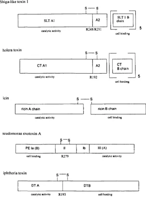
1.5 Common features of selected bacterial and plant toxins.
The Shiga toxin family share many structural elements with other bacterial and plant
toxins. This feature is important in structural studies of SL T as it allows predictions
to be made about the function of structural elements of SL T by analogy with other
The most striking similarities with SL T are between cholera toxin (CT) and E. coli heat labile enterotoxin (LT). Since cholera toxin is considered to be the prototype for
the heat labile enterotoxins (Finkelstein (1988)), only this toxin will be considered
here. Like SL T, cholera toxin is a bipartite toxin comprised of a single catalytic A
chain and a pentamer of cell binding B subunits. Cholera toxin has been crystallized
(Spangler (1989)) and the high resolution structure published (Zhang et at (1995)).
With the structures of CT and LT available (Sixma et at (1991) ; (1992)) the
structure of the holotoxin and the receptor binding sites can be seen. The overall
structural arrangement of the six subunits of CT is identical to that of SL T. Like
SLT, CT is proteolytically processed at the junction of the AI-A2 domains. The
location at which this processing occurs however is different, CT being nicked by a
protease produced by V. cholerae hemagglutinin / protease (Booth et at (1984)).
Hence under normal conditions, the toxin only encounters target cells in the nicked
form. This is in contrast to SLT which is secreted to the E.coli periplasm in the unnicked form and is thought to be processed later during transport in the target cell
(Burgess and Roberts (1993); Garred et al (1995a)). As with SLT, CT A interacts
with its cell binding pentamer via an A2 domain which inserts into a pore formed by
the B chain pentamer. Studies have shown that treatment of the nicked holotoxin
with reducing and mild denaturing agents can remove the CT A 1 portion leaving, the
CT A2-B5 portion intact (Mekalanos et al (1979)). This demonstrates that the CT Al
the B chain pentamer. In a later study the A2 portion of eT was expressed as a fusion
with bacterial alkaline phosphatase (BAP), maltose binding protein (MAP) or
13-lactamase (BLA) and all three fusion proteins were able to bind to GM1 ganglioside
(the eT receptor) and the enzyme markers retained activity. This provides further
evidence that the A2 portion of eT is responsible for the interaction with its B chain
and that the A I portion is not required to form any of the essential non-covalent
contacts (Jobling and Holmes (1992». When these fusion proteins are expressed in
Vibrio cholerae
they are found in the periplasm rather than being exported as normalto the external medium. This suggests that the Al domain may be involved in toxin
transport across the bacterial outer membrane. However, the possibility that the
foreign protein domains fused to eT prevents membrane translocation is also
probable. The function of the catalytic domain of eT is different to that of ST. eT
Al catalyses the ADP ribosylation of Gsa, and GTP-binding protein that functions to
regulate a component of the plasma membrane adenylate cyclase system, and
ultimately acts to activate adenylate cyclase (Gilman (1984». Despite their
difference in function and receptor specificity, the overall structural arrangements of
SL T and eT are extremely similar. A detailed comparison ofthe B chain pentamers
of SL T and heat labile enterotoxin was carried out by Sixma
et al
(1993). Despite analmost complete absence of sequence homology between the two pentamers,
structural features are highly conserved, with seven out of eight secondary structure
Pseudomonas exotoxin A is a bipartite A-B structure toxin from Pseudomonas
aeruginosa, the three dimensional structure for which was solved by Allured et al (1986). PE is translated as a single polypeptide that has both catalytic and cell
binding domains. During toxin entry the protein is separated into two domains by
proteolytic activation at a protease sensitive site which remain linked by a disulphide
bond (Ogata et at (1990)). Numerous mutagenic studies have identified the
functional domains of PE. Residues 1-252 (domain la) constitutes the cell binding
domain, deletion of these residues prevented binding to cells (Hwang et at (1987)). Domain 11 residues 253-364 contains a disulphide-bonded loop in which two
trypsin-sensitive arginines lie (Ogata et at (1990)). Mutation ofthese Arg residues reduces cytotoxicity by preventing intracellular processing of the toxin and release of a
catalytic portion capable of membrane translocation. A second site within domain II
is thought to be responsible for membrane translocation of a catalytic fragment
across an internal membrane. Alanine residues 339 and 343 are implicated in this
function because mutation to glycines still results in the generation of a catalytic
fragment during target cell entry but cytotoxicity is eliminated suggesting that the
mutant catalytic fragment can not reach its cytosolic target (Siegall et al (1991)).
Furthermore fusion of domains I and II of PE with the ribonuclease bamase results in a cytotoxic fusion protein presumably capable of translocation to the cytosol (Prior et
aI1992).The C-terminal domain of PE contains catalytic activity. Its function is the
ADP-ribosylation of cytosolic EF-2 thus preventing host cell protein synthesis
have analogous elements in the SLT structure. For example, a cell binding domain
analogous to that of the SL T B pentamer and a catalytic domain which must be
cleaved away from other elements of the toxin in order to be fully cytotoxic similar
to the Al domain in SLT. A translocation domain for SLT has not been identified.
Diphtheria toxin (DT) provides another example of a bipartite A-B structure toxin.
Like PE, DT is translated as a single polypeptide 62,000 Da (Gill and Dinius (1971);
Collier and Kandel (1971)). DT, like other A-B toxins, can be cleaved into two at a
surface exposed protease-sensitive loop subtended by a disulphide bond. The loop
contains three arginine residues and is sensitive to nicking by trypsin and bacterial
proteases (Michel et af (1972); DeLange et af (1976)). Sandvig and Olsnes (1981)
demonstrated that DT required proteolytic cleavage in order to express its full
cytotoxicity. The DT structure consists of three domains, the N-terminal catalytic
domain (C-domain), the transmembrane domain (T-domain) and the C-terminal
receptor binding domain (R-domain) (Choe et af (1992); Bennett et af (1994)). The
C-domain is the portion which enters the cytosol and ADP-ribosylates the side chain
of the residue diphthamide, a post-translationally-modified histidine of EF -2 (Van
Ness et af (1980)). After secretion from Corynebacterium diphtheriae and possible
extracellular cleavage within the disulphide bonded loop between the C and T
domains, DT binds to the DT receptor at the surface of target cells via a p-hairpin
loop in the R domain (Shen et af (1994)). Internalization to the acidic endosome
domain across the membrane. From solution of the crystal structure at 2.5
~
(Choe elal (1991» a model for membrane translocation has been proposed. It is thought that the T domain is able to penetrate the membrane in such a way as to form a pore
structure through which the catalytic domain can translocate. The charge of two
helices within the T domain is highly pH dependent, in the low pI I environment of
the endosome two of the helices in the T domain become neutral in charge due to
partial protonation. It is proposed that following exposure to low pH the tips of these
two helices become "membrane soluble daggers" which lead the apolar helix pairs
into the membrane. Further experimental evidence for this model was provided by
10hnson el al (1993) where mutation of proline 345, a residue known to undergo a
cis/trans isomerization reaction and thus thought to play a role in conformational
changes seen when OT enters a low pH environment, was seen to reduce the
cytotoxicity of the toxin by 100 fold.
The plant toxin ricin is a potent cytotoxin found in castor oil plant seeds (Stillmark
(1888». Ricin is isolated as a heterodimer, consisting of a 32 OOOOa A chain
glycoprotein linked by a disulphide bond to a 32 OOOOa B chain glycoprotein
(Olsnes and Pihl (1976». The B chain of ricin has lectin activity and can bind cell
surface galactosides (Olsnes and Pihl (1982». The A chain has catalytic activity and
Montfo11 et af (1987) and more recently refined by Rutenber et af (1991) to 2.52..
The A chain of ricin has some homology to that of SL T 1 (appendix 1 contains an
alignment of the active site residues of SL T and ricin). The region of homology lies
between the active sites of the two proteins. In a stretch of73 amino acids, the two
proteins have identical residues in 32% of positions and either identical or
chemically similar residues in 53% of positions. Using the Chou-Fasman algorithm,
virtually identical secondary structure predictions for the two proteins in the active
site region were made (Calderwood et af (1987)). Interestingly this was one early
indication of evolutionary relationships between prokaryotic and eukaryotic toxin
genes. The B chains of ricin and SL T 1 have no sequence homology. However both
serve the function of binding the toxin moiety to different cell surface molecules and
may also be involved in later translocation events. Proteolytic processing of mature
ricin A chain (RT A) has been reported by (Fiani et af (1993» in macrophages.
However, this work has not been repeatable (Argent (1995» and it is considered in
the absence of other data that R TA remains intact during intoxication. Ricin provides
another example of the bipartite A-B family of toxins to which SL T 1 conforms.
Several experiments have demonstrated that isolated domains from bacterial toxins
have specific functions. These isolated domains can be transferred from one protein
to another. For example the translocation domain of PE, domain 11. was fused with
proposed that domain 11 of PE was sufficient for translocation of toxin into the
cytosol of target cells (Prior et al (1992)). In a similar experiment Guidi-Rontani
(1992) fused domain 11 of PE with the catalytic A domain ofDT. This chimeric toxin
was cytotoxic demonstrating that domain 11 of PE is sufficient to direct and
translocate an enzymatically active heterologous polypeptide segment into the
cytosol of sensitive cells and that the A fragment of DT alone contains the catalytic
function. In SL T 1 residues involved in carbohydrate recognition were identified by
mutation of specific residues of SL T 1 B to the equivalent residues in SL T 11 v B
(Tyrrell et
at
(1992)). The resultant mutant SL T 1 toxin became specific for binding to the trisaccharide Gb4 as opposed to the natural SL T 1 receptor Gb). Theseexperiments and those discussed previously highlight the similarities between a
diverse group of toxins. By studying the toxins as a group a general theme emerges.
All the toxins discussed contain cell binding domains analogous to SL T 1 B and a
catalytic domain analogous to SLT AI. In addition in several cases a translocation
domain has also been identified. By analogy with the toxins discussed it seems
reasonable to assume that a translocation domain for SL T / ST is also present,
Figure 1.3 Schematic representation of the subunit compositions of
selected bacterial and plant toxins.
Shiga-like toxin 1
s - s I
I ;,
SLTAl
I
[,en]
cham5
catalytic activity R248/R251
cell binding
holera toxin
5 - 5
'---c.~-.:-t:-~-ctiv-it-Y ---'-RL!9-~,----'1
K
~~,;o
1\,
cell bmdmg
icin 5 - - 5
L-_ri_Ci_n _A_Ch_a_in _ _ _ _ _ _ _ _ '
-,I
!'
ricin B chaincatalytic activity
scudomonas exotoxin A
PE la (8)
cell binding
iphtheria toxin
DTA
catalytic activity
11
R279
5 - 5
I I
Rl93
Ib
I
DTB
cell binding
III (A)
catalytic activity
cell binding
Arginine residues within the trypsin sensitive regions are indicated.
1.6 Proteolytic processing of SLT/ST: Possible analogy with other
bipartite toxins.
All the toxins discussed in section 1.5 with the notable exception of ricin, which is
cleaved in the seed to the mature form, appear to show an absolute requirement for
proteolytic processing in order to generate a catalytic fragment which presumably is
capable of translocating an internal membrane. The need for proteolytic processing
of SL T 1 is the main area of investigation in the present study. The study of
intracellular processing during intoxication with bacterial toxins has been a field of
considerable interest in recent years. Observations that many of the bacterial toxins
isolated included at least a proportion of nicked toxin led to the hypothesis that these
toxins were extremely sensitive to proteases. Improved methods of purification and
advances in recombinant protein expression have led to the production of unnicked
samples of bacterial toxins and this has allowed their detailed study with respect to
the need for proteolytic activation. With the exception of cholera toxin, a single
mammalian protease has been implemented in the processing events which take
place during intoxication of eUkaryotic target cells with bacterial toxins. The protease
responsible is the ubiquitous, subtilisin-like endoprotease furin (for review see BaIT
Furin has a broad pattern of tissue expression and is conserved amongst eukaryotic
species (Schalken et al (1987); Barr et al (1991 b». The role offurin is to cleave
mammalian prohormones and proproteins such as human proinsulin and chicken
proalbumin (Brennan and Peach (1991 ». It is a dibasic endoprotease which cleaves
after the sequence R X RIK R (Barr (1991a» with a minimum requirement for
RXXR (Watanabe et at (1993». Furin has been localized primarily to the trans-Golgi
(Bresnahan et at (1990); Misumi et at (1991» however other reports indicate that
furin has a more extensive cellular location (Tsuneoka et at (1993); Klimpel et al
(1992»).
One well studied example of bacterial toxin processing is that of Pseudomonas
exotoxin A. Native PE is a proenzyme and to detect its ADP-ribosylation activity in biochemical assays it has to be treated with urea and dithiothreitol to partially unfold
the protein (Leppla et al (1989); Lory and Collier (1980». A study by Ogata et al
(1990) used radiolabelled
PE
to track the processing events which occur during intracellular uptake and translocation to the cytosol. Their results showed thatPE
is cleaved within domain 11 near Arg 279. This cleavage site is within the disulphideloop region and reduction is seen to follow proteolysis. A 37,000 Da fragment is
released when Swiss 3 T3 cells are incubated with radiolabelled toxin. They
determined that this fragment is generated from the C-terminus by comparing the
C-terminally labelled toxin. A mutant form of PE where Arg 276 is changed to Gly is
1000 fold less toxic to cells. Incubation of this mutant with cells revealed that it is
not proteolytically processed to the 37 kDa size. Further investigation showed that
the 37 kDa fragment is the only PE-related fragment which can be detected in the
cytosol following wild type PE intoxication. They conclude that proteolytic
processing of PE is required for cellular intoxication. This processing was said to
occur after Arg residues within the disulphide loop of domain 11 and, following
proteolysis, a 37 kDa fragment was believed to translocate to the cytosol.
More recent studies have revealed that furin is the proteolytic enzyme involved in PE
processing. Fryling et al (1992) showed that the protease responsible is associated
with membranes and is stimulated by divalent ions. This protease is independent of
A TP and has a pH optimum of 5.5. Inocencio et al (1994) have demonstrated that a
Chinese hamster ovary cell line resistant to PE is mutated within the furin gene. This
cell line can be made susceptible to PE by transfection of a mouse furin expressing
cDNA gene (Moehring et aI1993).
Diphtheria toxin has an identical catalytic mechanism to that of PE. Its activity in
unnicked DT and were able to show that nicked DT is more toxic and that it inhibits
protein synthesis more quickly than unnicked toxin. Furin-cleavage of DT was
demonstrated by Tsuneoka et al (1993), providing evidence that DT can be cleaved
in vitro by soluble furin. The cleavage site for furin is proposed to be between Arg 193-Ser 194. This site conforms to the minimal consensus recognition sequence for
furin (Watanabe et al (1992» and is located in a disulphide loop shown in the crystal
structure to be exposed on the protein surface (Choe et al (1992». Furthermore,
using an approach similar to that of Moehring with PE (Moehring et
at
(1993», Lo Vo cells, a human colon carcinoma cell line which lack functional furin, wereincubated with DT. Lo Vo cells were known to be resistant to intoxication with DT
whereas Lo Vo cells transfected with a mouse furin expressing cDNA were
susceptible. It has been reported that DT can also be nicked by furin at the cell surface and in the culture medium in addition to nicking in endosomes (Tsuneoka et
al (1993». These results challenge previous studies which indicate that furin is
located primarily in the trans-Golgi Network (Bresnahan et al (1990); Misumi et al
(1991» though it is not clear whether such external enzyme represents a very minor
fraction offurin which has escaped the TGN.
Perhaps the first evidence of the involvement offurin in toxin activation was
provided by the study of anthrax toxin protective antigen (P A). The causative agent
appropriately, form two toxins. P A and edema factor combine to form edema toxin
and P A and lethal factor form lethal toxin. The two toxins are known collectively as
anthrax toxin and both conform to the A-B structure toxin model (Leppla (1991 )).
For intoxication to take place, PA must bind to the cell surface where it is processed
by proteolytic cleavage. The bound, activated PAis then able to form ion-conducting
channels in cells (Milne and Collier (1993)). Release of a 20kDa fragment allows
binding ofLF or EF to form ET or LT (Leppla et al (1988)) which is then
internalized by receptor-mediated endocytosis (Freidlander (1986); Gordon et af
(1988)). The cleavage ofPA occurs after the sequence ArgI64-Lys-Lys-ArgI67
(Singh et af (1989)). Mutagenesis revealed that the toxin must contain the sequence
R-X-X-R i.e. Arg residues in the -I and -4 positions in order to be cytotoxic
(Klimpel et af (1992)). In the same study it was demonstrated that PAis a substrate
for furin in vitro and known inhibitors of furin prevent cell surface cleavage of P A.
The processing events described are thought to occur at the cell surface, another
indication that furin has a ubiquitous cellular location. They concluded that PAis
proteolytically-processed by a protease indistinguishable from furin.
A recent study by Gordon et al (1995) has provided evidence that P A and DT are
cleaved by at least one protease other than furin. It was showed that in contrast to the work of Tsuneoka et al (1993), LoVo cells are sensitive to DT using longer
furin-deficient, is also sensitive to DT and P A but not to a P A mutant with the
wild-type cleavage sequence R-K-K-R mutated to R-A-A-R (a cleavage site which does
not contain a paired dibasic sequence). They conclude that the sensitivity of this
mutant and of DT is due to proteolytic processing by a second enzyme distinct to that
of furin which is capable of recognizing paired dibasic sequences. So, although DT
can be cleaved by furin in vitro, furin may not exclusively nick this toxin in vivo.
Cholera toxin has a similar structure to that of heat labile enterotoxin from
Escherichia coli with a 80% homology between the A chains (Dallas and Falkow (1980». Despite a lack of sequence homology between CTIL T and SLT these two
toxins share remarkable homology with the tertiary structure of SL T 1. Both toxins
are seen to require proteolytic cleavage prior to target cell intoxication (Fisherman
(1982); Clements and Finkelstein (1979». However, in contrast to Escherichia coli
heat labile enterotoxin, cholera toxin is secreted from the bacterium in a cleaved form
(Booth et al (1984», whereas Escherichia coli heat labile enterotoxin (LT) is
produced largely in the unnicked form. By analogy with CT, studies have been
conducted to compare the activity ofunnicked LT with that of nicked LT. It is reported that nicked LT has greater activity in the skin permeability test than
unnicked toxin (Rappaport et al (1976». Experiments with LT intoxication of cells
have produced conflicting results. Unnicked toxin appears less toxic to YI adrenal
This evidence may suggest that Yl cells are disabled in their ability to process LT. A
more recent study demonstrates that mutation of Arg 192 to Gly (the residue thought
to be susceptible to trypsin cleavage in vitro) does not reduce the cytotoxicity to
CHO cells by a large amount. A slight decrease in the rate of intoxication and a 4
fold reduction in cytotoxicity between unnicked R192-G protein and trypsin nicked
wild-type protein was observed (Grant et al (1994)). These results do not rule out
processing which may occur at a site other than Arg192. In fact LT contains a furin
consensus motif at amino acid residues 141-146 which is highly conserved amongst
all LT variants (Pickett et al (1989)). The data regarding proteolytic activation of LT
remains ambiguous and more in vivo experiments are required to clarify its role in
toxin activation.
As discussed in section 1.4, ST and SL T 1 only display full protein synthesis
inhibition activity after separation of the cell binding B subunits and the A2 region
away from the catalytic Al domain. Like many other bacterial toxins, ST/SLT 1
display the structural features of typical A-B toxins. By analogy with other bacterial
toxins it seems reasonable to suggest that ST
ISL
T 1 also have a requirement forproteolytic activation. In common with PE, DT, PA and LT, SLT/ST have
disulphide-bonded protease-sensitive loops which contain minimal furin recognition
sequences. Kongmuang et al (1988) investigated the effect of nicking of SL T 1 on
cells, however no increase in cytotoxic activity between nicked and unnicked toxin
was reported. Whether toxin was nicked intracellulary however was not investigated.
SLT I contains the sequence R248-V-A-R25I in the exposed loop region. Burgess
and Roberts (1993) made a mutant SL T 1 R248, R25I ~G248,G251 which is more resistant to trypsin in vitro but is only 10 fold less toxic to Vero cells than the wild
type when incubated with cells for 3 hours or less. It was found that if the incubation
period is extended the mutant appears as toxic as wild-type protein. Garred et al
(1995a) mutated the same loop arginine residues of ST and converted them to
histidines. They demonstrated by direct visualisation that the mutant ST is still
cleaved during Vero cell intoxication and that the AI-sized fragment generated
appears identical in size to that generated from the wild-type toxin. In contrast to the
processing of ST wild-type, the mutant is not processed in cells pretreated with the
fungal metabolite brefeldin A. Brefeldin A (BF A) causes disassembly of the Golgi
apparatus and thus prevents transport through the Golgi (Fujiwara et al (1988);
Lippincott-Schwartz et al (1989)). They concluded that the mutant protein is
processed more slowly and that for this to occur the toxin must reach a later cellular
compartment. They also observed that the mutant toxin is not processed in the
presence of the membrane-permeable calpain inhibitor I where as the wild-type toxin
is unaffected by treatment of cells with this inhibitor. From these data they conclude
that in the absence of the most likely cleavage site in ST, the protein can still be
processed by an alternative protease which is inhibited by calpain inhibitor 1. It isn't
ubiquitous Ci+-requiring enzyme, calpain, nor is the cellular location of this enzyme
confirmed, though it is generally believed to be cytosolic (for a recent review see
Suzuki et af (1995». From the intoxication time-points used in the study by Garred
et af (1995a), namely two and four hours, it is conceivable that toxin has by this
stage reached the cytosol. That cleavage by calpain might occur in the cytosol to
activate the reduced Al fragment by causing its release from the A2-Bs complex,
suggests that holotoxin itself might be capable oftranslocating an internal
membrane. In the present investigation, shorter incubation times oftoxin mutants
with cells have been used in an attempt to distinguish any processing in the endocytic
pathway from possible processing in the cytosol.
In a separate study by Samuel and Gordon (1994), the loop arginines in SLT IIv were
mutated to glutamic acid or histidine residues, toxin labelled with 1251 was used to
trace toxin processing in target cells. Since the mutant toxins made in this study
remained cytotoxic but were processed less efficiently they concluded that
proteolytic toxin activation can be mediated by proteases with different specificities
to those which cleave the wild-type protein and that efficient cleavage is not required
Garred et al (1995b) demonstrated that wild-type ST can be a substrate for furin in
vitro and that Lo Vo cells nicked wild-type ST more slowly than Lo Vo cells
transfected with a furin expressing cDNA. LoVo cells pretreated with BFA or
calpain inhibitor I were not able to nick mutant ST (Garred et al (1995b )). This
report clearly demonstrates that furin is the enzyme normally responsible for efficient
cleavage of ST in Lo Vo cells but that other cellular proteases can also cleave the
toxin, albeit less efficiently.
In a report by Takao et al (1988) the molecular structures of SL T 1 and ST were
compared. By performing Edman degradation of trypsin-generated fragments of ST
it was observed that the toxin is nicked between Ala 253 and Ser 254. Since this
nicking could not have been performed by trypsin they concluded that the ST
preparation was pre-nicked at this site. The fragments generated by this nicking were
linked by a disulphide bond. Since mutation of the residues Arg 248 and Arg 251
(Garred et al (1995a)) did not result in a protein totally resistant to proteolytic attack
it is hypothesized that the Ala Ser residues in the loop region may constitute sites for
alternative processing. The disulphide loop of SL T 1 contains two pairs of Ala-Ser
residues either side of the proposed nicking site Arg248-Arg251. In this study the
possible relevance of these residues in the proteolytic activation of SL T 1 is
potential furin-recognition site located upstream at residues Arg 220-Arg223 is also
investigated.
Clearly in comparison with other bacterial toxins the number of reports on SL T 1ST processing are limited. The only report where protease-resistant mutants have been
constructed, expressed and visualized in cells has shown that the mutant protein
remains sensitive to host cell proteases (Garred et al (1995a). This report, and others
using either similar SLT mutants (Burgess and Roberts (1993); Samuel and Gordon
(1994)) or using protease-deficient cells (Garred et al (1995b)), have yielded
information about the physiological events which may occur during target cell
intoxication. However there is no literature to address whether the proteolytic
separation of the A2 domain away from Al is a absolute requirement for intoxication
and whether the A2 domain provides an efficient block to activity in vivo. In the
present study mutant SL T 1 proteins which are apparently resistant to host cell
proteases are described. The mutant proteins remain cytotoxic in the apparent
absence of intracellular proteolytic cleavage. The implications of this for the
membrane translocation of SL T 1 and ricin are discussed.
1.7 Endocytosis, intracellular trafficking and membrane
ST/SL T and ricin enter cells via receptor-mediated endocytosis (Sandvig et al
(1989); van Deurs et al (1986); van Deurs et al (1988». Receptor binding sites for
ST are located randomly on the cell surface. However following treatment of target
cells with ST at 37°C the binding sites become localized in coated pits. Horse radish
peroxidase conjugates and colloidal gold conjugates have been used to trace toxin
endocytosis and transport. Ricin appears to be endocytosed by clathrin-dependent
and clathrin-independent endocytosis (Sandvig et al (1982» whereas ST appears to
be internalized exclusively by the clathrin-dependent mechanism (Sandvig et al
(1989». The reason for ST being endocytosed by the clathrin-dependant mechanism
only is unclear. However this may be due to interactions of the multivalent receptor
binding sites on ST with the glycolipid receptors and with underlying coat and
plasma membrane proteins. These interactions may be strong enough to ensure that
the toxin remains in coated pits. The mechanisms by which clathrin-independent
endocytosis occurs are still unclear. A complete understanding of these processes
will inevitably reveal the significance between the two mechanisms.
Transport to or through the Golgi complex appears to be important for intoxication
of target cells with ST/SLT and ricin. Following endocytosis, toxin conjugates are
seen in endosomes, lysosomes and in the Golgi region (van Deurs et al (1986);
Sandvig (1989». In the case of ricin the pattern of labelling can be dependent on the