Manuscript version: Author’s Accepted Manuscript
The version presented in WRAP is the author’s accepted manuscript and may differ from the published version or Version of Record.
Persistent WRAP URL:
http://wrap.warwick.ac.uk/126697 How to cite:
Please refer to published version for the most recent bibliographic citation information. If a published version is known of, the repository item page linked to above, will contain details on accessing it.
Copyright and reuse:
The Warwick Research Archive Portal (WRAP) makes this work by researchers of the University of Warwick available open access under the following conditions.
Copyright © and all moral rights to the version of the paper presented here belong to the individual author(s) and/or other copyright owners. To the extent reasonable and
practicable the material made available in WRAP has been checked for eligibility before being made available.
Copies of full items can be used for personal research or study, educational, or not-for-profit purposes without prior permission or charge. Provided that the authors, title and full
bibliographic details are credited, a hyperlink and/or URL is given for the original metadata page and the content is not changed in any way.
Publisher’s statement:
Please refer to the repository item page, publisher’s statement section, for further information.
Structural basis of outer membrane protein insertion by the BAM complex
Yinghong Gu1*, Huanyu Li1*, Haohao Dong1*, Yi Zeng1*, Zhengyu Zhang1*, Neil G.
Paterson2*, Phillip J. Stansfeld3*, Zhongshan Wang1,4,5, Yizheng Zhang5, Wenjian
Wang6#, Changjiang Dong1#
1Biomedical Research Centre, Norwich Medical School, University of East Anglia,
Norwich Research Park, Norwich, NR4 7TJ, UK;
2Diamond Light Source, Harwell Science and Innovation Campus, Didcot,
Oxfordshire, OX11 0DE, UK;
3Department of Biochemistry, University of Oxford, South Parks Road, Oxford OX1
3QU, UK;
4Jiangsu Province Key Laboratory of Anesthesiology, Xuzhou Medical College,
Xuzhou 221004, China
5Key Laboratory of Bio-resources and Eco-environment, Ministry of Education,
Sichuan Key Laboratory of Molecular Biology and Biotechnology, College of Life
Sciences, Sichuan University, Chengdu, 610064, China
6Laboratory of Department of Surgery, the First Affiliated Hospital, Sun Yat-sen
University, 58 Zhongshan Road II, Guangzhou, Guangdong, 510080, China.
*These authors contributed equally to this work
#To whom correspondence should be addressed, [email protected] or
All Gram-negative bacteria, mitochondria and chloroplasts have outer
membrane proteins (OMPs), which perform many fundamental biological
processes. The OMPs in Gram-negative bacteria are inserted and folded into the
outer membrane (OM) by the β-barrel assembly machinery (BAM). The
mechanism involved is poorly understood due to the absence of a structure of the
entire BAM complex. Here we report two crystal structures of the E. coli BAM
complex in two distinct states: an inward-open and a lateral-open state. Our
structures reveal that the five polypeptide transport-associated domains of
BamA form a ring architecture with four associated lipoproteins, BamB-E in the
periplasm. Our structural, functional studies and molecular dynamics
simulations indicate that these subunits rotate with respect to the integral
membrane β-barrel of BamA to induce movement of the β-strands and promote
Outer membrane proteins (OMPs) play important roles in Gram-negative bacteria,
mitochondria and chloroplasts in nutrition transport, protein import, secretion, and
other fundamental biological processes1-3. Dysfunction of mitochondria outer
membrane proteins are linked to disorders such as diabetes, Parkinsons and other
neurodegenerative diseases4,5. The OMPs are inserted and folded correctly into the
outer membrane (OM) by the conserved OMP85 family proteins6-8, suggesting that
similar insertion mechanisms may be used in Gram-negative bacteria, mitochondria
and chloroplasts.
In Gram-negative bacteria, OMPs are synthesized in the cytoplasm, and are
transported across the inner membrane by SecYEG into the periplasm8,9. The
seventeen kilodalton (kDa) protein (Skp) and the survival factor A (SurA) chaperones
escort the unfolded OMPs across the periplasm to the β-barrel assembly machinery (BAM), which is responsible for insertion and assembly of OMPs into the OM10-12. In
Escherichia coli, the BAM complex consists of BamA and four lipoprotein subunits,
BamB, BamC, BamD and BamE. BamA is comprised of five N-terminal polypeptide
transport-associated (POTRA) domains and a C-terminal OMP transmembrane barrel,
while the four lipoproteins are affixed to the membrane by N-terminal lipid-modified
cysteines. Of these subunits, BamA and BamD are essential3,6. One copy of each of
these five proteins is required to form the BAM complex with an approximate
molecular weight of 200 kDa (Extended Data Fig. 1). In vitro reconstitution of the E.
coli BAM complex and functional assays showed that all five subunits are required to
obtain the maximum activity of BAM13-16. Individual structures of BamA17-20,
BamB21-25, BamC22,26, BamD22,27,28 and BamE22,29,30 have previously been reported, as
have complex structures of BamD with the N-terminal domain of BamC31, and BamB
insertion by the BAM complex is largely hampered by the lack of a complete
structure of the BAM complex11,32. Furthermore it is unknown how BAM manages to
insert OMPs into the OM without the use of ATP, proton motive forces or redox
potentials33,34.
Here we report two novel crystal structures of the E. coli BAM complex:
BamABCDE and BamACDE. The complexes reveal a unique ring architecture that
adopts two distinct conformations: an inward-open and a novel lateral-open.
Furthermore, comparison of the two complexes reveals that the periplasmic units are
rotated with respect to the barrel, which appears to be linked to significant
conformational changes in the β-strands β1C-β6C of the barrel. Taken together this
suggests a novel insertion mechanism whereby rotation of the BAM periplasmic ring
promotes insertion of OMPs into the OM. To our knowledge, this is the first reported
crystal structure of an intramembrane barrel with a lateral-open conformation.
Unique architecture of two E. coli BAM complexes
X-ray diffraction data of selenomethionine labelled crystals were collected to 3.9
Ångström (Å) resolution and the BAM structure was determined by
single-wavelength anomalous dispersion (SAD) and manual molecular replacement
(Methods, Extended Data Table 1). The first structure contained four proteins: BamA,
BamC, BamD and BamE (Fig. 1a-c), with the electron density and crystal packing
indicating that the BamB is absent in the complex. This was confirmed by
SDS-PAGE analysis of the crystals (Extended Data Fig. 1 and Supplementary Data Fig.
S1). In this model, BamA, BamC, BamD and BamE contain residues E22-I806,
C25-K344, E26-S243, and C20-E110, respectively. The machinery is approximately 115 Å
The architecture of BamACDE resembles a top hat with an opening in the crown.
This crown is formed by the BamA β-barrel with the encircling POTRA domains and
associated proteins forming the brim. The C-terminal β-barrel of BamA projects out of the complex and is fully immersed in the OM while the five POTRA domains of
BamA and the BamD form a ring in the periplasm (Fig. 1a-c). The other subunits of
the complex surround this central BamAD core. The coiled N-terminal loop of BamC
is bound to BamD, as is its N-terminal globular domain, which also interacts with
POTRA 1 of BamA. The C-terminal globular domain of BamC interacts with BamD
and POTRA 2. Interestingly, for one of the two BamACDE complexes in the
asymmetric unit cell no electron density was observed for the N-terminal and
C-terminal globular domains of BamC, which may indicate inherent flexibility
(Extended Data Fig. 2). This flexibility was also observed in the MD simulations.
Finally, BamE is found at the opposite end of the complex, coupling the C-terminal
domain of BamD to POTRA 4 and 5 of BamA, adjacent to the barrel (Fig. 1b, c).
In order to obtain a structure of the complex with all five subunits, we increased the
expression level of BamB (Methods). The structure of BamABCDE was determined
by co-crystallization with sodium iodine and SAD and manual molecular replacement
techniques to a resolution of 2.9 Å (Methods, Extended Data Table 1). The structure
we describe below is based on this BamABCDE complex unless otherwise
mentioned. The β-strands of BamA’s C-terminal barrel are named as β1C-β16C for
consistency with previous reports. The top hat architecture of BamABCDE is similar
to that of BamACDE with dimensions of around 120 Å in length, 98 Å in width and
140 Å in height, with the periplasmic ring structure retained (Fig. 1d-f). In the
BamABCDE structure, the opening in the crown of the top hat is now closed. This
bind to POTRA 2 and 3. Although SDS-PAGE analysis of the crystals showed BamC
is intact in the BamABCDE crystals (Extended Data Fig. 1), electron density is only
visible for the N-terminal loop (residues V35 to P88), bound to BamD. This indicates
that the rest of BamC is highly flexible. Molecular dynamics (MD) simulations of the
BamABCDE and BamACDE complexes suggests that both complexes are otherwise
stable and the periplasmic ring structure remains intact during the simulations
(Extended Data Fig. 3, Supplementary Data Video S1 and Supplementary Data Fig.
S2, 3 and 4).
Inward- and lateral-open conformations
In the structure of the BamABCDE complex, the extracellular loops (L-1 to L-8) cap
the pore of BamA to completely close it to the extracellular side, while the
periplasmic mouth is fully open to the periplasm (Fig. 2a, b). This conformation is
similar to all reported barrel structures of BamA, however, the POTRA domains are
significantly different (Extended Data Fig. 4). The POTRA domains of BamABCDE
appear locked through their interactions with BamD, which together form a ring
apparatus that may feed the unfolded OMP into the assembly machinery. It is worth
noting that the β16C of both Neisseria gonorrhoeae BamA and the BamABCDE
complex coils toward the inside of the barrel lumen, creating a gap between β1C and
β15C of the barrels (Fig. 1d, Fig. 2f and Extended Data Fig. 4), which may provide a
path for insertion of the OMPs.
In contrast, in the structure of BamACDE, extracellular loops L-1, L-2 and L-3 are
displaced from the pore, opening the barrel laterally between β1C and β16C. This
exposes the barrel lumen to both the extracellular leaflet of the OM and the outside of
T-4 completely plug the barrel (Fig. 2d and Extended Data Fig. 5). The barrel of BamA
in the BamACDE structure is therefore in a lateral-open conformation. The first 6 β
-strands, β1C to β6C, perform a scissor-like movement to rotate away from the pore to a maximum angle of around 65° and distance of about 15 Å (Fig. 2e and Extended
Data Fig. 5). The other strands of the barrel remain unchanged. These conformational
changes open the barrel laterally to the OM and the extracellular side, and, in
conjunction with POTRA 5, close the periplasmic mouth (Fig. 2c, d and Extended
Data Fig. 5). Such a mechanism of conformational changes between inward- and
outward-open conformations to transport small molecular substrates is common for α -helical inner membrane protein transporters35. However, to our knowledge, this is the
first crystal structure report to date of a β-barrel that may alternate between both inward- and lateral-open conformations. The novel architecture of the lateral-open
conformation is likely to facilitate the insertion of β-strands of the OMP into the OM, while permitting the interlinking extracellular loops to extend out of the cell upon
insertion.
It was suggested that lateral separation between β1C and β16C is required for normal BamA function by disulfide bond cross-linking36. To test the two solved
conformations, in vivo cross-links were designed to interlock BamA in one of the two
conformational states. Two double cysteine mutations, E435C/S658C and
E435C/S665C, were created to capture BamA in the inward-open conformation (Fig.
2f), and one double mutation, G393C/G584C, was produced to restrain BamA in the
lateral-open conformation (Fig. 2g). The single cysteine mutations do not affect cell
growth, while the double cysteine mutations G393C/G584C, E435C/S665C and
E435C/S658C are all lethal (Fig. 2h, i). In addition, the double cysteine mutants are
Tris(2-carboxyethyl)phosphine hydrochloride (TCEP) (Fig. 2j, k, Extended Data Fig.
5 and Supplementary Data Fig. S5), which breaks the disulfide bonds and therefore
unlocks the structure, providing strong evidence that the barrel can exist in the two
resolved conformations in the bacterial OM.
Essential interactions between BamA and BamD
Previous mutagenesis analysis has suggested that only POTRA 5 of BamA associates
with BamD37, however prior to this study no structures of this complex have been
solved. In our structures 12 residues of BamD interact with 17 residues of POTRA 5
(Fig. 3a, Extended Data Fig. 6 and Supplementary Data Table 1). In addition to
contacts with POTRA 5, our structures reveal that BamD also interacts with V480 and
D481 of periplasmic turn T-2 of the BamA barrel and also forms contacts with
POTRA 1 and 2. These interactions complete the ring structure (Extended Data Fig. 7
and Supplementary Data Table 1).
MD simulations of only the core BamAD periplasmic ring from both structures
retains the cyclic complex. Removal of BamD markedly increases the dynamics of
POTRA 1 and 2 in the BamACDE conformation (Extended Data Fig. 3,
Supplementary Data Video S1). In this instance, the POTRA domains rotate in a
counter-clockwise direction towards the OM. This rotation also causes POTRA 3 to
separate from the T-5 and T-6 periplasmic turns of the BamA barrel (Extended Data
Fig. 3 and Supplementary Data Video S1). In contrast, simulations of only BamA
from the BamABCDE structure, results in POTRA 2 coupling to POTRA 5, thereby
stabilizing POTRA 1 and 2. However, to achieve this configuration, POTRA 3 and 4
separate from the barrel, with a degree of deformation to POTRA 3, suggesting that
To test whether the BamA and BamD interactions are required for BAM function,
BamA POTRA 5 mutants E373K and R366E were generated. Functional assays
showed that R366E severely impairs cell growth, while E373K is lethal to the E. coli
cells (Extended Data Fig. 7). In the structures, BamA E373 and BamD R197 form a
salt-bridge (Fig. 3a). An R197L BamD mutation was able to rescue BamA E373K37.
BamB regulates BamA conformation
The most apparent differences between the two solved structures are the presence of
BamB in the BamABCDE complex, while BamC is more clearly resolved in the
BamACDE complex (Fig. 2a, c). In addition, the POTRA domains of BamA are
found in two distinct conformations, with a larger separation observed between
POTRA 3 and 5 in the BamACDE complex. Speculatively, this could be due to the
absence of BamB, which binds to POTRA 2 and 3 in the BamABCDE complex and is
known to have a regulatory role3,22,23. The overall interface between BamA and BamB
is around 1080 Å2, and is comprised of the three β-strands of POTRA 3 and a loop consisting of residues T245-K251 that anchors to the center of the BamB β-propeller
(Fig. 3b, Extended Data Fig. 7 and Supplementary Data Table 2). The BamB loops at
the BamA binding side adopt conformational changes to bind to POTRA 321,22
(Extended Data Fig. 7), consistent with the BamB structure in complex with POTRA
3 and 432. BamB also interacts with residues K135 and Y147 of POTRA 2 (Fig. 3b).
As a result, the binding of BamB appears to induce local conformational changes in
POTRA 2 and 3 with a root-mean-square deviation (RMSD) of 3.57 Å over 159 Cα
atoms (Extended Data Fig. 7).
Both periplasmic turns T-5 and T-6 of the BamA barrel are more ordered in the
BamACDE complex POTRA 3 separates from the periplasmic turns (Fig. 2c),
indicating that BamB may play a role in controlling the structural rearrangements of
the barrel through POTRA 3. It is worth noting that POTRA 5 also has extensive
contacts with the periplasmic turns T-1, T-2 and T-3 of the barrel domain in the
BamABCDE structure (Extended Data Fig. 6), and we speculate that the
conformational changes of POTRA domains may play a role in controlling the
barrel’s conformations.
In the MD simulations, the tight coupling between POTRA 3 and BamB was retained.
However, in the absence of BamB, the simulations reveal greater dynamics of
POTRA 3 and 4, with both domains moving away from the barrel and the membrane
(Supplementary Data Video S1). This suggests that BamB is important for coupling
of POTRA 3 at the appropriate height with respect to the barrel and OM.
BamE and BamC interactions with BamA and BamD
Previous studies suggested that BamE only binds to BamD directly 38,39. Surprisingly,
both BamABCDE and BamACDE structures show that BamE is not only positioned
between BamA and BamD, but also forms contacts with BamC (Fig. 4a, Extended
Data Fig. 6, 8 and Supplementary Data Table 3, 4). Interestingly, the BamE residues
P67 and F68 also interact with BamC residues M56 and I57 (Extended Data Fig. 8),
suggesting that BamC, BamD and BamE form a network to regulate the
conformations of BamA.
BamE residues I32, Q34, L63 and R78 interact with both BamA and BamD.
Mutations to any of these residues caused defects to the OM29. Additionally, single
D66, F74, V76, Q88 at BamE and BamD interface caused defects of the OM29. These
data suggest that BamE plays an important role in OMP assembly.
The whole BamC structure is revealed in the BamACDE complex. BamC forms
extensive contacts with BamD, with the average interface of 2686 Å2 (Fig. 4b,
Extended Data Fig. 8 and Supplementary Data Table 5). The N-terminal loop of
BamC, up to residue G94, is largely unstructured, coiling round BamD and forming a
cluster of contacts with BamE. The N-terminal globular domain of BamC interacts
with the N-terminal domain of BamD and POTRA 1 of BamA (Extended Data Fig.
8). The C-terminal globular domain interacts principally with POTRA 2, via their β
-sheets (Fig. 4b).
The C-terminal globular domain of BamC binds to POTRA 2 in one of the
BamACDE complexes. This likely enhances the periplasmic ring structure formed by
BamA and BamD, and plays a role in the conformational changes of BAM.
Comparison of the two BamACDE complexes in the asymmetric unit reveals
differences in the barrel domain, with an RMSD of 0.91 Å over 378 Cα atoms, while
the periplasmic ring is somewhat rotated, with respect to the barrel (Extended Data
Fig. 2). SDS-PAGE analysis confirmed that both BamACDE and BamABCDE
crystals contain full length BamC (Extended Data Fig. 1), suggesting that the two
globular domains of BamC are dynamic. MD simulations, with the addition of the
BamC globular domains to this structure, also show that these domains are not tightly
coupled to the complex. Indeed, in the simulations of both complexes BamC shows
the least stability of all five subunits. The total interface between BamC with BamA is
around 794 Å2 in the BamACDE structure (Fig. 4b, Extended Data Fig. 8 and
Supplementary Data Table 6). The MD simulations of BamACDE complex without
moves toward the barrel and engages with the periplasmic turns T-5 and T-6
(Extended Data Fig. 3). In the simulations of the inward-open BamABCDE complex
the absence of BamC globular domains has limited effects on the overall structure, as
BamD is more tightly coupled to the POTRA domains. Taken together, all four
lipoproteins BamB, C, D and E have direct contacts with BamA POTRA domains,
which may be important in terms of conformation and functional regulation. Analysis
of the BAM subunits reveal that conserved residues are mapped to those regions
involved in inter-subunit protein-protein interactions (Extended Data Fig. 9 and
Supplementary Data Fig. S6).
All the POTRA domains of BamA have the βααββ fold17. An NMR study suggested
that the β-sheet of the POTRA domains may bind substrate in a non-specific
manner40. Our structural studies show that both BamB and the C-terminal domain of
BamC bind to the POTRA domains 2 and 3 through their β-sheets. The three β
-strands of POTRA 5 adopt a significant conformational change, from 126° between
the β1C and the POTRA 5 β-sheet (β3) in the inward-open state to 165° in the
lateral-open state, aligning the β-sheet of POTRA 5 with β1C. Additionally, almost all
BamA of Gram-negative bacteria, SAM50 of mitochondria and OEP80 of
chloroplasts have the last POTRA domain, indicating that the POTRA 5 of BamA
may play an important role in the insertion of OMPs. To test this possibility, single
proline substitutions (K351P, R353P, T397P, D399P, D401P, V415P, K417P,
K419P), double proline substitutions (K351P/R353P, T397P/D399P, D399P/D401P,
T397P/D401P, V415P/K417P, K417P/K419P, V415P/K419P) and triple proline
substitution (T397P/D399P/D401P) were generated in β1, β2, and β3 of BamA
POTRA 5. Functional assays showed that all single mutants (except K351P) and
affect E. coli cell growth, but single substitution K351P at the β-strand 1, double
proline substitutions at the β-strand 1 (K351P/R353P) and the β-strand 3
(V415P/K417P, or K417P/K419P, or V415P/K419P) are lethal, and triple mutation at
β-strand 2 (T397P/D399P/D401P) impaired the cell growth (Fig. 5a, b). This strongly
suggests that the β-sheet, especially the β-strands 1 and 3 of POTRA 5, may play a
critical role in OMPs insertion, possibly by β-augmentation of the unfolded OMPs.
Mechanism and conclusion
In Gram-negative bacteria, outer membrane barrel proteins are inserted and assembled
into the OM by the BAM complex. Our studies have revealed the three-dimensional
architecture of the entire E. coli BAM complex, trapped in two distinct
conformational states. The structures suggest that a rotation of the periplasmic ring
(Extended Data Fig. 2, 5, Supplementary Data Video S1, 2) and conformational
changes of the POTRA domains and BamB-E (Extended Data Fig. 10) induces the
significant conformational changes to the barrel of BamA required for BAM-induced
OMP insertion (Fig. 2e). Considering all four lipoproteins subunits, BamB-E, directly
interact with POTRA domains, the ring architecture of the E. coli BAM complex may
be an efficient way to coordinate all BAM subunits and thereby promote OMPs
insertion into the OM. (Extended Data Fig. 10 and Supplementary Data Video 2).
To accomplish insertion, the OM periplasmic lipid head groups must be
circumnavigated by the unfolded or partially-folded OMPs41. A number of
mechanisms for OMP insertion have previously been described18,36, with the
“BamA-assisted model” and “the budding model” currently the two most favoured36.
Our structures reveal a 30° rotation of the periplasmic ring complex, which interacts
is likely coupled to the 65° tilting of strands β1C-6C of the BamA barrel and the
partial separation of the lateral gate, formed by β1C and β16C (Fig. 2c, e). This
exposes the barrel lumen to the core of the OM, whilst also inducing a degree of
membrane instability to facilitate OMP insertion. The BamA homologue, SAM50, in
mitochondria will likely use a similar scissor-like movement of the barrel strands to
promote OMPs insertion into the mitochondrial OM; however, this is performed in
absence of the periplasmic ring.
In summary, our structural, functional and molecular dynamics simulations have
revealed that the BAM complex has a unique ring architecture and is able to adopt
both inward-open and a lateral-open states. We hypothesize that these structures
represent the resting (BamABCDE) and post-insertion (BamACDE) states of the
complex. These findings shed an important light on how the BAM subunits work
together to insert unfolded OMPs into the OM without using ATP and sets up an
important platform for further studies of OM biogenesis and the potential
development of novel therapies, e.g. by inhibiting complex formation.
References
1 Walther, D. M., Rapaport, D. & Tommassen, J. Biogenesis of β-barrel membrane proteins in bacteria and eukaryotes: evolutionary conservation and
divergence. Cell. Mol. Life Sci. 66, 2789-2804 (2009).
2 Tommassen, J. Assembly of outer-membrane proteins in bacteria and
mitochondria. Microbiology 156, 2587-2596 (2010).
3 Wu, T. et al. Identification of a multicomponent complex required for outer
4. Sasaki K, et al. VDAC: Old protein with new roles in diabetes. Am. J. Physiol.
Cell Physiol. 303, C1055–C1060 (2012).
5. Bender A, et al. TOM40 mediates mitochondrial dysfunction induced by α -synuclein accumulation in Parkinson’s disease. PLoS One 8, e62277 (2013).
6 Knowles, T. J., Scott-Tucker, A., Overduin, M. & Henderson, I. R. Membrane
protein architects: the role of the BAM complex in outer membrane protein
assembly. Nat. Rev. Microbiol. 7, 206-214 (2009).
7 Hagan, C. L., Silhavy, T. J. & Kahne, D. β-barrel membrane protein assembly by the Bam complex. Annu. Rev. Biochem. 80, 189-210 (2011).
8 Ricci, D. P. & Silhavy, T. J. The Bam machine: a molecular cooper. Biochim.
Biophys. Acta 1818, 1067-1084 (2012).
9 Rigel, N. W. & Silhavy, T. J. Making a beta-barrel: assembly of outer membrane
proteins in Gram-negative bacteria. Curr. Opin. Microbiol. 15, 189-193 (2012).
10 Webb, C. T., Heinz, E. & Lithgow, T. Evolution of the β-barrel assembly
machinery. Trends. Microbiol. 20, 612-620 (2012).
11 Noinaj, N., Rollauer, S. E. & Buchanan, S. K. The β-barrel membrane protein
insertase machinery from Gram-negative bacteria. Curr. Opin. Struct. Biol. 31,
35-42 (2015).
12 Misra, R., Stikeleather, R. & Gabriele, R. In vivo roles of BamA, BamB and
BamD in the biogenesis of BamA, a core protein of the β-barrel assembly machine of Escherichia coli. J. Mol. Biol. 427, 1061-1074 (2015).
13 McMorran, L. M., Brockwell, D. J. & Radford, S. E. Mechanistic studies of the
biogenesis and folding of outer membrane proteins in vitro and in vivo: what
14 Hagan, C. L., Kim, S. & Kahne, D. Reconstitution of outer membrane protein
assembly from purified components. Science 328, 890-892 (2010).
15 Hagan, C. L., Westwood, D. B. & Kahne, D. Bam lipoproteins assemble BamA
in vitro. Biochemistry 52, 6108-6113 (2013).
16 Roman-Hernandez, G., Peterson, J. H. & Bernstein, H. D. Reconstitution of
bacterial autotransporter assembly using purified components. Elife 3, e04234
(2014).
17 Kim, S. et al. Structure and function of an essential component of the outer
membrane protein assembly machine. Science 317, 961-964 (2007).
18 Noinaj, N. et al. Structural insight into the biogenesis of β-barrel membrane proteins. Nature 501, 385-390 (2013).
19 Ni, D. et al. Structural and functional analysis of the β-barrel domain of BamA from Escherichia coli. FASEB J. 28, 2677-2685 (2014).
20 Albrecht, R. et al. Structure of BamA, an essential factor in outer membrane
protein biogenesis. Acta Crystallogr. D Biol. Crystallogr. 70, 1779-1789 (2014).
21 Noinaj, N., Fairman, J. W. & Buchanan, S. K. The crystal structure of BamB
suggests interactions with BamA and its role within the BAM complex. J. Mol.
Biol. 407, 248-260 (2011).
22 Albrecht, R. & Zeth, K. Structural basis of outer membrane protein biogenesis in
bacteria. J. Biol. Chem. 286, 27792-27803 (2011).
23 Dong, C., Yang, X., Hou, H. F., Shen, Y. Q. & Dong, Y. H. Structure of
Escherichia coli BamB and its interaction with POTRA of BamA. Acta
24 Kim, K. H. & Paetzel, M. Crystal structure of Escherichia coli BamB, a
lipoprotein component of the beta-barrel assembly machinery complex. J. Mol.
Biol. 406, 667-678 (2011).
25 Heuck, A., Schleiffer, A. & Clausen, T. Augmenting β-augmentation: structural
basis of how BamB binds BamA and may support folding of outer membrane
proteins. J. Mol. Biol. 406, 659-666 (2011).
26 Warner, L. R. et al. Structure of the BamC two-domain protein obtained by
Rosetta with a limited NMR data set. J. Mol. Biol. 411, 83-95 (2011).
27 Sandoval, C. M., Baker, S. L., Jansen, K., Metzner, S. I. & Sousa, M. C. Crystal
structure of BamD: an essential component of the β-barrel assembly machinery of gram-negative bacteria. J. Mol. Biol. 409, 348-357 (2011).
28 Dong, C., Hou, H. F., Yang, X., Shen, Y. Q. & Dong, Y. H. Structure of
Escherichia coli BamD and its functional implications in outer membrane protein
assembly. Acta Crystallogr. D Biol. Crystallogr. 68, 95-101 (2012).
29 Knowles, T. J. et al. Structure and function of BamE within the outer membrane
and the β-barrel assembly machine. EMBO Rep. 12, 123-128 (2011).
30 Kim, K. H. et al. Structural characterization of Escherichia coli BamE, a
lipoprotein component of the β-barrel assembly machinery complex.
Biochemistry 50, 1081-1090 (2011).
31 Kim, K. H., Aulakh, S. & Paetzel, M. Crystal structure of β-barrel assembly machinery BamCD protein complex. J. Biol. Chem. 286, 39116-39121 (2011).
32 Jansen, K. B., Baker, S. L. & Sousa, M. C. Crystal structure of BamB bound to a
33 Hagan, C. L. & Kahne, D. The reconstituted Escherichia coli Bam complex
catalyzes multiple rounds of β-barrel assembly. Biochemistry 50, 7444-7446 (2011).
34 Rigel, N. W., Ricci, D. P. & Silhavy, T. J. Conformation-specific labeling of
BamA and suppressor analysis suggest a cyclic mechanism for β-barrel assembly in Escherichia coli. Proc. Natl Acad. Sci. USA 110, 5151-5156 (2013).
35 Noinaj, N. & Buchanan, S. K. Structural insights into the transport of small
molecules across membranes. Curr. Opin. Struc. Biol. 27, 8-15 (2014).
36 Noinaj, N., Kuszak, A. J., Balusek, C., Gumbart, J. C. & Buchanan, S. K. Lateral
opening and exit pore formation are required for BamA function. Structure 22,
1055-1062 (2014).
37 Ricci, D. P., Hagan, C. L., Kahne, D. & Silhavy, T. J. Activation of the
Escherichia coli β-barrel assembly machine (Bam) is required for essential components to interact properly with substrate. Proc. Natl Acad. Sci. USA 109,
3487-3491 (2012).
38 Rigel, N. W., Schwalm, J., Ricci, D. P. & Silhavy, T. J. BamE modulates the
Escherichia coli beta-barrel assembly machine component BamA. J. Bacteriol.
194, 1002-1008 (2012).
39 Tellez, R., Jr. & Misra, R. Substitutions in the BamA β-barrel domain overcome
the conditional lethal phenotype of a ΔbamB ΔbamE strain of Escherichia coli.
J. Bacteriol. 194, 317-324 (2012).
40 Bennion, D., Charlson, E. S., Coon, E. & Misra, R. Dissection of β-barrel outer
membrane protein assembly pathways through characterizing BamA POTRA 1
41 Gessmann, D. et al. Outer membrane β-barrel protein folding is physically
controlled by periplasmic lipid head groups and BamA. Proc. Natl Acad. Sci.
USA 111, 5878-5883 (2014).
Supplementary Information is linked to the online version of the paper at
www.nature.com/nature.
Acknowledgements
We thank Dr. Harris D Bernstein for providing HDB150 strain and plasmid pJH114
and Prof. Thomas J. Silhavy for providing JCM166 cells. We appreciate the staff at
I24, I02 and I03 of Diamond Light Source UK for beamtime (proposal mx9475) and
their assistance with data collection. CJD is a recipient of the Wellcome Trust
investigator award (WT106121MA) and supported by Medical Research Council
(G1100110/1). WJW thanks for the support of Science and technology program of
Guangzhou, China (201510010040) and China National Natural Science Foundation
of Guangdong (2015A030313152).
Author Contributions
CJD, WJW and YHG conceived and designed the experiments. CJD and WJW
supervised the project. YHG, YZ, ZSW and YZZ designed primers and generated the
constructs for protein expression and functional assays. YHG, YZ, HD, HYL, ZYZ
and ZSW expressed, purified and crystallized the BamABCDE and BamACDE
complexes. YHG and YZ optimized crystallization, harvested crystals and performed
site-directed mutagenesis, functional assays, heat-modifiability assays and western
NP and CJD did the structure refinements. PJS performed the molecular dynamics
simulations. YHG, CJD, PJS and NP prepared tables and figures. CJD, YHG, WJW,
PJS and NP wrote and revised the manuscript.
Author Information
The atomic coordinates and structure factors of BamABCDE and BamACDE are
deposited at the Protein Data Bank under access code 5D0O and 5D0Q. Reprints and
permissions information is available at www.nature.com/reprints. The authors declare
no financial competing interests. Correspondence and requests for materials should be
addressed to CJD ([email protected]) or WJW ([email protected]).
Figure legends
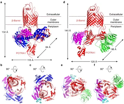
Figure 1 | Structure of two complexes of E. coli β-barrel assembly machinery.
Two structures of E. coli BAM: BamABCDE and BamACDE. The BamA (red)
C-terminal barrel is embedded in the OM, while the N-C-terminal domain of BamA is in
the periplasm, forming a novel circular structure with lipoproteins BamB (green),
BamC (blue), BamD (magenta) and BamE (cyan). a, b and c, Cartoon representation
of the structure of BamACDE complex, viewed for the membrane plane, extracellular
and periplasm, respectively. BamD interacts with POTRA 1, 2 and 5 to form a ring
structure in the periplasm, while BamC binds to both BamD and POTRA 1 and 2 of
BamA. BamE forms contacts with both BamA and BamD. The dimensions of
BamACDE were measured at the widest points of the outer surfaces of the complex.
d, e and f, Cartoon representation of BamABCDE structure, viewed from the
POTRA 2 and 3, while only N-terminal loop of BamC forms contacts with BamD.
The dimensions of BamABCDE were measured as in a.
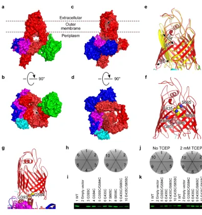
Figure 2 | Inward- and lateral-open conformations of BAM. BamA-E are in the
same colors as in Figure 1. a, Membrane view of the molecular surface of
BamABCDE. The pore of BamA is completely sealed at the extracellular side by the
extracellular loops. b. Periplasmic view of BamABCDE. The barrel is open to the
periplasm (indicated by the arrow). c, Membrane view of the molecular surface of
BamACDE. The barrel is open laterally to the OM and the extracellular side
(indicated by the arrow). d, Periplasmic view of BamACDE surface structure. The
barrel is completely closed to the periplasm. e, The significant conformational
changes of the BamA barrel domain between the inward-open (red) and the
lateral-open (yellow) conformations. The barrel strands β1C-β6C of BamA have been
rotated about 65° with the distance around 15 Å to laterally open the barrel from the
inward-open state. f, The double mutation E435C/S665C or E435C/S658C is
expected to lock the barrel in the inward-open conformation. Residues I806-K808 of
the β16C of BamA coils toward the inside of the barrel lumen. g, The G393C/G584C mutant is expected to lock the barrel in the lateral-open conformation. h, The
functional assays of the mutants. The single residue mutations do not affect the E. coli
cell growth, but the double cysteine mutations kill the bacteria. 1, 2, 3, 4, 5, 6, 7, 8, 9,
and 10 represent the wild type BamA, the vector without BamA, BamA mutants
G393C, G584C, G393C/G584C, E435C, S665C, S658C, E435C/S665C and
E435C/S658C, respectively. i, The protein expression levels of BamA mutants in the
OM were checked by western blotting. j, The reducing reagent TCEP could rescue the
cysteine mutants in absence and in present of TCEP were checked by western
blotting.
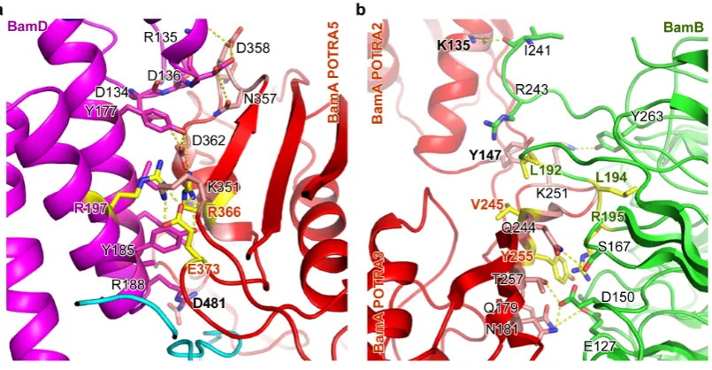
Figure 3 | BamA interacts with BamD and BamB in BamABCDE complex.
BamD contacts POTRA domains 1, 2 and 5 to form a ring structure. a, BamA
POTRA 5 interacts with the C-terminal domain of BamD. BamA residues R366 and
E373 and BamD residue R197 are important for the two protein interactions, and their
carbon atoms are colored in yellow. b. BamA and BamB interaction. Both POTRA 2
and 3 involve in BamB interaction. BamA residues V245, Y255 and BamB residues
L192, L194 and R195 play important roles in BamA and BamB interactions.
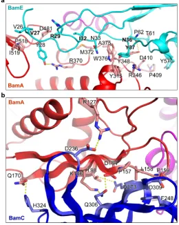
Figure 4 | BamE and BamC interact with BamA and BamD. a, The interface
between BamE and BamA in the BamABCDE complex. BamE forms contacts with
POTRA 5 residues, BamA periplasmic turns T-2 and T-3, and POTRA 4 in the
BamACDE complex (Extended Data Fig. 6). b, The C-terminal globular domain of
BamC interacts with BamA POTRA 2 at the β-sheets in BamACDE. Residues in the
two β-sheets that are involved in the BamC and BamA interactions are shown.
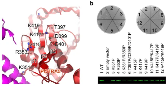
Figure 5 | The function of BamA POTRA 5. a, The three β-strands of BamA
POTRA 5. The residues selected for functional assays are shown: residues K351 and
R353 on β1, T397, D399 and D401 on β2 and V415, K417 and K419 on β3. b, The
β-strands of POTRA 5 are critical for the bacterial survive. The single mutant K351P,
the double mutant K351P/R353P at β1, the double mutants V415P/K417P,
K417P/K419P and V415P/K419P at β3 kill the bacteria, while the triple mutant
wild-type and mutants. 1, 2, 3, 4, 5, 6, 7, 8, 9, 10, 11 and 12 represent the wild type
BamA, the vector without BamA, BamA mutants K351P, R353P, K351P/R353P,
T397P/D399P/D401P, V415P, K417P, K419P, V415P/K417P, K417P/K419P and
V415P/K419P, respectively.
Figure legends for Extended Data Figures:
Extended Data Figure 1 | BamABCDE and BamACDE complexes and electron
density maps contoured at 1 σ. a, Schematic diagram of the five BAM subunits. P-1
to P-5 represent the five BamA POTRA domains.b, SDS-PAGE analysis of the BAM complex from crystals. M, 1 and 2 are protein molecular weight marker, crystals of purified the BAM complex expressed by construct pYG120 and pJH114, respectively (Supplementary Data Figure 1). The BamABCDE crystals contain the full length BamA-E. The crystals were washed five times in fresh reservoir solution, and then dissolved in SDS-PAGE loading buffer. The results showed that the BamB is absent in the BamACDE crystals, while the BamC is complete in both the BamABCDE and BamACDE crystals. c, SDS-PAGE analysis of the purified BAM complex. M, 1 and 2 are protein molecular weight marker, purified BAM protein complex expressed by construct pYG120 and pJH114, respectively. The BAM complexes expressed from pJH114 is a mixture of BamABCDE and BamACDE complexes (Supplementary Data Figure 1). d, 2FoFc electron density map of BamA residues W576-K580 of BamACDE contoured at 1 σ. e, 2FoFC electron density map of BamD residues Y177-W191 of BamACDE contoured at 1 σ. f, 2FoFc electron density map of BamA residues Y504-Y509 and F490-F494 of BamABCDE complex contoured at 1σ. g, 2FoFc electron density map of BamB residues Y345-W348 contoured at 1 σ.
Extended Data Figure 2 | Superimposition of the two BamACDE complexes in
with some conformational changes in the β-strands of barrel and extracellular loops
with root-mean-square deviation (RMSD) of 0.908 Å over 378 Cα atoms, while the
periplasmic circular structure has some rotation (see arrows) with a RMSD of 4.706 Å over 385 Cα atoms. b, Periplasmic view of the superimposition of the two structures.
The periplasmic circular structure has some rotations when the C-terminal global domain binds on the POTRA 2. c. Superimposition of the barrels of the two complexes. d, Superimposition of the two BamCs. The N-terminal coil structures superimpose well with a RMSD of 0.807 Å over 86 Cα atoms.
Extended Data Figure 3 | Molecular dynamics simulation of BAM complexes. a, BamABCDE and b, BamACDE structures modelled with all subunits present and embedded in a model E. coli outer membrane (grey). Phosphate atoms are shown in orange spheres. Lipid-modified cysteine residues of BamB, BamC, BamD and BamE are shown in yellow spheres. c, Both complexes are stable in MD simulations, showing limited deviation from the starting configuration (shown in the background). d, Simulations of the complexes of only BamA and BamD subunits retain the ring structure. Without BamC present POTRA 1 (black circle) moves towards the membrane, while POTRA 3 (black arrow) moves towards and interacts with the periplasmic loops of the barrel. The dynamics of POTRA 3 appear to be modulated by BamB. e, Simulations of BamA show enhanced dynamics of the POTRA domains, with POTRA 1 and 2 rotating towards the membrane in an anti-clockwise direction (blue arrow). This separates POTRA 3 from the barrel (black arrow). This conformation of the POTRA domains is unable to form the BAM ring, highlighting the essential nature of BamD and its interactions with BamA in maintaining the ring structure.
Extended Data Figure 4 | BamA of the BamABCDE complex is superimposed onto the other published BamA structures. All the published BamA structures are in the inward-open conformation. In all cases the BamA from BamABCDE is shown in red. a, The BamA of BamABCDE complex is superimposed onto BamA of N. gonorrhoeae (grey) (protein data bank access code 4K3B)18. The two barrel structures are similar with a RMSD of 3.803 Å over 385 Cα atoms, but the conformations of the
five POTRA domains are quite different. The dotted circle indicates the hydrophobic gap between β1C and β15C. b, BamA of E. coli (magenta) (PDB access code
4N75)19. The two barrel structures superimpose well with a RMSD of 0.644 Å over 385 Cα atoms, but differences are observed for the β16C terminal residues. The
C-terminal residues in BamA of BamABCDE move toward to the lumen of the barrel. c, BamA of E. coli (yellow) (PDB access code 4C4V)20 with a RMSD of 1.382 Å over 365 barrel Cα atoms. The conformations of the POTRA 5 are quite different. d,
BamA of H. ducreyi (green) (PDB access code 4K3C)18. The barrel structures are similar with a RMSD of 2.376 Å over 365 barrel Cα atoms, but the conformations of
POTRA 4 and 5 are quite different.
the 379 barrel Cα atoms and a maximum RMSD of 20 Å . The POTRA domains align
with an RMSD of 5.764 Å over 384 Cα atoms with maximum 15 Å. The
BamABCDE complex is in the same colour scheme as Figure 1. The BamACDE complex is in yellow. The barrel strands β1-6C rotate around 65° from BamABCDE
to BamACDE, while the BAM periplasmic unit rotates around 30° in a
counter-clockwise direction from BamABCDE to BamACDE. a, Membrane view of the superimposition of the BamABCDE and BamACDE complexes. The conformations of BamA POTRA domains, BamB, BamC and BamD are significantly different between the two complexes. b, The periplasmic view of the superimposition of BamABCDE and BamACDE. The circular units rotate around 30° between the two
BAM complexes. c, The residues involved in closing the barrel at the periplasmic side in the BamACDE structure. d, Heat-modifiability assays of the BamA double cysteine mutants. SDS-PAGE/western blot analysis of the wild type BamA, BamA G393C/G584C, E435C/S665C and E435C/S658C mutants showed the heat-modifiability, indicating that the three double cysteine BamA mutants were correctly folded into the OM. U, F indicate unfolded and folded, respectively (Supplementary Data Figure S5).
Extended Data Figure 6 | Periplasmic loops bind to BamA POTRA 3, 5, BamD and BamE. In the BamABCDE complex, the BamA barrel interacts with POTRA 3, 5, BamE and BamD through the periplasmic turns T-1, -2, -3, -5, -6 and -7. a, In the BamABCDE complex, the residues of T-1, -2 and -3 are involved in the interactions with POTRA 5, BamD and BamE. b, Residues in T-5, -6 and -7 interact with POTRA 3 in the BamABCDE complex. c, In the BamACDE complex no interactions are observed between the periplasmic turns and POTRA 3. The figure shows that the residues in T-1, -2 and -3 interact with residues in POTRA 5, BamD and BamE. These structural data may suggest that BamB, C, D and E either directly or indirectly control the conformation of the barrel through its periplasmic turns.
Extended Data Figure 7 | BamA and BamD interactions, and Superimposition of
the BamB structures and the conformational changes of POTRA2 and 3. a,
BamA POTRA 1 and 2 interact with the N-terminal domain of BamD. The interacting residues from both BamA and BamD are shown. b, Functional assays of the BamA interaction with BamD. The mutation BamA E373K is lethal, while mutant R366E impairs the bacterial growth, suggesting these residues may play an important role in the BAM complex. 1, 2, 3, and 4 represent the wild type BamA, the vector without BamA, BamA mutants E373K and R366E, respectively. c, Protein expression levels of BamA mutations were detected by western blotting. d, Periplasmic view of BamB of the BamABCDE complex (green) superimposed onto the free BamB structure (orange) (PDB code 3Q7N)21 with a RMSD of 1.81 Å over 351 C
α atoms with the
maximum deviation of 12 Å at loop 19. Loops 15, 19, 23 and 27 of BamB adopt conformational changes to bind to POTRA 2 and 3. e, BamB of the BamABCDE complex superimposed onto BamB in complex with POTRA 3 and 4 (magenta) (PDB code 4PK1)32. The two BamB structures are very similar with a RMSD of 0.5860 Å over 341 Cα atoms. f, Superimposition of BamABCDE and BamACDE at POTRA 2
and 3 with a RMSD of 3.57 Å over 159 Cα atoms. In the BamACDE structure the
Extended Data Figure 8 | BamE interacts with BamD and BamC, and BamC interactions with BamD. BamE interacts with BamA, BamD and BamC. BamC binds extensively to the C-terminal domain of BamD. a. BamE interacts with BamD in the BamACDE complex. BamE contacts the C-terminal domain residues of BamD in the BamACDE complex. b, BamE forms hydrophobic interactions with BamC in the BamACDE complex. BamE residues P67, F68 and BamC residues M56 and I57 are shown. c, BamC forms contacts with BamA POTRA 1 in BamACDE. BamA residues F31, Q35, V39 and BamC residues G94 and R96 are shown. d, BamC interacts with the C-terminal domain of BamD. The interacting residues are shown as sticks. e, BamC interacts with the N-terminal domain of BamD.
Extended Data Figure 9 | Conserved residues analysis of BAM complex. Consurf residue conservation scores (1-9), plotted onto the molecular surfaces as a colour scale for BamA (red), BamB (green), BamC (blue), BamD (purple) and BamE (cyan), for the BamABCDE structure. Regions of white/grey indicate poorly conserved residues, whereas a more intense colour indicates highly conserved residues. Black dashed circles represent the interaction points on removal of BamC (a), BamD (b), BamE (c) and BamB (d). For each interaction patch a high density of conserved residues is apparent.
Extended Data Figure 10 | Conformational differences of the BAM subunits between the BamABCDE and BamACDE complexes, and BAM complex interacts with lipid of the OM. The subunits of BamABCDE are colored in the same colours as Figure 1, while the BamACDE subunits are in yellow. a, Superimposition of the BamA subunits onto the barrel domain with an RMSD of 4.85 Å over the 379 barrel Cα atoms and an RMSD of 5.76 Å over the 384 Cα atoms of the POTRA
domains. The BamA barrel has significant conformational changes in β1C-β6C. The
periplasmic POTRA domains rotate about 30° from BamABCDE complex to
BamACDE complex, suggesting a novel rotation mechanism to facilitate OMP insertion into the OM. b, Superimposition of the BamC structures. The BamC structures have some conformational changes with a RMSD of 2.102 Å over 47 Cα
atoms of the BamC N-terminal loop. The N-terminal loop C25-V35 becomes more ordered in BamACDE complex. Particularly, the N-terminal domain and the C-terminal domain are ordered and bind to POTRA 1, 2 and the N-C-terminal domain of BamD in BamACDE complex. The N-terminal loops of the BamC structures superimpose well between residues V35 to P88. c, Superimposition of the BamD structures with an RMSD of 1.201 Å over 203 Cα atoms. The α-helices are
conserved, but the loops have some conformational changes, especially loop 6 (residues D121-D136) between α-helix 5 and α-helix 6. d, Superimposition of BamE
structures with a RMSD of 1.721 Å over 81 Cα atoms. The β strands and α-helices of
BamABCDE complex by molecular modelling, using the solved domain from the companion complex. BamABCDE complexe was inserted into the OMP, with lipid anchors designed (Methods).
Extended Data Table 1 | Data collection and refinement statistics.
Online Content Full methods, along with any additional Extended Data display items and Source Data, are available in the online version of the paper; references unique to these sections appear only in the online paper.
METHODS
Cloning, expression and purification of BAM complex
Expression plasmid pJH114 containing the five E. coli bamABCDE genes which were under the control of a trc promoter, and with an octa-‐‑histidine (8×His) tag at the C-‐‑terminus of bamE was initially used for overexpression of BamABCDE complex in E. coli HDB150 cells16. Expression of the native BamABCDE complex was induced with 100 μmol l-‐‑1 Isopropyl-‐‑β-‐‑D-‐‑1-‐‑thiogalactopyranoside (IPTG;
Formedium) at 20°C overnight when the optical density of the cell culture at 600 nm reached 0.5-‐‑0.8. The selenomethionine-‐‑labeled BAM complexes were expressed in M9 medium supplemented with selenomethionine Medium Nutrient Mix (Molecular Dimensions) and 100 mg l-‐‑1 L-‐‑(+)-‐‑selenomethionine (Generon Ltd) using the similar conditions as the native BamABCDE.
Systems Ltd) at 30 kpsi. The lysate was centrifuged to remove the cell debris and unbroken cells, and the supernatant was ultracentrifuged to pellet the membranes at 100,000g for 1 h. The cell membranes were resuspended in solubilization buffer containing 20 mM Tris-‐‑HCl, pH 8.0, 300 mM NaCl, 10 mM imidazole and 1~2% n-‐‑Dodecyl-‐‑β-‐‑D-‐‑Maltopyranoside (DDM; all detergents were purchased from Anatrace) and rocked for 1 h at room temperature or overnight
at 4°C. The suspension was ultracentrifuged and the supernatant was applied to
a 5 mL pre-‐‑equilibrated HisTrap HP column (GE Healthcare). The column was washed with wash buffer containing 20 mM Tris-‐‑HCl, pH 8.0, 300 mM NaCl and 35 mM imidazole and eluted with elution buffer containing 300 mM imidazole.
The eluent was applied to HiLoad 16/600 Superdex 200 prep grade column (GE healthcare) pre-‐‑equilibrated with gel filtration buffer containing 20 mM Tris-‐‑HCl, pH 7.8, 300 mM NaCl and detergents. Different detergents were used in protein purification procedures.
The purified BamABCDE complex was analyzed by SDS-‐‑PAGE (Extended Data
Figure 1 and Supplementary Data Figure 1), which indicated that BamB is not enough in the complex, and BamB is absent in the determined structure. We therefore decided to generate a new plasmid to express the BamABCDE complex. Additional copy of E. coli bamB gene was introduced into pJH11416 after the 8×His tag to generate a new expression plasmid pYG120 using a modified
CTCTAGAGGATCTTAGTGGTGATGATGGTG-‐‑3’), and PF_EBB_SLIC (5’-‐‑ TCATCACCAC-‐‑TAAGATCCTCTAGAGAGGGACCCGATGCAATTGC-‐‑3’) and
PR_EBB_SLIC (5’-‐‑CTTGC-‐‑
ATGCCTGCAGGTCGATTAACGTGTAATAGAGTACACGGTTCC-‐‑3’), respectively. Gel extracted fragments were digested by T4 DNA polymerase (Fermentas) at 22°C for 35 min followed by 70°C for 10 min, and then placed on ice immediately. The
digested fragments were annealed in an annealing buffer (10 mM Tris, pH 8.0, 100 mM NaCl and 1 mM EDTA) by incubating at 75°C for 10 min and decreasing by 0.1°C every 8 seconds to 20°C. The mixture was transformed into E. coli DH5α for plasmid preparation. The DNA sequences were confirmed by sequencing. For the purification of the BamABCDE complex from the pYG120 construct, the
wash buffer, elution buffer and gel filtration buffer were supplemented with different detergent combinations. A second gel filtration was performed to change detergents with gel filtration buffer containing 1 CMC n-‐‑Octyl-‐‑β-‐‑D-‐‑ Glucopyranoside (OG) and 1 CMC n-‐‑Dodecyl-‐‑N,N-‐‑Dimethylamine-‐‑N-‐‑Oxide
(LDAO). For BamABCDE complex purification from construct pJH114, the wash buffer, elution buffer and gel filtration buffer were supplemented with 2 CMC n-‐‑ Nonyl-‐‑β-‐‑d-‐‑glucoside (β-‐‑NG) and 1 CMC Tetraethylene Glycol Monooctyl Ether (C8E4). The peak fraction was pooled and concentrated using Vivaspin 20 centrifugal concentrator (Sartorius, molecular weight cut off: 100 kDa). The
selenomethionine-‐‑labeled proteins were purified in the same way as the native proteins of BamABCDE complex.
Crystallization, data collection and structure determination
The purified proteins were concentrated to 8~12 mg ml-‐‑1 for crystallization. For
crystallizations were carried out by sitting-‐‑drop vapour diffusion method in the MRC 96 well crystallization plates (Molecular Dimensions) at 22 °C. The protein solution was mixed in a 1:1 ratio with the reservoir solution using the Gryphon crystallization robot (Art Robbins Instruments). The best NaI co-‐‑crystallized crystals were grown from 150 mM HEPES, pH 7.5, 30% PEG6000 and CYMAL®-‐‑4 in MemAdvantageTM (Molecular Dimensions) as additive. The best native crystals
were grown from 150 mM HEPES, pH 7.5 and 27.5% PEG6000. The best selenomethionine-‐‑labeled crystals were grown from 100 mM Tris, pH 8.0, 200 mM MgCl2∙6H2O, 24% PEG1000 MME and OGNG in MemAdvantageTM as additive. The crystals were harvested, flash-‐‑cooled and stored in liquid nitrogen for data collection. The data sets of selenomethionine labelled BAM complex were
collected on the I03 beamline at Diamond Light Resources (DLS) at a wavelength of 0.9795 Ångström (Å). All data were indexed, integrated and scaled using XDS43. The crystals belong to space group of P42212, with the cell dimensions a = b =
254.16 Å, c = 179.22, α = β = γ =90°. There are two complexes in the asymmetric
unit. The structure was determined to 3.9 Å resolution (Extended Data Table 1) using ShelxD44,45. Fifty-‐‑six selenium sites were found, which gave a FOM of 0.32. Following density modification using DM46, the BamACDE complex was clearly visible in the electron density map, but without BamB. Using the individual high-‐‑ resolution models, the BamACDE complex was built using Coot47 by
reference model secondary structure restraints from higher resolution models. Weights were automatically optimised by PHENIX48.
To obtain the BamABCDE complex structure, the new construct was used to produce sufficient BamB to form the BamABCDE complex. The data sets of BamABCDE complex were collected on the I02 beamline at DLS. The crystals belong to space group P41212, with the cell dimensions a = b = 116.69 Å, c =
435.19 Å, α=β=γ=90°. There is one complex molecule in the asymmetric unit.
Although the crystals diffracted to 2.90 Å, the crystal structure of BamABCDE could not be determined by molecular replacement. BamABCDE complex was crystallized in presence of 0.2 M sodium iodide, and SAD datasets were collected
at a wavelength of 1.8233 Å. 4 x 360 degree datasets were collected on different regions of the same crystal of NaI co-‐‑crystallization then combined. The phases were determined by ShelxD44,45 at 4 Å resolution. Eleven iodide sites were found, which gave a FOM of 0.28. The phases were extended to 2.90 Å by DM46, and the model was built using Coot47 by skeletonizing the electron density map and
docking the individual high-‐‑resolution subunits in the electron density map and rigid body fit this model into the higher resolution native dataset while retaining and extending the free R set from the iodide dataset. The BamABCDE complex was refined using PHENIX48. TLS groups were automatically determined using PHENIX48 and used for refinement along with individual B-‐‑factors. Weights were
automatically optimised and secondary structure restraints were used.
Site-‐‑directed mutagenesis and functional assays
An E. coli bamA expression plasmid was constructed for functional assays using
DNA Polymerase (New England BioLabs), and plasmid pJH114 as template and
primers PF_bamA_SLIC (CCATCATCATCATCATCATC-‐‑
ATCATGAAGGGTTCGTAGTGAAAGATATTCATTTCGAAG) and PR_bamA_SLIC (AGA-‐‑CTCGAGTTACCAGGTTTTACCGATGTTAAACTGGAAC). Vector backbone was amplified from a modified pRSFDuet™-‐‑1 vector (Novagen, Merck Millipore) containing an N-‐‑terminal pelB signal peptide coding sequence with primers
PF_RSFM_SLIC (CGGTAAAACCTGGTAACTC-‐‑GAGTCTGGTAAAGAAACCGCTGC) and
PR_RSFM_SLIC (ATGATGATGATGATGATG-‐‑
ATGATGGTGATGGGCCATCGCCGGCTG). Plasmids were prepared using GeneJET Plasmid Miniprep Kit (Thermo Scientific). Site-‐‑directed mutagenesis was performed according to a previously described protocol49 with slight
modification (PCR conditions and the sequences of the primers are available upon request). The sequences of the wild type and all mutant constructs of bamA were confirmed by sequencing. E. coli JCM166 cells3 transformed with the wild-‐‑ type bamA or its mutants were plated on LB agar plates supplemented with 50
μg ml-‐‑1 kanamycin and 100 μg ml-‐‑1 carbenicillin in the presence or absence of 0.05% L-‐‑(+)-‐‑arabinose and grown overnight at 37°C. Single colonies grown on arabinose-‐‑containing plates were inoculated in 10 ml LB medium supplemented with 50 μg ml-‐‑1 kanamycin, 100 μg ml-‐‑1 carbenicillin and 0.025% L-‐‑(+)-‐‑arabinose, and incubated at 200 rpm at 37°C for 16 h. For plate assays, the cells were
pelleted and resuspended in fresh LB medium supplemented with 50 μg ml-‐‑1 kanamycin and 100 μg ml-‐‑1 carbenicillin, and diluted to an absorbance (A600 nm) of ~0.3 and streaked onto LB agar plates supplemented with 50 μg ml-‐‑1 kanamycin, 100 μg ml-‐‑1 carbenicillin in the presence or absence of 0.05% L-‐‑(+)-‐‑
Western blot
Western blotting was performed to examine protein expression levels of BamA in the membrane. 50 ml of overnight cultures of transformed JCM166 cells with respective wild-‐‑type or each mutant of BamA were pelleted. The cells were resuspended in 25 ml 20 mM Tris-‐‑HCl (pH 8.0), 150 mM NaCl and sonicated. The cell debris and unbroken cells were removed by centrifugation at 7,000 g for 30
min. The supernatant was centrifuged at 100,000 g for 60 min and the membrane fraction was collected. The membrane fraction was suspended in 5 ml buffer containing 20 mM Tris-‐‑HCl (pH 8.0), 150 mM NaCl and 1% 3-‐‑(N,N-‐‑ Dimethylmyristylammonio) propanesulfonate (Sigma) and solubilized for 30 min at room temperature. Samples were mixed with 5 × SDS-‐‑PAGE loading
buffer, heated for 5 min at 90 °C, cooled for 2 min on ice and centrifuged. Ten microliters of each sample was loaded onto 4-‐‑20% Mini-‐‑PROTEAN® TGX™ Gel (Bio-‐‑Rad) for SDS-‐‑PAGE and then subjected to immunoblot analysis.
The proteins were transferred to PVDF membrane using Trans-‐‑Blot® Turbo™
Transfer Starter System (Bio-‐‑Rad) according to the manufacturer's instructions. The PVDF membranes were blocked in 10 mL Protein-‐‑free T20 (TBS) blocking buffer (Fisher) overnight at 4°C. The membranes were incubated with 10 mL His•Tag® Monoclonal Antibody (diluted, 1:1000) (Millipore) for 1 h at room temperature followed by washed with PBST for 4 times and incubated with
IRDye 800CW goat anti-‐‑mouse IgG (diluted, 1:5000) (LI-‐‑COR) for 1 h. The membrane was washed with PBST for 4 times and PBS for 2 times. Images were acquired using LI-‐‑COR Odyssey (LI-‐‑COR).