Original Article
Knockdown of TMEM45A inhibits the proliferation,
migration and invasion of glioma cells
Wei Sun1*, Guanzhong Qiu1*, Yongxiang Zou1, Zheng Cai1, Peng Wang2, Xianbin Lin1, Jinxiang Huang1, Lei Jiang1, Xuehua Ding1, Guohan Hu1
1Department of Neurosurgery, Shanghai Institute of Neurosurgery, Shanghai Changzheng Hospital, Second
Military Medical University, Shanghai, China; 2Department of Radiology, Shanghai Changzheng Hospital, Second
Military Medical University, Shanghai, China. *Equal contributors.
Received August 13, 2015; Accepted September 22, 2015; Epub October 1, 2015; Published October 15, 2015
Abstract: Gliomas are the most common and aggressive type of primary adult brain tumor. Although high expression and prognostic value of TMEM45A has been recently reported in various types of human tumors, the association of TMEM45A expression and glioma is still unknown. Here, we reported that TMEM45A was significantly overexpressed in glioma tissues compared to non-tumorous brain tissues. Furthermore, TMEM45A mRNA levels were gradually in-creased with the increasing severity of histological grade of glioma. Moreover, high TMEM45A expression level was correlated with short survival time of glioma patients. Down-regulation of TMEM45A in two glioma cell lines, U251 and U373 by transected with TMEM45A siRNA resulted in a significant reduction of cell proliferation and G1-phase arrest. Additionally, we found that suppressing of TMEM45A expression in glioma cells remarkably suppressed cell migration and cell invasion. More importantly, TMEM45A siRNA treatment significantly down-regulated the proteins promoting cell cycles transition (Cyclin D1, CDK4 and PCNA) and cell invasion (MMP-2 and MMP-9), which indicted a possible mechanism underlying its functions on glioma. In summary, our study suggests that TMEM45A may work as an oncogene and a new effective therapeutic target for glioma treatment.
Keywords: TMEM45A, glioma, proliferation, invasion
Introduction
Gliomas are the most common and aggressive type of primary adult brain tumor and persist
as serious clinical and scientific problems [1].
There are three types of gliomas: astrocyto-
ma, oligodendroglioma and ependymoma [2].
Current therapies are not effective in treating gliomas. Therefore, glioma remains as one of the leading causes of cancer deaths worldwide
[3, 4]. The median survival of patients with
glioblastoma multiforme, the most malignant glioma, remains less than one year, even with aggressive surgery, radiation and
chemothera-py [3]. New prognostic indicators and effective
therapeutic targets for gliomas still needed to
be identified, which urges a better understand -ing of the molecular mechanisms govern-ing dis-ease manifestation and progression.
Transmembrane protein 45A (TMEM45A), a multi-pass membrane protein, belongs to the
large family of genes encoding predicted trans-membrane (TMEM) proteins. TMEM45A has been reported to be involved in epidermal
kera-tinization [5, 6]. Recently, high expression of
TMEM45A has been linked to poor prognostic
in patients with different types of tumors [7-10].
In liver and breast cancer cells, TMEM45A ex- pression is implicated in protecting liver and breast cancer cells from drug-induced
apopto-sis [7]. Lee et al. reported that TMEM45A inhib -its the progression of ductal carcinoma into
invasive breast cancer [11]. However, how alter -ations of TMEM45A expression are involved in the malignancies remains to be solved.
In the present study, we explored the role of TMEM45A in gliomas and sought to identify the involved mechanisms. TMEM45A mRNA
level was significantly higher in glioma tissues
histo-logical grades of gliomas. Our in vitro experi-ments indicated that TMEM45A was involved in multiple cellular progress including cell prolif-eration, cell cycle progression, migration and invasion. Furthermore, the mRNA and protein levels of cell cycle related-genes and invasion related-genes were decreased in TMEM45A knockdown cells. Collectively, these data sug-gest that TMEM45A is a potent oncogene in glioma and it may be an effective therapeutic target for this disease.
Materials and methods
Tissue samples and gene expression data
Fresh frozen samples of 45 glioma tissues and 11 non-neoplastic brain tissues from surgical procedures for epilepsy were obtained from Shanghai Changzheng Hospital. In 45 patients with glioma enrolled in this study, 10 patients were with WHO (World Health Organization) Grade II glioma, 12 were with Grade III glioma, and 23 were with Grade IV glioma. Informed consent was obtained from all patients. This study was approved by the ethics committee of Second Military Medical University.
The Cancer Genome Atlas (TCGA) glioblastoma (GBM) dataset of 529 patients and 10 normal brain tissues (version: 2014-08-22) were down-loaded from TCGA website (https://tcga-data. nci.nih.gov/tcga/).
bovine serum (FBS, Life Technologies, Carlsbad, CA, USA), 100 mg/ml penicillin G, and 50 g/ml streptomycin (Life Technologies). U251 and SHG44 cells were cultured in DMEM (Life Technologies), while U87, U373 and T98G were cultured in Eagle’s Minimum Essential Medium (Life Technologies).
Silencing of TMEM45A by small interfering RNA
Three siRNAs targeting human TMEM45A mRNA were synthesized (TMEM45A-siRNA1: 5’-GGCCUUUAUCUUCUACAACUU-3’; TMEM45A-siRNA2: 5’-GUUCCUUGUUCGGAACAAUUU-3’; TMEM45A-siRNA2: 5’-AGUGUACUGUUUGCAU-
UUCUU-3’). A non-specific scramble siRNA
sequence was used as negative control (NC: 5’-UUGUACUACACAAAAGUACUG-3’). The siR-NAs were transiently transfected into U251 and U373 cells using Lipofectamine 2000 (Invitrogen) according to the manufacture’s instruction. Assays were performed 48 h after transfection.
Reverse transcription and Real-Time PCR
Total RNA was extracted from cultured cells or tissue samples using TRIzol Reagent (Invitrogen) according to the manufacturer’s instructions.
Total RNA (1 μg) was reverse-transcribed using
[image:2.612.89.377.83.319.2]M-MuLV Reverse Transcriptase (Thermo Fisher
Table 1. Primers sequences for quantitative PCR
Primer Primer sequence Size (bp)
TMEM45A (NM_018004.1) F: 5’-CTCCTGGGCTGGCATCATTTC-3’ 167 R: 5’-CGGCCATGAGTGTGGTTGTAG-3’ Cyclin D1 (NM_053056) F: 5’-GCTGCGAAGTGGAAACCATC-3’ 135
R: 5’-CCTCCTTCTGCACACATTTGAA-3’ CDK4 (NM_000075) F: 5’-ATGGCTACCTCTCGATATGAGC-3’ 124
R: 5’-CATTGGGGACTCTCACACTCT-3’ CDK2 (NM_001798) F: 5’-CCAGGAGTTACTTCTATGCCTGA-3’ 90
R: 5’-TTCATCCAGGGGAGGTACAAC-3’ PCNA (NM_002592.2) F: 5’-GCCTGACAAATGCTTGCTGAC-3’ 223
R: 5’-GCTGTTCTTGGCCTCAGTC-3’
MMP-2 (NM_004530) F: 5’-TACAGGATCATTGGCTACACACC-3’ 90 R: 5’-GGTCACATCGCTCCAGACT-3’
MMP-9 (NM_004994) F: 5’-TGTACCGCTATGGTTACACTCG-3’ 97 R: 5’-GGCAGGGACAGTTGCTTCT-3’
GADPH (NM_001256799.1) F: 5’-CACCCACTCCTCCACCTTTG-3’ 110 R: 5’-CCACCACCCTGTTGCTGTAG-3’
Immunohistochemical analy-sis
Immunohistochemistry (IHC) was performed as previously
described [12]. The results of
IHC staining were evaluated independently by two trained pathologists without knowl-edge of clinical data.
Cell lines
Scientific, Rockford, IL, USA). The resulting
cDNA was used as template in real-time quanti-tative PCR by using a standard SYBR Green PCR kit (Thermo) on an ABI 7300 Thermocycler Real-Time PCR machine (Applied Biosystems, Foster City, CA, USA). GAPDH was used as con-trol of the input RNA level. Primer sequences were listed in Table 1. All reactions were con-ducted using the following cycling parameters, 95°C for 10 min, followed by 40 cycles of 95°C for 15 s, 60°C for 45 s. GAPDH was served as an internal control. The gene expression was
calculated using the ΔΔ Ct method. All data
represent the average of three replicates.
Immunoblotting
Treated and untreated cells were lysed in radio-immunoprecipitation assay buffer (JRDUN Biotehnology, Shanghai, China) and protein concentration was measured by BCA protein
assay kit (Thermo Fisher Scientific) [13]. Protein
isolates were then subjected to SDS gel elec-trophoresis and analyzed by western blot using enhanced chemiluminescence system (ECL, Millipore). Antibodies against TMEM45A, PCNA and MMP-9 were purchased from Abcam (Cambridge, MA, USA). Anti-Cyclin D1 was from Santa Cruz Biotechnology (Santa Cruz, CA, USA). Antibodies against CDK4 and GAPDH were purchased from CST Biotech. (Danvers, MA, USA). Anti-MMP-2 was from Epitmics (Burlingame, CA, USA).
Cell proliferation assay
Treated and untreated cells were seeded at a density of 5×10³ cells per well in a 96- well plate. Cell proliferation was detected by using the Cell Count Kit-8 (CCK-8, Dojindo Laboratories) according to the manufacturer’s instructions. At indicated time point, CCK8 solution was added to each well and incubated for 1 h. Absorbance was detected at a wave-length of 450 nm by a microplate reader (Bio-Rad Laboratories Inc., Hercules, CA, USA). Three wells were measured for cell viability in each treatment group, and all independent treatments were performed in triplicate.
Cell cycle distribution analysis
The cell cycle was evaluated by flow cytometry using propidium iodide (PI) staining on a flow
cytometer (BD Biosciences, San Jose, CA, USA).
Briefly, treated and untreated cells were har
-vested, re-suspended in PBS and fixed ice-cold 70% ethanol at -20°C for at least 2 h. The fixed
cells were washed with PBS, incubated with ribonuclease A (Sigma) and PI (0.05 mg/ml, Sigma, St. Louis, MO, USA) at room tempera-ture in the dark for 30 min. DNA content was
then analyzed using a FACScan flow cytometry.
The percentage of cells in the G0/G1, S, and G2/M phases was determined by the FlowJo software (Tree Star). Experiments were per-formed in triplicate and 3×10⁴ cells were ana-lyzed per sample.
Boyden chamber assay for migration and inva-sion
Quantitative cell migration and invasion assays were performed using Boyden chambers
con-taining polycarbonate filters with a pore size of 8 μm (Coring Incorporated, NY, USA).
For transwell migration assay, treated and untreated cells were serum starved for 24 h, 5×10⁴ cells were plated into the upper well of the Boyden chambers with serum-free medium in the top chamber and medium containing 10% FBS in the lower chamber. After 24 h of incubation, the cells on the upper surface of
the filter were completely removed by wiping
with a cotton swab. The remaining migrated
cells were washed with PBS, fixed in 4% para -formaldehyde and stained with 0.2% crystal violet. The migrated cells were observed under
Leica inverted microscope (Deerfield, IL, USA). Cell number was counted in 10 random fields
for each condition. The experiments were per-formed in triplicate.
For in vitro invasion assay, the upper wells of the Boyden chambers were coated with Matrigel (BD Biosciences) at 37°C in a 5% CO2 incubator for 1 h. The rest of the assay was performed as described above.
Statistical analysis
The two-tailed Student’s t-test was used to evaluate statistical differences between two
Results
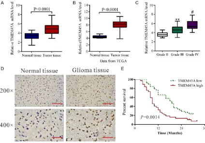
Elevated expression of TMEM45A was cor-related with the histological grade of primary human glioma
We first compared TMEM45A mRNA level in
glioma tissues (n=45) and normal brain tissues (n=11) by using real-time PCR. Statistical analy-sis with student’s t-test suggested that TMEM-
45A expression was significantly elevated in
glioma tissues compared with that in normal tissues (Figure 1A, P<0.0001). Then we re-ana -lyzed high throughput RNA-sequencing data of the glioblastomas cohort of The Cancer Genome Atlas (TCGA) and found that TMEM45A
expression was significantly increased in glio -ma tissues compared with nor-mal brain tissues (Figure 1B, P<0.0001). We also assessed the
expression level of TMEM45A by immunohisto-chemistry and found that TMEM45A protein
level was also elevated in glioma tissues (Figure 1D).
To assess the clinical relevance of TMEM45A, we compared its mRNA expression among a series of gliomas of different grades (WHO (World Health Organization) grades II, III and IV) using real-time PCR data. As shown in Figure 1C, TMEM45A mRNA levels were gradually increased with the increasing grade of disease severity, which indicates the role of TMEM45A in the progression of glioma. Moreover, Kaplan-Meier analysis showed that the overall survival time of lower-TMEM45A-expressing was nota-bly higher than that of higher-TMEM45A-ex- pressing patients (Figure 1E).
Silencing of TMEM45A expression by RNA in-terference (RNAi)
We evaluated the protein and mRNA levels of
TMEM45A in five glioma cell lines, U87, SHG44,
[image:4.612.93.517.73.369.2]U251, T98G and U373 by western blot and real-time PCR, respectively. Two cell lines, U251 and U373 cells, showed higher TMEM45A mRNA and protein expression than the other three cell lines, U87, SHG44 and T98G cells (Figure 2A). As high levels of TMEM45A were related to severity of glioma, we wonder whether TMEM- 45A might be an oncogene in glioma. We thus knockdown the expression of TMEM45A in U251 and U373 cells, which expressed high levels of TMEM45A by siRNA transfection. Three siRNAs targeting human TMEM45A (TMEM45A-RNAi) and negative control (NC, a
non-specific scramble siRNA) were
synthe-sized. As shown in Figure 2B and 2C, all si-
RNAs were able to efficiently suppress
endo-genous TMEM45A expression in both glioma cells, whereas TMEM45A expression remained unaffected in NC cells. TMEM45A-RNAi-2 was the most effective one and used for the follow-ing assays.
Depletion of TMEM45A suppressed the prolif-eration and induced G1-phase arrest of glioma cells
To examine the effects of TMEM45A silencing on the proliferation of glioma cells, we assessed the proliferation of TMEM45A knockdown glio-ma cells by using CCK-8 assay. In U251 (Figure 2D) and U373 cells (Figure 2E) with TMEM45A
silenced, cell growth was significantly inhibited at 24 h, 48 h and 72 h (***P<0.001). These
results indicated the proliferation-promoting role of TMEM45A in glioma cells.
To determine whether TMEM45A influences the
cell cycle of glioma cells, cell cycle distribution was assessed in TMEM45A knockdown cells. Flow cytometry analysis revealed that the popu-lation of G0/G1 phase cells in U251 (Figure 2F)
transfected with TMEM45A siRNA was signifi
-cantly increased by 40.6% (**P<0.01), and S
phase cells was decreased by 30.9%,
com-pared with NC and WT cells. Similar results were obtained in U373 cells (Figure 2G).
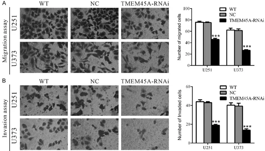
Silencing of TMEM45A inhibited the metasta-sis of glioma cells
To investigate the involvement of TMEM45A in cell motility, transwell assays were carried out to quantitatively determine the effect of TMEM45A knockdown on cell migration. Suppressing of TMEM45A expression brought
about a significant reduction in the migration
of U251 and U373 cells. As shown in Figure 3A, similar numbers of WT and NC-transfected cells migrated to the lower face of Boyden chambers (U251: WT, 76 ± 1; NC, 75 ± 1; U373: WT, 62 ± 2; NC, 61 ± 2), whereas a strongly inhibited motility was observed in TMEM45A knockdown cells (U251: 46 ± 1; U373: 27 ± 1). We also investigated whether TMEM45A affect-ed the invasive ability of glioma cells. In vitro invasion assay was performed in Boyden cham-bers with the upper wells coated with Matrigel to mimic the extracellular matrix. In sharp con-trast to control cells, TMEM45A knockdown cells showed dramatically reduced invasive ability (Figure 3B). The number of invaded cells was 44.2% and 35.9% of that of the control cells in U251 and U373 cells, respectively (U251: WT, 44 ± 1; NC, 43 ± 1; RNAi, 19 ± 1; U373: WT, 40 ± 1; NC, 39 ± 2; RNAi, 14 ± 1). These data suggested a role of TMEM45A in the promotion of glioma metastasis.
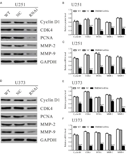
TMEM45A siRNA down-regulated the expres-sion of cell cycle related and invaexpres-sion related genes in glioma cells
2 and MMP-9 in U251 cells with TMEM45A silenced were 71.9%, 50.5%, 21.6%, 53.2% and 35.3% of that in the control cells, respec-tively. The mRNA levels of detected genes were also decreased in TMEM45A treated U251 cells (Figure 4C). Similar results were obtained in U373 cells (Figure 4D-F).
Discussion
High expression of TMEM45A was reported in
breast cancer [7], bladder cancer [9] and ovar
-ian cancer [10, 14] and may serve as poor prog -nostic factor for these malignancies. Here, we found that TMEM45A mRNA levels were elevat-ed in glioma tissues, which suggestelevat-ed a role of TMEM45A in glioma (Figure 1A and 1B). Further analysis revealed that its expression was pro-gressively increased with the increasing sever-ity of histological grade (Figure 1C). Survival of patients with glioma depends heavily on the histological grade of the tumor, thus TMEM45A expression may be a useful prognosis factor for glioma.
Then we investigated the functions of TMEM45A in glioma cells by suppressing its expression. Deregulated proliferation has profound effects on the malignant phenotype of cancer. Here, we found that knockdown of TMEM45A in
glio-ma cells significantly reduced cell growth by
inhibiting cell cycle progression (Figure 2). Cyclin D1-CDK4 complex are key players for
G1/S transition [15, 16]. Therefore, we then
evaluated the expression of the above genes. As shown in Figure 4, the expression of Cyclin D1, CDK4 and PCNA was notably decreased in TMEM45A knockdown cells, which further dem-onstrated the role of TMEM45A in the G1/S transition.
Previous studies indicate that gliomas tend to recur soon after resection of the initial tumor mass because of the highly invasive ability of glioma cells to invade adjacent areas by degrad-ing extracellular matrix (ECM) components and
diffusing into normal brain tissue [17]. MMP-2
[image:8.612.90.520.73.318.2]and recurrence [18, 19]. Here, our data indi -cated that TMEM45A siRNA notably inhibited the migration and invasion of glioma cells
[image:9.612.94.522.69.589.2]invasion-promoting function through regulating MMP-2 and MMP-9.
A recent study reported that inhibition of TMEM45A impaired cell proliferation, cell cycle transition and reduced cell invasion of ovarian
cancer cells [14]. These data matched well with
our results of in vitro experiments, suggesting that TMEM45A is a potential oncogene regard-less of location and might be a therapy target for multiple cancers.
In summary, our study demonstrated for the
first time that TMEM45A was overexpressed in
glioma tissues and TMEM45A expression was associated with the histological tumor grade. In addition, our data indicated that TMEM45A played a key role in the proliferation and metas-tasis of glioma cells. Whether TMEM45A can be used as a therapeutic target for glioma remains to be further investigated.
Acknowledgements
This study was supported by grant no. 81172- 398 from the National Natural Science Found- ation of China.
Disclosure of conflict of interest
None.
Address correspondence to: Dr. Guohan Hu, Depart- ment of Neurosurgery, Shanghai Institute of Neuro- surgery, Shanghai Changzheng Hospital, Second Military Medical University, 415 Fengyang Road, Shanghai 200003, China. E-mail: huguohan6504@ sina.com
References
[1] Sathornsumetee S and Rich JN. New treat-ment strategies for malignant gliomas. Expert Rev Anticancer Ther 2006; 6: 1087-1104. [2] Kleihues P and Cavenee WK. Pathology and
genetics of tumours of the nervous system. International Agency for Research on Cancer, 2000.
[3] Davis FG, Freels S, Grutsch J, Barlas S and Brem S. Survival rates in patients with primary malignant brain tumors stratified by patient age and tumor histological type: an analysis based on Surveillance, Epidemiology, and End Results (SEER) data, 1973-1991. J Neurosurg 1998; 88: 1-10.
[4] Penas-Prado M and Gilbert MR. Molecularly targeted therapies for malignant gliomas:
ad-vances and challenges. Expert Rev Anticancer Ther 2007; 7: 641-661.
[5] Hayez A, Malaisse J, Roegiers E, Reynier M, Renard C, Haftek M, Geenen V, Serre G, Simon M, de Rouvroit CL, Michiels C and Poumay Y. High TMEM45A expression is correlated to epi-dermal keratinization. Exp Dermatol 2014; 23: 339-344.
[6] Mattiuzzo NR, Toulza E, Jonca N, Serre G and Guerrin M. A large-scale multi-technique ap-proach identifies forty-nine new players of ke-ratinocyte terminal differentiation in human epidermis. Exp Dermatol 2011; 20: 113-118. [7] Flamant L, Roegiers E, Pierre M, Hayez A,
Sterpin C, De Backer O, Arnould T, Poumay Y and Michiels C. TMEM45A is essential for hy-poxia-induced chemoresistance in breast and liver cancer cells. BMC Cancer 2012; 12: 391. [8] Jinawath N, Chamgramol Y, Furukawa Y,
Obama K, Tsunoda T, Sripa B, Pairojkul C and Nakamura Y. Comparison of gene expression profiles between Opisthorchis viverrini and non-Opisthorchis viverrini associated human intrahepatic cholangiocarcinoma. Hepatology 2006; 44: 1025-1038.
[9] Urquidi V, Goodison S, Cai Y, Sun Y and Rosser CJ. A candidate molecular biomarker panel for the detection of bladder cancer. Cancer Epidemiol Biomarkers Prev 2012; 21: 2149-2158.
[10] Crijns AP, Fehrmann RS, de Jong S, Gerbens F, Meersma GJ, Klip HG, Hollema H, Hofstra RM, te Meerman GJ, de Vries EG and van der Zee AG. Survival-related profile, pathways, and transcription factors in ovarian cancer. PLoS Med 2009; 6: e24.
[11] Lee S, Stewart S, Nagtegaal I, Luo J, Wu Y, Colditz G, Medina D and Allred DC. Differentially expressed genes regulating the progression of ductal carcinoma in situ to invasive breast can-cer. Cancer Res 2012; 72: 4574-4586. [12] Hayez A, Malaisse J, Roegiers E, Reynier M,
Renard C, Haftek M, Geenen V, Serre G, Simon M and Rouvroit CL. High TMEM45A expression is correlated to epidermal keratinization. Exp Dermatol 2014; 23: 339-344.
[13] Ding DF, Li XF, Xu H, Wang Z, Liang QQ, Li CG, Wang YJ. Mechanism of resveratrol on the pro-motion of induced pluripotent stem cells. J Integr Med 2013; 11: 389-396.
[14] Guo J, Chen L, Luo N, Yang W, Qu X and Cheng Z. Inhibition of TMEM45A suppresses prolifera-tion, induces cell cycle arrest and reduces cell invasion in human ovarian cancer cells. Oncol Rep 2015; 33: 3124-3130.
[16] Dietrich DR. Toxicological and pathological ap-plications of proliferating cell nuclear antigen (PCNA), a novel endogenous marker for cell proliferation. Crit Rev Toxicol 1993; 23: 77-109.
[17] Giese A and Westphal M. Glioma invasion in the central nervous system. Neurosurgery 1996; 39: 235-252.
[18] Forsyth PA, Wong H, Laing TD, Rewcastle NB, Morris DG, Muzik H, Leco KJ, Johnston RN, Brasher PM, Sutherland G, Edwards DR. Gelatinase-A (MMP-2), gelatinase-B (MMP-9) and membrane type matrix metalloprotein-ase-1 (MT1-MMP) are involved in different as-pects of the pathophysiology of malignant glio-mas. Br J Cancer 1999; 79: 1828-35.