Original Article
Cellular transcriptomics: gelsolin negatively regulates
the expression of apoptosis-associated genes and
inhibits apoptosis in hepatocarcinoma cells
Yi Zhou1,4, Xiaofang Deng2, Xiaoxiao Ma3, Ning Zang1,4, Hongtao Li1, Gang Li1, Danrong Li1,4, Cuiping Li2,
Wendong Huang3, Min He2,4
1Medical Scientific Research Center of Guangxi Medical University, Nanning 530021, P. R. China; 2School of Public Health, Guangxi Medical University, Nanning 530021, P. R. China; 3Department of Diabetes and Metabolic Diseases, Beckman Research Institute of The City of Hope, Duarte, CA 91010, USA; 4Key Laboratory of
High-Incidence-Tumor Prevention & Treatment (Guangxi Medical University), Ministry of Education, Nanning 530021, P. R. China
Received September 10, 2015; Accepted October 22, 2015; Epub November 1, 2015; Published November 15, 2015
Abstract: Gelsolin (GSN), which is a Ca2+-dependent actin filament severing and capping protein, plays a critical role in the cancer progress and has the potential for providing a novel thread for cancer therapy. In current study, we demonstrate the roles of GSN on anti-apoptosis of hepatocarcinoma cells by transcriptome RNA-seq method. Then flow cytometry (FCM), in-cell immunoblotting and transmission electron microscopy (TEM) were used to ex -amine the GSN regulatory cell apoptosis. The results revealed GSN significantly suppresses apoptosis-associated functional categories through down-regulating apoptosis-associated genes in 5 apoptosis terms and 6 relevant KEGG pathways. FCM showed a significant lower apoptotic rate in GSN-SMMC7721 (P<0.05). In-cell immunoblot -ting detected discrepant expression of the apoptosis factors among GSN expressed/shRNA transfectants (P<0.05). TEM observed the discernible apoptosis morphology. Above results suggest a negative relationship between GSN expression and hepatocarcinoma cell apoptosis. GSN overexpression suppresses apoptosis while down-regulated GSN promotes apoptosis. The possible mechanism could be associated with the regulation of GSN on the apoptosis-associated pathways and the apoptosis factors caspase 3 and bcl-2.
Keywords: Hepatocarcinoma, transcriptomics, RNA-seq, gelsolin, anti-apoptosis
Introduction
Hepatocellular carcinoma (HCC) is the most
common form of primary cancer of liver. In
terms of numbers, HCC is the sixth most malig-nant cancer worldwide (estimated 711,000
cases annually); however, due to its poor prog -nosis (survival rates 3-5% in cancer registries), HCC is the third most common cause of cancer
death [1, 2]. In our previous study, we revealed
that GSN is a potential HCC serum biomarker
through iTRAQ-MALDI-MS/MS technology [3].
Moreover, other reports also suggested GSN
plays important roles in the biological process -es of cancerization [4, 5]. As for the correla-tions between GSN and HCC, however, were still unclear.
As a Ca2+-regulated actin filament severing and
capping, gelsolin (GSN) is a widespread, poly -functional regulator of cell structure and metab-olism [6]. Research data showed that GSN was
ubiquitously expressed in various kinds of cells
[7-11], in spite of the variations of expression levels during cell differentiation [12, 13] and carcinogenesis [5, 14-24]. A recent
understand-ing of the functions and regulatory mechanisms
of GSN might lead to new considerations of this protein as a potential biomarker and/or thera-peutic target of tumor. High levels of GSN expression were thought to be an independent marker for tumor recurrence and progression
[25]. Actually, increased GSN expression was
Gelsolin inhibits apoptosis in hepatocarcinoma cells
grade in renal cell carcinoma [28]. In human hepatocarcinoma HepG2 cells, overexpression of GSN inhibited the nuclear localization of
p53, repressed apoptosis and negatively regu
-lated transcriptional activity of a reporter con
-struct in the cytoplasm [29]. Intriguingly, in uro -thelial and oral carcinomas, GSN exhibits a
biphasic expression profile, being down-regu -lated in premalignant lesions but increased in higher grade lesions. 24Furthermore, the
co-expression of GSN with erb-B2 and epidermal
growth factor receptor (EGFR) is a predictor of poor prognosis in breast cancer [30]. It is likely
that the role of GSN differs during the course of tumor progression, and in more advanced
dis-ease GSN may cooperate with other oncogenic
factors to accelerate disease progression. Recent development of the next-generation
sequencing technologies makes RNA-seq become a powerful approach in defining the molecular mechanism of specific diseases with the advantage of analyzing abnormal transcrip -tome at genome-wide level [31, 32]. Previous
applications of RNA-seq with greater efficiency
and higher resolution [33] than other
expres-sion profiling technologies included yeast [34],
viruses [35], tissues[36] and cell lines [37]. For cancer expression profiling, the reprogramming
of the transcriptome leads to aberrant cellular
behavior and thus directly contributes to can -cer progression [38]. Monitoring the can-cer
transcriptome not only enables us to fill in the
gap between gene regulation and cancer cell
behavior, but also allows us to identify the underlying mechanism [39, 40].
Despite the prevalence of using RNA-seq to study various cancer pathogenetic mecha -nisms [41-43], the deep transcriptome descrip-tions of HCC and the roles of GSN in HCC are
scarcely mentioned. We therefore applied RNA-seq technology to analyze the transcriptomics
and to determine the roles of GSN in
hepatocel-lular carcinoma. Through bioinformatics analy -sis, we revealed the role of GSN on suppressing HCC apoptosis and explored the possible
regu-latory mechanism.
Materials and methods
Cell culture and recombinant plasmid DNA
HCC cell line SMMC7721 cells were purchased
from the Institute of Biochemistry and Cell Biology, Chinese Academy of Sciences. Stable
SMMC7721 cell lines in which GSN were either
of GSN cDNA or down-regulated by the short
hairpin RNA recombinant plasmids, and their
control transfectants induced by empty vector plasmids were all established by our institute.
RT-PCR and in-cell immunoblotting were used to determine the GSN expression levels and were carried out in triplicate.
Transcriptome sequencing
Stable GSN expressed cells (GSN-SMMC7721) and parent SMMC7721 were used to explore the cell transcription. RNA was extracted from about 1×10⁷ cells respectively, according to the
manufacturer’s instruction, and quantified by NanoDrop 2000 (Thermo-Fisher Scientific). The whole transcriptome RNA-seq was constructed using Ion Total RNA-Seq Kit v2, Ion PI™ Chip kit v2, Ion PI™ Template OT2 200 Kit v2, and Ion PI™ Sequencing 200 Kit v2 based on the Life
Technologies Corporation’s guide. We per-formed two runs of sample repetition. In brief,
mRNA was purified using oligo-dT beads from 100 μg of total RNAs for each sample and frag -mented into small fragments. The cleaved RNA
fragments were reverse-transcribed into first strand cDNA, followed by second-strand cDNA synthesis. After end-repair procedure, a single
‘A’ base was added to cDNA fragments at 3’end. The cDNAs were then ligated to adapters,
enriched by PCR to generate the final cDNA library. After amplifying sequencing template, RNA-seq was performed using Ion proton sys -tem (Life Technologies Corporation) with the standard protocol.
FCM analysis
GSN overexpressed (GSN-SMMC7721), shRNA down-regulated (shGSN-SMMC7721), and their
empty vector transfected cells
(NC1-SM-MC7721 and NC2-SM(NC1-SM-MC7721) were used to
examine the GSN-related apoptosis. After tryp
-sinized, approximately 1×10⁶ cells were
collect-ed by centrifugation at 1200 g for 5 min, and then washed in PBS, followed by resuspension
in apoptosis detection buffer for about 0.5 h in
dark. Cells were stained by propidium iodide (PI) and Annexin-V, and analyzed by FCM using Cell quest software (BD Biosciences). The
experiment was carried out in triplicate.
In-cell immunoblotting
Gelsolin inhibits apoptosis in hepatocarcinoma cells
Gelsolin inhibits apoptosis in hepatocarcinoma cells
phase, then the cells were immediately fixed,
permeabilized and blocked at room
tempera-ture. After incubated with primary antibodies overnight at 4°C, the cells were sequentially
incubated with corresponding second infrared labeled antibodies for 2 h in dark place.
Odyssey Infrared Imaging System (LI-COR
Biosciences GmbH) was used to obtain the
image and analyze the target protein expres -sion, which was calculated as the ratio of the
intensity of target protein to that of GAPDH. The
experiments were carried out in triplicate.
TEM observations
GSN down-regulated cells induced by shRNA
transfection were used to observe the
[image:6.629.98.532.79.541.2]detector for determining the apoptosis
mor-phology of cells. The observed cells were
dropped on a copper mesh coated with an
amorphous carbon film for TEM observations.
The experiment was carried out in triplicate.
Statistical analysis
Results are expressed as means ± standard
error. Statistical analyses were performed
using SPSS statistics software (SPSS). P-value
<0.05 is considered statistically significant.
Results
Transcriptome sequencing gained superior sequences
GSN expression recombinant plasmid was transfected into hepatocarcinoma SMMC7721
cell line and then treated with puromycin for the
selection of stable cell lines. We performed real-time PCR and in-cell immunoblotting to
confirm the overexpression of GSN. The experi -ments were carried out in triplicate. The target GSN expression was calculated as the ratio of
the intensity of target to that of GAPDH. As
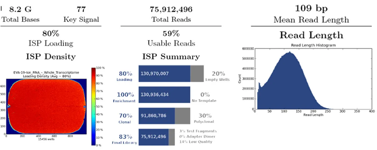
expected, GSN level was increased in the GSN-SMMC7721 cell line (Figure 1A, 1B). Then,
RNA-seq was carried out to analyze the tran -scriptional differences. We performed two runs of sample repetition. On completion, 75.9
mil-lion 109 bp long sequencing reads and 88.4 million 119 bp long sequencing reads were
generated in two runs of repetition, and that corresponded to an 8.2G and 10.5G raw
sequence data respectively (Figure 1CI, 1CII). Samples in both above repetitive runs of
tran-scriptome RNA-seq gained >50 M superior sequences that could be utilized in downstream analysis.
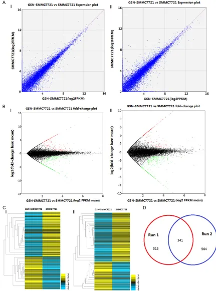
Enrichment analysis confirmed 341 mutual DEGs
To estimate gene expression abundance, we
conducted an enrichment analysis to estimate the gene expression and identify significantly dysregulated genes. The results showed that
approximate 50000 genes were detected in each run and the DEGs were 515 and 546
respectively. The normalized gene expression was measured by Fragments Per Kilobase of exon per Million fragments mapped (FPKM)
(Figure 2A, 2B). In addition, the clustering
anal-ysis indicated that the transcriptome of GSN
overexpressed SMMC7721 cells was distinct from the parental control (Figure 2C). The over-lapping of the mutual DEGs between two runs reached up to 341 and was shown as a Venn diagram in Figure 2D.
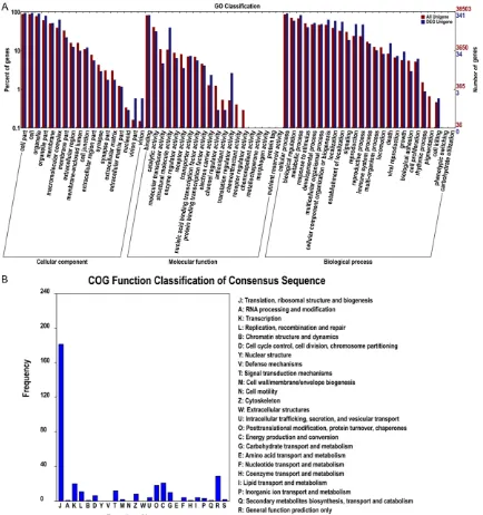
DEGs involved in 116 functional categories.
To better understand the function of DEGs, we
conducted an enrichment analysis of Gene Ontology for the dysregulated genes. To identify the functional categories, we first performed
GO categories using online tools from WEGO.
Three hundred forty one DEGs involved in 63 GO classifications including cellular compo -nent, molecular function, and biological pro-cess were enriched (Figure 3A). A further analy
-sis of functional annotation was achieved by studying the enrichment of DEGs in Cog catego
-ries, which revealed 20 function classifications of consensus sequences (Figure 3B). For GO
categories provided informative DEGs in each
gene ontology term and liable to cancer-specif
-ic analysis, we counted the signif-icantly up- and
down-regulated DEGs in terms from the
compo-nent ontology with corrected p-value better than 1. In total, all DEG were categorized into
approximately 116 functional categories, con -taining 40 up-regulated GO categories and 28 down-regulated GO categories (Figure 4).
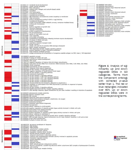
Cancer-specific functional filter highlighted a GSN effect on cell apoptosis
Interestingly, although we identified approxi -mate numbers of up- and down-regulated terms
in the functional enrichment analysis, we observed clues of significant GO categories for
GSN overexpressed SMMC7721 cells,
suggest-ing that the increased GSN might functionally important for cancer progression. For example,
the DEGs expression in the GO terms for
“apop-totic process” is mainly significantly sup -pressed, the same trend can also be found in the gene catalog of “DNA damage response,
signal transduction by p53 class mediator
resulting in induction of apoptosis”, “JNK cas-cade”, “cellular response to interleukin-3”, and
“cysteine-type endopeptidase activator activity
involved in apoptotic process”, which indicates that GSN might affect apoptosis of cancer cells. Then we focused on the apoptosis-related
Gelsolin inhibits apoptosis in hepatocarcinoma cells
to seek for the evidences of GSN’s regulatory
effect in HCC apoptosis. The results showed that in all the 13 mutual apoptosis-related DEGs of two runs, 9 pro-apoptotic genes
includ-ing PSME2, PTK2B, FOS, JUN, ITGB1, MAP2K7,
MAP3K4, MAP3K12 and Rac1 were down-regu-lated, and RRM2B, as a anti-apoptotic gene was up-regulated (Table 1). Above 10 apopto-sis-associated DEGs participated in the
pro-cess of anti-apoptosis, and simultaneously plays important roles in 6 apoptosis-related pathways (antigen processing and presenta
-tion, natural killer cell mediated cytotoxicity, p53 signaling pathway, pathways in cancer, Jak-STAT signaling pathway and MAPK signaling pathway) (Table 2).
We further paid attention to mutual association
between the above apoptosis pathways. The
results suggested that among above 6
apopto-sis-related pathways, except antigen process
-ing and presentation pathway, Natural killer cell mediated cytotoxicity, JAK-STAT signaling path
[image:8.629.95.532.70.569.2]cancer all interacts with MAPK-JNK signaling
pathway. We also noticed that the regulation of MAPK-JNK pathway for apoptosis is carried out through P53 signaling pathway, which finally
affects apoptosis through terminal apoptosis-associated factors such as caspase 3, bcl-2
and cytochrome C.
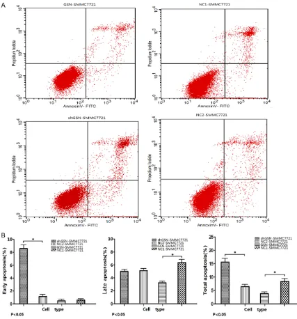
GSN inhibited hepatocarcinoma cell apoptosis
We then generated the stable GSN overexpres-sion cell line (GSN-SMMC7721) and stable GSN knock down cell line(shGSN-SMMC7721), as
0.72±0.11, 3.48±0.25, and 4.20±0.18
respec-tively in GSN-SMMC7721. The early, late and
total apoptosis rates were 9.97±1.01, 5.44±
0.98, 15.41±1.26 respectively in
shGSN-SMMC7721. The late and total apoptosis rates in GSN overexpressed SMMC7721 cells were lower than those of the NC1-SMMC7721
(P<0.05). And the early and total apoptosis
rates in shGSN-SMMC7721 cells were higher
than those of the NC2-SMMC7721 (P<0.05)
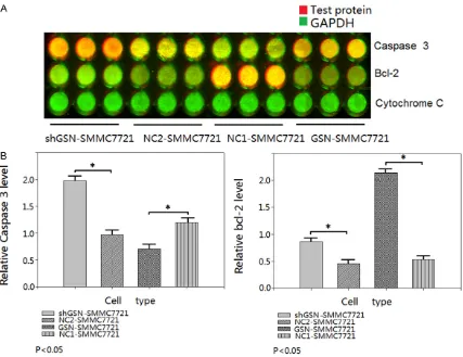
(Figure 5). In-cell immunoblotting showed the expression of caspase 3 was down-regulated
[image:9.629.98.536.94.431.2]0.625± 0.133 (P<0.05), and the expression of
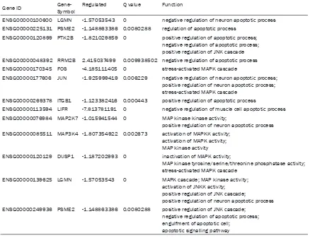
Table 1. List of mutual apoptosis-related genes in DEGs annotation Gene ID Gene- Symbol Regulated Q value Function
ENSG00000100600 LGMN -1.57053543 0 negative regulation of neuron apoptotic process ENSG00000225131 PSME2 -1.148863386 0.0080288 regulation of apoptotic process
ENSG00000120899 PTK2B -1.821029859 0 positive regulation of apoptotic process; negative regulation of apoptotic process; positive regulation of JNK cascade ENSG00000048392 RRM2B 2.415037499 0.009938502 negative regulation of apoptotic process ENSG00000170345 FOS -4.185111405 0 stress-activated MAPK cascade
ENSG00000177606 JUN -1.925999419 0.006229 negative regulation of neuron apoptotic process; positive regulation of neuron apoptotic process; stress-activated MAPK cascade
ENSG00000269378 ITGB1 -1.123382416 0.000443 positive regulation of apoptotic process
ENSG00000113594 LIFR -7.813781191 0 negative regulation of muscle cell apoptotic process ENSG00000076984 MAP2K7 -1.015941544 0 MAP kinase kinase activity;
positive regulation of neuron apoptotic process ENSG00000085511 MAP3K4 -1.807354922 0.002673 activation of MAPKK activity;
activation of MAPK activity; MAP kinase activity
ENSG00000120129 DUSP1 -1.187202993 0 inactivation of MAPK activity;
MAP kinase tyrosine/serine/threonine phosphatase activity; stress-activated MAPK cascade
ENSG00000139625 LGMN -1.57053543 0 MAPK cascade; MAP kinase activity; activation of JNKK activity; positive regulation of JNK cascade;
positive regulation of neuron apoptotic process ENSG00000249936 PSME2 -1.148863386 0.0080288 positive regulation of JNK cascade;
negative regulation of apoptotic process; engulfment of apoptotic cell;
apoptotic signalling pathway
Table 2. Apoptosis-related KEGG pathway
Pathway P-value Pathway ID
Antigen processing and presentation 4.9392e-01 ko04612 Natural killer cell mediated cytotoxicity 7.8074e-01 ko04650 P53 signaling pathway 9.4388e-01 ko04115
pathways in cancer 9.7949e-01 ko05200
JAK-STAT signaling pathway 9.8563e-01 ko04630 MAPK-JNK signaling pathway 5.5878e-01 ko04010
well as their corresponding control cell lines (NC1- and NC2-SMMC7721) to performed apoptosis studies in vitro so as to evaluate the roles of GSN in
hepatocarcinoma cells. FCM, in-cell
immunoblotting and TEM were per-formed, and were carried out in triplicate.
The results of FCM showed the early,
[image:9.629.100.352.462.555.2]Gelsolin inhibits apoptosis in hepatocarcinoma cells
bcl-2 was up-regulated 4.138±0.857 (P<0.05)
in GSN-SMMC7721 while comparing to NC1-SMMC7721. The expression of caspase 3 was
up-regulated 1.908±0.257 (P<0.05), and the
expression of bcl-2 was down-regulated
0.411±0.122 (P<0.05) in shGSN-SMMC7721
while comparing to NC2-SMMC7721. But the
expression of cytochrome C was invariable
while GSN was overexpressed/inhibited (Figure
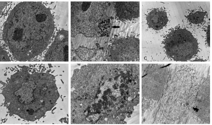
6). Finally, TEM confirmed the discernible apop
-tosis morphology in the GSN shRNA transfec
-tant, including apoptotic body, autophagy, early
apoptosis, late apoptosis and oncosis (Figure 7).
[image:10.629.98.525.79.537.2]Overall, above results suggest a negative rela-tionship between GSN expression and hepato-carcinoma cell apoptosis. Overexpressed GSN
inhibits apoptosis and the knockdown of GSN promotes apoptosis.
Discussion
Transcriptome bridges the gap between the genetic codes and the functional molecule
pathways. By comparing transcriptomics
between normal and cancerous cells, it is able to determine what genes are turned on or off in disparate biological process and gain a deeper
understanding of what constitutes a specific cell type and how changes in transcriptional activity may reflect or contribute to disease pro -gression. Comparative transcriptomics has
been applied to many human pathological stud -ies such as neurodegenerative disease [44], retina defection [45], prostate cancer [46], and
colorectal cancer [47]. For hepatocarcinoma
transcriptome, Huang et al performed
tran-scriptome analyses for 10 matched pairs of
cancer and non-cancerous tissues from HCC patients on Solexa/Illumina GAII platform [48].
About 21.6 million sequencing reads and 10.6
million aligned reads were obtained and 1,378
significantly DEGs were annotated. Additionally, comprehensive functional analysis indicated
that cell growth-related, metabolism-related
and immune-related pathways are most signifi
-cantly enriched by DEGs, pointing to a complex
mechanism for HCC carcinogenesis. In another
study, David et al detected five hundred DEGs from HCC cells, and the further gene ontology analysis indicated that the over-expressed genes were associated with inflammation, drug
[image:11.629.103.530.78.407.2]resistance and lipid metabolism [49].
Gelsolin inhibits apoptosis in hepatocarcinoma cells
Our study was mainly focusing on GSN regula
-tory HCC transcription through next-generation RNA-seq technology. The results revealed a
regulator role of GSN in hepatoma cell apopto-sis. We noticed 5 functional categories with 13 relevant DEGs, which relate to cell apoptosis,
are mainly restrained and might play important roles in 6 apoptosis-related pathways. This
observation provide hint for the potential mech-anism of how GSN regulate apoptosis.
Interestingly, among all of the 13 mutual apop -tosis-related DEGs, 7 DEGs including PTK2B,
FOS, JUN, RAC1, MAP2K7, MAP3K4, MAP3K12 enriched in MAPK-JNK signaling pathway are
down-regulated.
Meanwhile, except antigen processing and
pre-sentation pathway, all other 4 apoptosis-relat
-ed pathways such as Natural killer cell m-ediat
-ed cytotoxicity, JAK-STAT signaling pathway, P53 signaling pathway and pathways in cancer all interacts with MAPK-JNK signaling pathway. Both observations strongly suggest that GSN
might suppress apoptosis through the
cross-linking of above pathway, while MAPK-JNK path
-way might played central roles in the
GSN-regulated apoptosis.
We also noticed that the regulation of
MAPK-JNK pathway for apoptosis is carried out
through P53 signaling pathway, which finally
affects apoptosis through terminal apoptosis-associated factors such as caspase 3, bcl-2
and cytochrome C. We detected the expression
of above apoptosis factors through in-cell immunoblotting. The results suggest the dis-crepant expression of caspase 3 and bcl-2 in GSN up-/down-regulated hepatocarcinoma
cells. But the expression of cytochrome C was
not changed in the test. Previous reports eluci-dated the role of GSN as an anti-apoptotic pro-tein in mitochondrial-dependent cell death due to the inositol lipids PI (4, 5) P2 and PI (3, 4) P2 prevent caspase 3 cleavage of GSN in vitro. Through the formation of a stable PI (4, 5) P2-GSN-Caspase 3 complex, PI (4, 5) P2-GSN
strongly inhibited caspase-3 activity [50].
Except caspase 3, GSN affects actin-depen-dent VDAC, which is thought to be a target of
bcl-2 family members [51]. Though the release of cytochrome C is considered as an important
factor for mitochondria initiated apoptosis, and
GSN is thought to be an apoptosis inhibitor by
blocking mitochondrial membrane potential
loss and cytochrome c release, we did not detect the expression change of cytochrome C.
We guessed the result might be accord with the
theory that the release of cytochrome C could
[image:12.629.99.534.78.334.2]not be relative to its expression level.
The structural changes of cytoskeletal proteins
(such as GSN) in apoptosis circumstance leads
to the transmogrification of cells, for instance,
chromatin condensation, nuclear fragmenta-tion, cell membrane blebbing, and apoptotic
body formation. Early studies showed that
there is a tight connection between skelemin
change and the apoptosis morphology [52-55]. In our study, we observed the GSN-derived morphology of apoptosis through TEM.
According to the previous reports, dissociation of GSN products a NH2-extreme of 352aa, which induces cells turned round, fallen off, and splintered in a Ca2+ independent mode.
The observation of TEM suggests that as the regulator and effector of apoptosis, GSN might regulate cell apoptosis through acting on the
cytoskeleton and actin activity.
In conclusion, GSN overexpression suppresses apoptosis while down-regulated GSN promotes apoptosis in human HCC cells. The regulation of GSN in the apoptosis of hepatoma cell
main-ly involves several important pathway such as natural killer cell mediated cytotoxicity, p53 sig
-naling pathway, pathways in cancer, Jak-STAT signaling pathway and MAPK signaling path
-way. Caspase 3 and bcl-2 are two pivotal apop
-tosis factors regulated by GSN. These findings
suggest a role for GSN in HCC and a potential of using GSN for the treatment of this
malignancy.
Acknowledgements
This research was supported by the National Natural Science Foundation of China, (No. 81260445, 30960332). The Fund of Key Laboratory of High-Incidence-Tumor Prevention & Treatment (No. GK2013-13-A-01 -02, GK2014-ZZ04). The Natural Science Foun-dation of Guangxi (No. 2013GXNSFBA019183).
Disclosure of conflict of interest
None.
Address correspondence to: Min He, School of Public Health, Guangxi Medical University, Nanning 530021, P. R. China; Key Laboratory of High-Incidence-Tumor Prevention & Treatment (Guangxi Medical University), Ministry of Education, Nanning 530021, P. R. China. E-mail: [email protected]
References
[1] Huang G, Lau WY, Wang ZG, Pan ZY, Yuan SX, Shen F, Zhou WP, Wu MC. Antiviral therapy im
-proves postoperative survival in patients with hepatocellular carcinoma: a randomized con-trolled trial. Ann Surg 2015; 261: 56-66. [2] Ochiai T, Ogino S, Ishimoto T, Toma A,
Yamamoto Y, Morimura R, Ikoma H, Otsuji E. Prognostic impact of hepatectomy for patients with non-hepatitis b, non-hepatitis c hepatocel-lular carcinoma. Anticancer Res 2014; 34: 4399-410.
[3] He X, Wang Y, Zhang W, Li H, Luo R, Zhou Y, Liao CL, Huang H, Lv X, Xie Z, He M. Screening differential expression of serum proteins in AFP-negative HBV-related hepatocellular carci -noma using iTRAQ-MALDI-MS/MS. Neoplasma 2014; 61: 17-26.
[4] Mullauer L, Fujita H, Ishizaki A, Kuzumaki N. Tumor-suppressive function of mutated gelso-lin in ras-transformed cells. Oncogene 1993; 8: 2531-6.
[5] Winston JS, Asch HL, Zhang PJ, Edge SB, Hyland A, Asch BB. Down regulation of gelsolin correlates with the progression to breast carci-noma. Breast Cancer Res 2001; 65: 11-21. [6] Li GH, Arora PD, Chen Y, McCulloch CA, Liu P.
Multifunctional Roles of Gelsolin in Health and Diseases. Med Res Rev 2012; 32: 999-1025. [7] Chaponnier C, Kocher O, Gabbiani G.
Modulation of gelsolin content in rat aortic smooth muscle cells during development, ex-perimental intimal thickening and culture. An immunohistochemical and biochemical study. Eur J Biochem 1990; 190: 559-65.
[8] Scholz A, Hinssen H. Biphasic pattern of gelso-lin expression and variations in gelsogelso-lin-actin interactions during myogenesis. Exp Cell Res 1995; 219: 384-91.
[9] Kwiatkowski DJ. Predominant induction of gel-solin and actin-binding protein during myeloid differentiation. J Biol Chem 1988; 263: 13857-62.
[10] Tanaka M, Mullauer L, Ogiso Y, Fujita H, Moriya S, Furuuchi K, Harabayashi T, Shinohara N, Koyanagi T, Kuzumaki N. Gelsolin: A candidate for suppressor of human bladder cancer. Cancer Res 1995; 55: 3228-32.
[11] Chaponnier C, Gabbiani G. Gelsolin modula-tion in epithelial and stromal cells of mammary carcinoma. Am J Pathol 1989; 134: 597-603. [12] Paunio T, Kangas H, Kiuru S, Palo J, Peltonen
L, Syvanen AC. Tissue distribution and levels of gelsolin mRNA in normal individuals and pa-tients with gelsolin-related amyloidosis. FEBS Lett 1997; 406: 49-55.
[13] Ahn JS, Jang IS, Kim DI, Cho KA, Park YH, Kim K, Kwak CS, Chul Park S. Aging-associated in-crease of gelsolin for apoptosis resistance. Biochem Biophys Res Commun 2003; 312: 1335-41.
gelso-Gelsolin inhibits apoptosis in hepatocarcinoma cells
lin in ras-transformed cells. Oncogene 1993; 8: 2531-6.
[15] Gay F, Estornes Y, Saurin JC, Joly-Pharaboz MO, Friederich E, Scoazec JY, Abello J. In colon car -cinogenesis, the cytoskeletal protein gelsolin is down-regulated during the transition from ad-enoma to carcinoma. Hum Pathol 2008; 39: 1420-30.
[16] Kim JH, Choi YK, Kwon HJ, Yang HK, Choi JH, Kim DY. Downregulation of gelsolin and retino-ic acid receptor beta expression in gastrretino-ic can-cer tissues through histone deacetylase 1. J Gastroenterol Hepatol 2004; 19: 218-24. [17] Tanaka M, Mullauer L, Ogiso Y, Fujita H, Moriya
S, Furuuchi K, Harabayashi T, Shinohara N, Koyanagi T, Kuzumaki N. Gelsolin: A candidate for suppressor of human bladder cancer. Cancer Res 1995; 55: 3228-32.
[18] Dosaka-Akita H, Hommura F, Fujita H, Kinoshita I, Nishi M, Morikawa T, Katoh H, Kawakami Y, Kuzumaki N. Frequent loss of gelsolin expres -sion in non-small cell lung cancers of heavy smokers. Cancer Res 1998; 58: 322-7. [19] Lee HK, Driscoll D, Asch H, Asch B, Zhang PJ.
Downregulated gelsolin expression in hyper -plastic and neo-plastic lesions of the prostate. Prostate 1999; 40: 14-9.
[20] Visapaa H, Bui M, Huang Y, Seligson D, Tsai H, Pantuck A, Figlin R, Rao JY, Belldegrun A, Horvath S, Palotie A. Correlation of Ki-67 and gelsolin expression to clinical outcome in renal clear cell carcinoma. Urology 2003; 61: 845-50.
[21] Ni XG, Zhou L, Wang GQ, Liu SM, Bai XF, Liu F, Peppelenbosch MP, Zhao P. The ubiquitinpro -teasome pathway mediates gelsolin protein downregulation in pancreatic cancer. Mol Med 2008; 14: 582-9.
[22] Noske A, Denkert C, Schober H, Sers C, Zhumabayeva B, Weichert W, Dietel M, Wiechen K. Loss of gelsolin expression in hu-man ovarian carcinomas. Eur J Cancer 2005; 41: 461-9.
[23] Kim CS, Furuya F, Ying H, Kato Y, Hanover JA, Cheng SY. Gelsolin: A novel thyroid hormone receptor-beta interacting protein that modu-lates tumor progression in a mouse model of follicular thyroid cancer. Endocrinology 2007; 148: 1306-12.
[24] Shieh DB, Chen IW, Wei TY, Shao CY, Chang HJ, Chung CH, Wong TY, Jin YT. Tissue expression of gelsolin in oral carcinogenesis progression and its clinicopathological implications. Oral Oncol 2006; 42: 599-606.
[25] Rao J, Seligson D, Visapaa H, Horvath S, Eeva M, Michel K, Pantuck A, Belldegrun A, Palotie A. Tissue microarray analysis of cytoskeletal actin-associated biomarkers gelsolin and E-cadherin in urothelial carcinoma. Cancer 2002; 95: 1247-57.
[26] Kimura K, Ojima H, Kubota D, Sakumoto M, Nakamura Y, Tomonaga T, Kosuge T, Kondo T. Proteomic identification of the macrophage-capping protein as a protein contributing to the malignant features of hepatocellular carcino-ma. J Proteom 2013; 78: 362-73.
[27] Shieh DB, Godleski J, Herndon JE 2nd, Azuma T, Mercer H, Sugarbaker DJ, Kwiatkowski DJ. Cell motility as a prognostic factor in stage I non small cell lung carcinoma: The role of gel-solin expression. Cancer 1999; 85: 47-57. [28] Visapää H, Bui M, Huang Y, Seligson D, Tsai H,
Pantuck A, Figlin R, Rao JY, Belldegrun A, Horvath S, Palotie A. Correlation of Ki-67 and gelsolin expression to clinical outcome in renal clear cell carcinoma. Urology 2003; 61: 845-50.
[29] An JH, Kim JW, Jang SM, Kim CH, Kang EJ, Choi KH. Gelsolin negatively regulates the activity of tumor suppressor p53 through their physical interaction in hepatocarcinoma HepG2 cells. Biochem Biophys Res Communicat 2011; 412: 44-9.
[30] Thor AD, Edgerton SM, Liu SQ, Moore DH, Kwiatkowski DJ. Gelsolin as a negative prog-nostic factor and effector of motility in erbB-2-positiveepidermal growth factor receptor-positive breast cancers. Clin Cancer Res 2001; 7: 2415-24.
[31] Mardis ER. The impact of next-generation se-quencing technology ongenetics. Trends Genet 2008; 24: 133-41.
[32] Wang Z, Gerstein M, Snyder M. RNA-Seq: a revolutionary tool for transcriptomics. Nat Rev Genet 2009; 10: 57-63.
[33] Metzker ML. Sequencing technologies-the next generation. Nat Rev Genet 2010; 11: 31-46. [34] Nagalakshmi U, Wang Z, Waern K, Shou C,
Raha D, Gerstein M, Snyder M. The transcrip -tional landscape of the yeast genome defined by RNA sequencing. Science 2008; 320: 1344-9.
[35] Walter NM and Joan AS. Genome-wide analy -ses of Epstein-Barr virus reveal conserved RNA structures and a novel stable intronic se-quence RNA. BMC Genomics 2013; 14: 543-558.
[36] Mortazavi A, Williams BA, McCue K, Schaeffer L, Wold B. Mapping and quantifying mammali -an tr-anscriptomes by RNA-Seq. Nat Methods 2008; 5: 621-8.
[37] Sultan M, Schulz MH, Richard H, Magen A, Klingenhoff A, Scherf M, Seifert M, Borodina T, Soldatov A, Parkhomchuk D. A global view of gene activity and alternative splicing by deep sequencing of the human transcriptome. Science 2008; 321: 956-60.
-quencing and beyond. Annu Rev Genomics Hum Genet 2011; 12: 407-30.
[39] Ozsolak F, Milos PM. RNA sequencing: advanc -es, challenges and opportunities. Nat Rev Genet 2011; 12: 87-98.
[40] Wang Z, Gerstein M, Snyder M. RNA-Seq: a revolutionary tool for transcriptomics. Nat Rev Genet 2009; 10: 57-63.
[41] Maher CA, Kumar-Sinha C, Cao X, Kalyana-Sundaram S, Han B, Jing X, Sam L, Barrette T, Palanisamy N, Chinnaiyan AM. Transcriptome sequencing to detect gene fusions in cancer. Nature 2009; 458: 97-101.
[42] Pflueger D, Terry S, Sboner A, Habegger L, Esgueva R, Lin PC, Svensson MA, Kitabayashi N, Moss BJ, MacDonald TY, Cao X, Barrette T, Tewari AK, Chee MS, Chinnaiyan AM, Rickman DS, Demichelis F, Gerstein MB, Rubin MA. Discovery of non-ETS gene fusions in human prostate cancer using next-generation RNA se-quencing. Genome Res 2011; 21: 56-7. [43] Shah SP, Köbel M, Senz J, Morin RD, Clarke BA,
Wiegand KC, Leung G, Zayed A, Mehl E, Kalloger SE, Sun M, Giuliany R, Yorida E, Jones S, Varhol R, Swenerton KD, Miller D, Clement PB, Crane C, Madore J, Provencher D, Leung P, DeFazio A, Khattra J, Turashvili G, Zhao Y, Zeng T, Glover JN, Vanderhyden B, Zhao C, Parkinson CA, Jimenez-Linan M, Bowtell DD, Mes-Masson AM, Brenton JD, Aparicio SA, Boyd N, Hirst M, Gilks CB, Marra M, Huntsman DG. Mutation of FOXL2 in Granulosa-Cell Tumors of the Ovary. N Engl J Med 2009; 360: 2719-29.
[44] Courtney E, Kornfeld S, Janitz K, Janitz M. Transcriptome profiling in neurodegenerative disease. J Neurosci Methods 2010; 193: 189-202.
[45] Farkas MH, Grant GR, Pierce EA. Transcriptome analyses to investigate the pathogenesis of RNA splicing factor retinitis pigmentosa. Adv Exp Med Biol 2012; 723: 519-25.
[46] Ren S, Peng Z, Mao JH, Yu Y, Yin C, Gao X, Cui Z, Zhang J, Yi K, Xu W. RNA-seq analysis of prostate cancer in the Chinese population identifies recurrent gene fusions, cancer-asso -ciated long non coding RNAs and aberrant al-ternative splicings. Cell Research 2012; 22: 806-21.
[47] Castellarin M, Warren RL, Freeman JD, Dreolini L, Krzywinski M, Strauss J, Barnes R, Watson P, Allen-Vercoe E, Moore RA, Holt RA. Fusobacteriumnucleatum infection is preva -lent in human colorectal carcinoma. Genome Res 2012; 22: 299-306.
[48] Huang Q, Lin B, Liu H, Ma X, Mo F, Yu W, Li L, Li H, Tian T, Wu D, Shen F, Xing J, Chen ZN. RNA-Seq analyses generate comprehensive tran -scriptomic landscape and reveal complex tran-script patterns in hepatocellular carcinoma. Plos One 2011; 6: e26168.
[49] Ho DW, Yang ZF, Yi K, Lam CT, Ng MN, Yu WC, Lau J, Wan T, Wang X, Yan Z, Liu H, Zhang Y, Fan ST. Gene expression profiling of liver can -cer stem cells by RNA-sequencing. PLos One 2012; 7: e37159.
[50] Azuma T, Koths K, Flanagan L, Kwiatkowski D. Gelsolin in complex with phosphatidy-linositol4,5-bisphosphate inhibits Caspase-3 and -9 to retard apoptotic progression. J Biol Chem 2000; 275: 3761-66.
[51] Kusano H, Shimizu S, Koya RC, Fujita H, Kamada S, Kuzumaki N, Tsujimoto Y. Human gelsolin prevents apoptosis by inhibiting apop -totic mitochondrial changes via closing VDAC. Oncogene 2000; 19: 4807-14.
[52] Jordan M, Wilson L. Microtubules as a target for anticancer drugs. Nat Rev Cancer 2004; 4: 253-65.
[53] Holy TE, Ieibler S. Dynamic instability of micro -tubules as an efficient way to search in space. Proc Natl Acad Sci U S A 1994; 91: 5682-5. [54] Shen ZY, Xu LY, Li EM, Li JT, Chen MH, Shen J,
Zeng Y. Ezrin, acin and cytoskeleton in apopto -sis of esophageal epithelial cells induced by arsenic trioxide. Int J Mole Med 2003; 12: 341-7.