Int J Clin Exp Pathol 2014;7(9):5909-5915 www.ijcep.com /ISSN:1936-2625/IJCEP0001398
Original Article
Effect of adriamycin on BRCA1 and PARP-1 expression
in MCF-7 breast cancer cells
Hui Wang1, Changqing Lu1, Yan Tan1, Jun Xie1, Jingting Jiang2
1Department of Pathology, The Third Affiliated Hospital of Soochow University, 185 Juqian Street, Changzhou
213003, P. R. China; 2Department of Tumor Biological Treatment, The Third Affiliated Hospital of Soochow Univer
-sity, Changzhou 213003, P. R. China
Received July 13, 2014; Accepted August 21, 2014; Epub August 15, 2014; Published September 1, 2014
Abstract: To study the effects of adriamycin on the expression of BRCA1 and PARP-1 in BRCA1 wild-type MCF-7 cells. We used Western blotting to detect BRCA1 and PARP-1 levels in MCF-7 cells treated with adriamycin, and used flow cytometry to detect apoptotic MCF-7 cells. Results showed that adriamycin can increase PARP-1 activa-tion in a dose- and time-dependent manner. BRCA1 levels were also increased upon treatment with a high dose of adriamycin, but gradually decreased over time. Treatment of MCF-7 cells with 3-ABA inhibited PARP-1 activity, but had no effect on BRCA1 levels. Compared to adriamycin and 3-ABA treatment alone, the co-treatment can increase the apoptosis of MCF-7 cells. Compared to BRCA1-defective HCC1937 cells, adriamycin combined with 3-ABA can further induce apoptosis of MCF-7 cells (P < 0.05). All of these suggested that adriamycin can affect the PARP-1 acti-vation and the expression of BRCA1. Combined with 3-ABA has an additive effect on the rate of apoptosis observed.
Keywords: MCF-7, adriamycin, BRCA1, PARP-1
Introduction
Chemotherapy is important for breast cancer treatment and drug resistance can inhibit the effectiveness of these agents. The emergence of drug resistance is closely related to the dys-regulation of the DNA-damage repair proteins [1]. Both breast cancer susceptibility 1 (BRCA1) and poly (ADP-ribose) polymerase (PARP-1) play an important role during DNA-damage repair [2].
Germline mutations in the breast cancer sus-ceptibility genes BRCA1 have been linked to the development of breast cancer [3]. Recent studies suggest that BRCA1 and PARP-1 maybe function in the sensing and/or repair of DNA
damage. The first PARP enzyme was discovered
over 40 years ago and PARP-1 is the most
abundant and best-characterized member of the PARP family of enzymes [4]. PARP-1 has a
key role in the repair of single-strand breaks (SSBs), resulting from oxidative stress via the base excision repair/SSB repair (BER/SSBR) pathway [5]. It has been reported that ADR can induce decreases in BRCA1 protein and/or
mRNA in MCF-7 [6]. However, in this study, we found that the DNA-damaging agent, adriamy-cin, can upregulate BRCA1 with low does, but down-regulate BRCA1 with high does, and indu- ce PARP-1 activation.
Materials and methods
Cell culture
MCF-7 (wild BRCA1) and HCC1937 (mutant BRCA1) breast cancer cell lines were purchased from the Cell Bank of Type Culture Collection of Chinese Academy of Sciences, Shanghai, China. The cells were cultured in High glucose Dul-
becco’s modified Eagle’s medium (DMEM), con -taining 10% heat-inactivated fetal bovine serum (FBS), 100 U/mL penicillin, 100 U/ml strepto-mycin, and 2 mmol/L L-glutamine, at 37°C in a
humidified 5% CO2 atmosphere.
Cell treatments
μmol/L ADR, respectively, for 24 hours, fol -lowed by 12 h of recovery. Cells were then col-lected, and total proteins were extracted with 1 × cell lysis buffer (20 mM Tris-HCl pH 7.5, 150 mM NaCl, 1 mM Na2EDTA, 1 mM EGTA, 1% Triton, 2.5 mM sodium pyrophosphate, 1 mM
β-glycerophosphate, 1 mM Na3VO4, and 1 μg/
mL leupeptin). The expression levels of BRCA1, PARP-1 and PARP activation were detected by Western blot with corresponding antibodies. The control cells were handled similarly without ADR treatments. Each treatment had 3 repli-cates for each experiment. Cells treated with 1
μM ADR for 24 h and recovered for 6, 12, 24,
and 48 h were collected for Western blot analy-sis. The control cells had 0 h recovery. Each treatment had 3 replicates for each experi-ment. All cells were collected for Western blot analysis to detect the expression levels of relat-ed proteins. Each treatment had 3 replicates for each experiment. Cells were plated and
treated with 2.5, 5, 10, and 20 μM PARP-1 inhi-bitor (3-Aminobenzamide, 3-ABA) (Amresco) dissolved in DMSO for 24 h, followed by 12 h of
recovery. Cells were collected for Western blot analysis of BRCA1 and PARP-1 proteins. Cells without 3-ABA treatment were used as control. Each treatment had 3 replicates for each experiment. Flow cytometry was used to detect
cell apoptosis after the treatment of 5 μM
3-ABA with 1 μM ADR for 24 h, followed by 12 h
of recovery.
Western blot analysis
Cells were detached from the culture dish,
washed with PBS, and lysed in 100 μL lysis buf -fer for 10 min on ice. The pellet was removed, and sample buffer was added to the superna-tant. Proteins were resolved by SDS/PAGE (8%
gel) and transferred onto poly-vinylidene difluo -ride (PVDF) membrane. The blot was blocked with 5% (w/v) non-fat powdered milk in PBS containing 0.1% Tween for 1 h, washed with PBS/Tween, incubated overnight with
antibod-ies against BRCA1 (Santa Cruz), PARP-1 (Cell
Signaling), PAR (SAB) and a-tubulin (Sigma), and incubated for 2 h with appropriate
second-ary antibody. Protein bands were visualized by
ECL-plus (Amersham Biosciences), and pictures were taken with the Chemi-Doc XPS imaging system.
Detection of apoptosis
The treated cells were washed twice with cold PBS, before being harvested and resuspended
in 1 × binding buffer. Five μL FITC Annexin V and 5 μL PI (propidium iodide) were added to the
[image:2.612.92.522.76.245.2]suspension and incubated for 10 min at RT in
Change of BRCA1 and PARP-1 after ADR treatment
the dark, before 400 µL 1 × binding buffer was
added to each tube for flow cytometry analysis
(Becton Dickinson) within 1 h.
Statistical analysis
Image-Pro plus 6.0 was used to quantify the bands from western blotting. Results were pre-sented as means ± S.D. Thet-test was per-formed using the Statistical Package for Social Science (SPSS for Windows package release 17.0) and indicated in Results and Figure leg-ends. P < 0.05 was considered as statistically
significant. *P < 0.05; **P < 0.01.
Results
ADR treatment induces PARP-1 activation and decreases BRCA1 expression in MCF-7 cells
It has been shown in many different cells that infringing DNA damage activates PARP-1 [7]. The impact of DNA damaging agent, ADR, on PARP-1 activity remains unknown. Additionally, BRCA1 has been implicated in the process of apoptosis and both BRCA1 and BRCA2 may be involved in DNA repair [8]. We examined the expression of BRCA1 and PARP-1 following treatment of MCF-7 human breast cancer cells with ADR. Cells were treated with 0, 0.05, 0.1,
0.5, 1, 2 μM ADR for 24 h, and examined by
[image:3.612.92.522.73.249.2]Western blot. No obvious cytotoxicity was observed in cells treated with ADR. ADR initially increased BRCA1 levels, but its level was
Figure 2. PARP-1 activation and the change of BRCA1 expression after 1 μmol/L ADR treatment with recovery dif-ferent time. After treatment with 1 μmol/L ADR (24 h) and recovery difdif-ferent time (0, 6 12, 24, 48 h), cells were harvested and analyzed by Western blot for PAR formation and BRCA1expression. A. Decreased level for BRCA1 were detected as early as 6 h after ADR treatment and recovery, but these alterations were much more prominent after long times. PARP-1 activation showed time dependence, but with a prominent increase observed by 6 h after ADR treatment and recovery. B. Relative expression levels of the three proteins determined by Image-Pro plus 6.0 in MCF-7. *P < 0.05, compared with the 0 μmol/L doxorubicin group & the 0 recovery time group (BRCA1); *P < 0.05,
compared with the 0 μmol/L doxorubicin group & the 0 recovery time group (PAR); P > 0.05, compared with the 0 μmol/L doxorubicin group & the 0 recovery time group (113 kDa PARP-1); n = 3.
[image:3.612.91.288.374.556.2]reduced over time. Low BRCA1 expression was induced using high dose ADR compared to low dose ADR (Figure 1A). We hypothesized that
the activation of PARP-1 (PAR) may counteract the ADR-induced cytotxicity by promoting DNA repair. First, we demonstrated that ADR induced PARP-1 activation, PAR (P < 0.05) (Figure 1A), but did not effect levels of full-length and cleaved PARP-1 (P > 0.05) (Figure 1A).
ADR treatment decreases BRCA1 and induces PARP-1 activation
MCF-7 cells were treated with ADR (1 μM) and cells were collected at different time points to assess BRCA1 level and PARP-1 activation. Decreased levels of BRCA1 were detected as early as 6 h after ADR treatment and recovery, but these alterations were much more promi-nent with time (Figure 2A). PARP-1 activation occurred in a time-dependent manner, with a prominent increase by 6 h after ADR treatment and recovery (Figure 2A). In MCF-7 cells con-taining WT p53, signifcant down-regulation of BRCA1 was observed by 6 h and with time after ADR treatment and recovery. Signifcant
up-reg-ulation of PARP-1 activation was also observed. In all treatment conditions, PARP-1 cleavage
fragments were observed without significant
changes in full-length PARP-1 (P > 0.05) (Figure 2A).
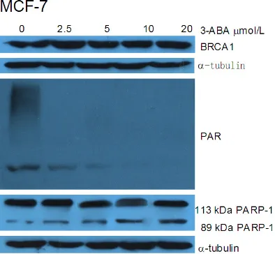
No effects of PARP-1 inhibitor on BRCA1 expression
PARP-1 inhibitor, 3-aminobenzamide (3-ABA),
inhibits the activity of poly (ADP-ribose)
trans-ferase enzymes, and can prevent ADR-induced
PARP-1 activation. PARP-1 inhibitor has poten-tial clinical application [9]. We used 2.5, 5, 10,
and 20 μM 3-ABA to treat MCF-7 cells for 24 h,
followed by 12 h of recovery. As shown in Figure 3, 3-ABA inhibited the expression of PAR but did not affect the expression of BRCA1 and the full-length PARP-1.
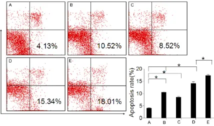
Co-treatment of MCF-7 and HCC1937 cells with doxorubicin and 3-ABA potentiates apop -tosis cell death
MCF-7 cells normally express WT BRCA1. To determine the different in the ability of
DNA-Figure 4. Effect of PARP-1 inhibition on cell apoptosis after ADR treatment. Cell apoptosis of MCF-7 cell lines, after co-treatment 24 h with 1 μM ADR and 5 μM 3-ABA, and 12 h recovered, measured by FCM. D. The PARP-1 inhibitor 3-ABA significantly increased the cell apoptosis induced by ADR inMCF-7 cells; E. and 3-ABA can further increase the HCC1937 cells apoptosis compared with MCF-7 cells induced by ADR. *P < 0.05, compared with the control
group (MCF-7); *P < 0.05, compared with the ADR MCF-7 cell lines group & the control group; *P < 0.05, compared
[image:4.612.92.520.72.324.2]Change of BRCA1 and PARP-1 after ADR treatment
damage repair between BRCA1 and PARP-1, we compared HCC1937 cells with mutant BRCA1 (BRCA1-/-) [10]. We further determined whether changes in BRCA1 and PARP-1 activation induced by adriamycin might be directly related to its cytotoxicity. MCF-7 and HCC1937 cells
were co-treated with 5 μM 3-ABA and 1 μM
ADR for 24 h and recovered for 12 h, and apop-tosis was detected by FCM. We observed that the degree of apoptosis was greater in MCF-7/ WT BRCA1 cells than in HCC1937 parental cells (Figure 4).
Discussion
Breast cancer is one of the most common and important diseases affecting women. More than 40%-50% of the hereditary breast can-cers are linked to germ-line breast cancer (BRCA1/2) gene mutations [11]. The breast cancer susceptibility gene, BRCA1, an impor-tant determinant of response to DNA-damaging agents, has been is linked to the resistance of
tumor cells to ionizing radiation (IR) or other
DNA-damaging agents due to up-regulation of DNA-damage repair and damage-induced che- ckpoint controls [12]. BRCA1 is a tumor sup-pressor gene. It has been shown that upregula-tion of BRCA1 expression leads to increased resistance to CDDP in ovarian cancer cells, but it was different to DNA helix intercalator and topoisomerase II inhibitor ADR [13]. Poly (ADP-ribose) polymerases (PARPs) play a key role in the repair of base damage via the base excision repair pathway [14]. Pharmacological inhibition of PARP induces cell death in tumors with muta-tions in certain DNA repair pathways, such as the BRCA pathways of double-strand break repair, and when combined with chemothera-pies that cause DNA damage [15]. ADR has long been considered one of the most active agents in the treatment of breast cancer [16]. ADR causes DNA double strand and single strand breaks by inhibiting topoisomerase II [17]. It is also a powerful iron chelator. Iron induces cell death by binding to DNA and pro-ducing free radicals that immediately cleave DNA and disrupt cell membranes.
Although, it has been reported that ADR can induce decreases in BRCA1 protein and/or
mRNA in MCF-7 [6, 18], in this study, we first
demonstrated that BRCA1 levels decrease upon treatment with high dose cytotoxic agents ADR but increase upon treatment with low does
ADR in MCF-7 cells. Time-dependent decrease of BRCA1 was detected as early as 6 h after 1
μmol/L ADR treatment and recovery, but these
alterations were much more prominent over time. Dose and time dependent up regulation
of PARP activation protein were first observed
as early as 6 h after treatment and 6 h recov-ery. The ADR-induced down-regulation of BRC- A1 did not appear to be directly related to the induction of apoptotic DNA fragmentation or cell death in HCC1937 cells (BRCA1-/-). Promi- nent apoptosis of MCF-7 cells with wild-type BRCA1 was observed with ADR.
BRCA1 encodes the tumor suppressor protein that acts as a negative regulator of tumor growth [19]. The functional BRCA1 protein is present in normal breast epithelial tissue and is reduced or absent in some breast tumors. Therefore, BRCA1 may function in monitoring genomic integrity and in DNA repair [20]. Many chemotherapeutic agents including ADR induce DNA damage in mammalian cells and eventu-ally deregulate the growth control or cause
dis-tractions in DNA metabolisms. Our results
show that ADR can down-regulation BRCA1 at
high dose, but with time dependent at 1 μmol/L
ADR treatment and increased PARP-1 activa-tion in a dose- and time-dependent manner. Low expression of BRCA1 is associated with decrease DNA-damage repair activity which may allow cells to be more susceptible to undergo apoptosis. PARP-1 activation can enhance DNA-damage repair and decrease cell apoptosis. For ADR treatment, compared with DNA repair, DNA damage plays a more impor-tant role. Thus, if BRCA1 is mutated or deleted, PARP-1 activation would play a more important role in DNA damage repair. Inhibiting PARP-1 activation will increase tumor cells apoptosis. Poly (ADP-ribose) polymerase inhibition enhan- ces DNA damage responses induced by DNA damaging agent, especially with BRCA1 muta-tion or delemuta-tion. PARP inhibitors are being inve- stigated as a monotherapy for the treatment of patients with BRCA1/2 mutations [21]. It is also investigated for triple-negative breast can-cers because of its molecular similarities to BRCA1-mutated malignancies [22]. PARP inhib-itors may be a potential therapeutic strategy to potentiate the DNA-damaging effects of che-motherapy and radiation [23].
of BRCA1 and up-regulation PARP-1 activation in MCF-7 cell lines. The sensitivity of MCF-7 cells with BRCA1-/- to adriamycin-induced cell apoptosis. Abrogating the cell-cycle arrest induced by PARP inhibition plus chemothera-pies may be a strategy in the treatment
of BRCA-proficient cancer. Further research is
required to establish the mechanism(s) and
functional significance of down-regulation of
BRCA1 and up-regulation of PARP-1 activation.
Acknowledgements
This research project was supported by Na- tional Natural Science Foundation of China (No. 81171653, 30972703), Natural Science Found- ation of Jiangsu Province (BK2011246, BK20- 11247), the Project of Six Batch of Major Talent Summit (BRA2010037), Society Development Plans, Department of Science and Technology
Changzhou (CE20135048, CJ20112020,
CZ20-110024, CS20102020).
Disclosure of conflict of interest
None.
Address correspondence to: Dr. Jingting Jiang, De- partment of Tumor Biological Treatment, The Third Affiliated Hospital of Soochow University, Changzhou 213003, China. Tel: 86-519-68870978; Fax: 86- 519-86621235; E-mail: jjtnew@163.com
References
[1] Chitikova ZV, Gordeev SA, Bykova TV, Zubova SG, Pospelov VA and Pospelova TV. Sustained activation of DNA damage response in irradi-ated apoptosis-resistant cells induces revers-ible senescence associated with mTOR down-regulation and expression of stem cell markers. Cell Cycle 2014; 13: 1424-1439.
[2] Jacot W, Thezenas S, Senal R, Viglianti C, Labe-renne AC, Lopez-Crapez E, Bibeau F, Bleuse JP, Romieu G and Lamy PJ. BRCA1 promoter hy-permethylation, 53BP1 protein expression and PARP-1 activity as biomarkers of DNA repair deficit in breast cancer. BMC Cancer 2013; 13: 523.
[3] Gabaldo Barrios X, Sarabia Meseguer MD, Alonso Romero JL, Marin Vera M, Marin Zafra G, Sanchez Henarejos P, Sanchez Bermudez AI and Ruiz Espejo F. Novel BRCA1 deleterious mutation (c.1918C>T) in familial breast and ovarian cancer syndrome who share a com-mon ancestry. Fam Cancer 2014; [Epub ahead of print].
[4] Morales J, Li L, Fattah FJ, Dong Y, Bey EA, Patel M, Gao J and Boothman DA. Review of poly (ADP-ribose) polymerase (PARP) mechanisms of action and rationale for targeting in cancer and other diseases. Crit Rev Eukaryot Gene Expr 2014; 24: 15-28.
[5] Tinker AV and Gelmon K. The role of PARP in-hibitors in the treatment of ovarian carcino-mas. Curr Pharm Des 2012; 18: 3770-3774. [6] Andres JL, Fan S, Turkel GJ, Wang JA, Twu NF,
Yuan RQ, Lamszus K, Goldberg ID and Rosen EM. Regulation of BRCA1 and BRCA2 expres-sion in human breast cancer cells by DNA-damaging agents. Oncogene 1998; 16: 2229-2241.
[7] Weaver AN and Yang ES. Beyond DNA Repair: Additional functions of PARP-1 in cancer. Front Oncol 2013; 3: 290.
[8] Rosen EM and Pishvaian MJ. Targeting the BRCA1/2 tumor suppressors. Curr Drug Tar-gets 2014; 15: 17-31.
[9] Sinha G. Downfall of iniparib: a PARP inhibitor that doesn’t inhibit PARP after all. J Natl Can-cer Inst 2014; 106: djt447.
[10] Tassone P, Blotta S, Palmieri C, Masciari S, Quaresima B, Montagna M, D’Andrea E, Eramo OP, Migale L, Costanzo F, Tagliaferri P and Venuta S. Differential sensitivity of BRCA1-mu-tated HCC1937 human breast cancer cells to microtubule-interfering agents. Int J Oncol 2005; 26: 1257-1263.
[11] Bernard-Gallon DJ, Dechelotte PJ, Le Corre L, Vissac-Sabatier C, Favy DA, Cravello L, De La-tour MP and Bignon YJ. Expression of BRCA1 and BRCA2 in male breast cancers and gyne-comastias. Anticancer Res 2003; 23: 661-667.
[12] Thompson LH. Recognition, signaling, and re-pair of DNA double-strand breaks produced by ionizing radiation in mammalian cells: the mo-lecular choreography. Mutat Res 2012; 751: 158-246.
[13] Tassone P, Tagliaferri P, Perricelli A, Blotta S, Quaresima B, Martelli ML, Goel A, Barbieri V, Costanzo F, Boland CR and Venuta S. BRCA1 expression modulates chemosensitivity of BR-CA1-defective HCC1937 human breast cancer cells. Br J Cancer 2003; 88: 1285-1291. [14] Das BB, Huang SY, Murai J, Rehman I, Ame JC,
Sengupta S, Das SK, Majumdar P, Zhang H, Bi-ard D, Majumder HK, Schreiber V and Pommi-er Y. PARP1-TDP1 coupling for the repair of topoisomerase I-induced DNA damage. Nucle-ic Acids Res 2014; 42: 4435-4449.
Change of BRCA1 and PARP-1 after ADR treatment
C-terminal binding protein 2. Neoplasia 2013; 15: 600-608.
[16] Mittal N, Gupta M and Singla M. Cutaneous ad-verse drug reactions notified by pharmacovigi-lance in a tertiary care hospital in north India. Cutan Ocul Toxicol 2014; [Epub ahead of print]. [17] Lage H, Aki-Sener E and Yalcin I. High antineo-plastic activity of new heterocyclic compounds in cancer cells with resistance against classi-cal DNA topoisomerase II-targeting drugs. Int J Cancer 2006; 119: 213-220.
[18] Su J and Ciftci K. Changes in BRCA1 and BRCA2 expression produced by chemothera-peutic agents in human breast cancer cells. Int J Biochem Cell Biol 2002; 34: 950-957. [19] Elia AE and Elledge SJ. BRCA1 as tumor
sup-pressor: lord without its RING? Breast Cancer Res 2012; 14: 306.
[20] Savage KI, Matchett KB, Barros EM, Cooper KM, Irwin G, Gorski JJ, Orr KS, Vohhodina J, Ka-vanagh JN, Madden AF, Powell A, Manti L, Mc-Dade SS, Park BH, Prise KM, McIntosh S, Salto-Tellez M, Richard DJ, Elliot CT and Harkin DP. Brca1 Deficiency Exacerbates Estrogen In-duced DNA Damage and Genomic Instability. Cancer Res 2014; 74: 2773-84.
[21] Horton JK and Wilson SH. Strategic Combina-tion of DNA-damaging agent and PARP inhibi-tor results in enhanced cytotoxicity. Front On-col 2013; 3: 257.
[22] Al-Ejeh F, Shi W, Miranda M, Simpson PT, Var-gas AC, Song S, Wiegmans AP, Swarbrick A, Welm AL, Brown MP, Chenevix-Trench G, Lakhani SR and Khanna KK. Treatment of tri-ple-negative breast cancer using anti-EGFR-di-rected radioimmunotherapy combined with radiosensitizing chemotherapy and PARP in-hibitor. J Nucl Med 2013; 54: 913-921. [23] Gilardini Montani MS, Prodosmo A, Stagni V,