Original Article
Overexpression of MAGE-D4 in colorectal cancer is a
potentially prognostic biomarker and
immunotherapy target
Qing-Mei Zhang1*, Shu-Jia He1*, Ning Shen2*, Bin Luo1, Rong Fan1, Jun Fu1, Guo-Rong Luo1, Su-Fang Zhou1,
Shao-Wen Xiao3, Xiao-Xun Xie1
1Department of Histology and Embryology, School of Pre-clinical Medicine, 3Department of Surgery, First Affiliated Hospital, Guangxi Medical University, Nanning 530021, Guangxi Zhuang Autonomous Region, China; 2 Depart-ment of Oral and Maxillofacial Surgery, The People’s Hospital of Guangxi Zhuang Autonomous Region, Nanning 530021, Guangxi Zhuang Autonomous Region, China. *Equal contributors.
Received May 20, 2014; Accepted June 27, 2014; Epub June 15, 2014; Published July 1, 2014
Abstract: Melanoma-associated antigen D4 (MAGE-D4)is a novel member of MAGE family. This study aimed to examine the expression and immunogenicity of MAGE-D4in colorectal cancer (CRC) to determine its potential as a prognosis and immunotherapeutic target. The expression of MAGE-D4 mRNA and protein was determined by RT-PCR and immunohistochemistry (IHC) in CRCs with paired adjacent non-tumor tissues, colorectal adenomas and normal colorectal tissues, respectively. Sera from 64 CRC patients were tested for MAGE-D4antibody by ELISA. MAGE-D4 mRNA was more frequently expressed in CRCs (76.7%, 46/60) than in adjacent non-tumor tissues (15.0%, 9/60). MAGE-D4 protein was detected in all the CRC tissues tested, 70.0% of which showed high expression. There was no MAGE-D4 protein detected in any paired adjacent non-tumor tissue. No MAGE-D4 expression was found in colorectal adenomas and normal colorectal tissues by either RT-PCR or immunohistochemistry. Patients with high
MAGE-D4 protein expression had significantly shorter overall survival than those with low MAGE-D4 protein expres -sion (median, 68.6 vs 122.2 months; P=0.030). Furthermore, multivariate analysis exhibited high MAGE-D4 protein expression had a trend toward an independent prognostic factor (hazard ratio: 6.124; P=0.050). Humoral immunity to MAGE-D4 was detected in 12 of 64 (18.8%) CRC patients’ sera but not in 77 healthy donors. There was no
cor-relation between MAGE-D4 expression, serum antibody and clinicopathological parameters. These findings suggest
MAGE-D4 may serve as a potentially prognostic biomarker and an attractive target of immunotherapy in CRC.
Keywords: Melanoma-associated antigen, MAGE-D4, colorectal cancer, serum immunoreactivity
Introduction
Colorectal cancer (CRC) is the third most com-mon malignancy and fourth leading cause of cancer mortality worldwide, with more than a million individuals diagnosed and about half million deaths annually [1, 2]. Although there are many established therapeutic strategies including surgery, chemotherapy and radiother-apy, its prognosis remains unsatisfactory due to late diagnosis [3]. Thus, novel therapeutic strategies are urgently needed for this malig-nancy. Immunotherapy is an attractive approach among novel therapeutic strategies [4, 5]. This approach requires the identification of tumor specific antigens. Currently, a number of such antigens are encoded by the genes of
melanoma-associated antigen (MAGE) family [6, 7].
iden-tification of other MAGE antigens with high expression in CRC is essential.
MAGE-D4, originally named MAGE-E1, is a novel member of MAGE family. It has been reported restricted expression in normal tissues except brain and ovary and overexpression in some human malignancies, including lung, liver, oral, kidney, breast, esophagus cancer and glioma [19-25]. A MHC class I ligand from MAGE-D4 presented by HLA-A on tumor tissue was also reported by Kramer et al [20]. Several previous studies have shown that MAGE-D4 contributes to proliferation, migration, and invasion of tumor cells in breast cancer and oral squamous cell carcinoma [22, 23]. Recent studies have shown that MAGE-D4 is a marker of poor prog-nosis in hepatocellular, esophagus and breast carcinoma [23-25]. To some extent, these pre-liminary findings have been suggested that MAGE-D4 may play an important role in the pro-gression of tumors and may be a potentially promising target for tumor prognosis and treatment.
Current knowledge about MAGE-D4 gene expression in CRC was only based on mRNA level from one previous report [26]. However, MAGE-D4 protein expression has not been elu-cidated including its immunogenicity. In the present study, we examined the expression of MAGE-D4 at mRNA and protein levels in CRC tissues, as well as the serum antibody against MAGE-D4 in a subset of CRC patients. Furthermore, whether MAGE-D4 protein can be a prognostic factor was analyzed, and the cor-relation among MAGE-D4 expression, serum antibody and clinicopathological parameters in CRC patients were also investigated.
Materials and methods
Tissue and serum samples
All tissue and serum samples were collected from the First Affiliated Hospital of Guangxi Medical University with the informed consent of patients and approved by Hospital Ethic Review Committee. A total of 154 tissue samples, including 60 primary CRCs with adjacent non-tumor tissues (42 men and 18 women; mean age, 56.37±14.74 years; age ranging, 30-86 years), 24 colorectal adenomas and 10 normal colorectal tissues, were analyzed by RT-PCR. 82 of paraffin-embedded tissue sections for immunohistochemical analysis was
construct-ed from 30 primary CRCs with pairconstruct-ed adjacent non-tumor tissues (17 men and 13 women; mean age, 56.45±11.43 years; age ranging, 35-76 years), 12 colorectal adenomas and 10 normal colorectal tissues. CRC patients were followed up for 1-130 months (mean, 79.6±52.6 months). Overall survival was defined as the time from diagnosis to the date of death or last follow-up. All CRC and paired non-tumor tissues were surgically removed without undergoing preoperative treatment including chemothera-py and radiation. The colorectal adenomas were available from the patients by endoscopic polypectomy and normal colorectal tissues were obtained at autopsy. Sera were collected from 64 patients with CRC (47 men and 17 women; mean age, 56.03±14.96 years; age ranging, 26-93 years) at diagnosis prior of ther-apy and sera of 77 healthy donors from routine physical examination of students of Guangxi Medical University were used as controls. All tumor samples were classified according to the TNM classification of the Union for International Cancer Control [27]. Tumor samples were coded and assessed in a blinded manner.
RT-PCR analysis
Total RNA was prepared by Trizol reagent (Invitrogen, CA) and 2.5 μg RNA was reverse transcribed into cDNA with RevertAid™ First Strand cDNA Synthesis Kit (Fermentas, USA) according to the manufacturer’s instructions. cDNA was then tested for integrity by amplifica-tion of Glyceraldehyde-3-phosphate dehydroge-nase (GAPDH) gene. RT-PCR was carried out with MAGE-D4 specific primers as previously reported [19]. The cycling parameters were as following: initial denaturation at 94°C for 5 min followed by 30 sec at 94°C, 30 sec at (64°C for MAGE-D4, 55°C for GAPDH), and 30 sec at 72°C for 35 cycles, and a final extension for 10 min at 72°C. Target bands were visualized on a 1.5% agarose gel with ethidium bromide stain-ing. The expression of MAGE-D4 was counted as positive, only if the RT-PCR reaction repeat-ed at least twice with same result.
Immunohistochemistry (IHC)
eth-Figure 1. RT-PCR analysis of MAGE-D4 mRNA expression in CRC tissues (T) with paired adjacent non-tumor tissues (Nt), colorectal adenomas (A) and normal colorectal tissues (N). GAPDH was used as internal control for the parallel PCR analysis of the same sample. P, positive control (glioma); Ne, negative control (no cDNA template).
ylene diamine tetraacetic acid (EDTA, pH8.0) for antigen retrieval. After the inactivation of endogenous peroxidase, the sections were treated with normal goat serum for blocking and then immunostained with anti-MAGE-D4 polyclonal antibody (1:500 dilution, Santa Cruz Biotechnology, USA) overnight at 4°C. Negative controls using rabbit serum collected before immunization were also incubated in parallel. Then horseradish peroxidase-conjugated goat anti-rabbit IgG (ZSGB-BIO, China) was added as the secondary antibody. Immunoreactivity was visualized with 3, 3’-diaminobenzidine (DAB) (Maixin Biotechnology, China) followed by he- matoxylin counterstain.
Positive immunoreactivity was assessed by two independent pathologists who did not know patients’ clinical information and recorded semi-quantitatively according to the staining intensity, in combination with the percentage of positive cells. The staining intensity was quanti-fied using the following scores: 0=no staining, 1=weak staining, 2=moderate staining, 3=strong staining. The percentage of positive tumor cells was defined as follows: 0=0-5%, 1=6-25%, 2=26-50%, 3=51-75%, 4=76-100%. According to the sum of both points, each sec-tion was assigned as no MAGE-D4 expression when the sum score was 0, low MAGE-D4 expression when the sum score was between 1 to 4, and high MAGE-D4 expression when the sum score was more than 4 [21, 29].
ELISA
MAGE-D4 recombinant protein was generated according to a previous report by He et al [30].
ELISA was performed as previous report by Zhou et al [31]. In brief, MAGE-D4 protein (1 μg/ml) was coat-ed on the 96-well plates (Corning, USA) at 4°C over-night. Maltose binding pro-tein (MBP) propro-tein was used as a blank control. The plates were blocked with 5% nonfat milk 37°C for 1 h, then 1:800 diluted serum were added and incubated at 37°C for 1 h. Streptavidin-biotinylated horseradish peroxidase complex (KPL, USA) was used as secondary antibody. Detection was accomplished using tetramethylbenzidine sub-strate, followed by adding sulfuric acid to stop the reaction. The absorbance at 450 nm with 630nm as reference filter was determined with a microplate reader (Bio-Rad, USA). All serum samples were performed from triplicates and indicated as mean. An optical density (OD) value that exceeds three standard deviations (SDs) above the mean OD value of sera from healthy donors was defined as positive. Specificity of each positive serum sample was examined by testing reactivity after pre-incu-bating with recombinant MAGE-D4 protein.
Statistical analysis
Statistical analyses were conducted by SPSS software (version 16). Statistical significance was defined as P<0.05. Relationships between MAGE-D4 expression and antibody response with clinicopathological parameters were test-ed by χ2 test or Fisher’s exact test. Overall
sur-vival rates were calculated using the Kaplan– Meier method, and differences in survival curves were compared using the log-rank test. Multivariable regression analysis was per-formed to detect prognostic factors using Cox proportional hazards models, and variables with a two-sided P value of <0.05 were entered into the final model.
Results
Expression of MAGE-D4 mRNA
[image:3.612.94.385.72.199.2]Figure 2. Representative immunohistochemical staining of MAGE-D4 protein in CRC tissues (A, C), paired adjacent non-tumor tissues (B, D), colorectal adenomas (E) and normal colorectal tissues (F). Low and high immunoreactivity of MAGE-D4 protein immunostaining were shown in (A and C) respectively, using polyclonal MAGE-D4 antibody. No positive reactivity was observed in paired adjacent non-tumor tissues (B, D), colorectal adenomas (E) and normal
colorectal tissues (F). Original magnification, ×100 (×400 in the right down corner).
Figure 3. Kaplan–Meier analysis of MAGE-D4 protein in 30 patients with CRC, categorized as having low and high expression of MAGE-D4. Patients with high MAGE-D4 protein expression in CRC tissues had
sig-nificantly shorter overall survival than patients with
low MAGE-D4 protein expression.
normal colorectal tissues by RT-PCR. MAGE-D4 mRNA was detected in 46 of 60 (76.7%) CRCs and 9 of 60 (15.0%) of adjacent non-tumor tis-sues (Figure 1). Statistical analysis revealed
that there was significant difference between CRCs and adjacent non-tumor tissues (P= 0.000). No positive expression was detected in any of both colorectal adenomas and normal colorectal tissues (Figure 1).
Expression of MAGE-D4 protein
[image:4.612.91.288.427.595.2]Table 1. Prognostic factors in 30 patients with CRC
Variable n Univariate Multivariate
HR (95% CI) P value HR (95% CI) P value Gender (Male) 17 1.416 (0.426-4.708) 0.567 -
-Age ≥56 (years) 18 0.669 (0.212-2.116) 0.490 -
-Tumor location (Colon) 24 1.407 (0.307-6.444) 0.658 - -Tumor size ≥5 cm) 17 1.037 (0.334-3.244) 0.950 - -CEA (>5 ng/mL) 17 5.296 (1.153-24.323 0.016* 4.891 (0.930-25.728) 0.174 Depth of tumor Invasion (T4) 5 2.237 (0.601-8.325) 0.216 - -Lymph node metastasis 23 4.514 (0.582-35.030) 0.113 - -Distant metastasis 12 4.951 (1.470-16.672) 0.004* 2.663 (0.582-12.173) 0.010* TNM stage (III+IV) 23 4.514 (0.582-35.030) 0.113 - -Histological type (Non-mucin-producing) 27 0.040 (0.00-62.396) 0.179 - -Histological grade (Moderate and poor) 22 1.587 (0.347-7.254) 0.546 - -MAGE-D4 protein expression (High) 21 7.001 (0.900-54.451) 0.030* 6.124 (0.780-48.095) 0.050
Univariate analysis was performed using the log-rank test. Multivariate analysis was performed using the Cox proportional hazards model. *Statistically significant (P<0.05). HR, Hazard ratio; 95 percent CI, 95 percent confidence interval for relative risk; CEA, carcinoembryonic antigen.
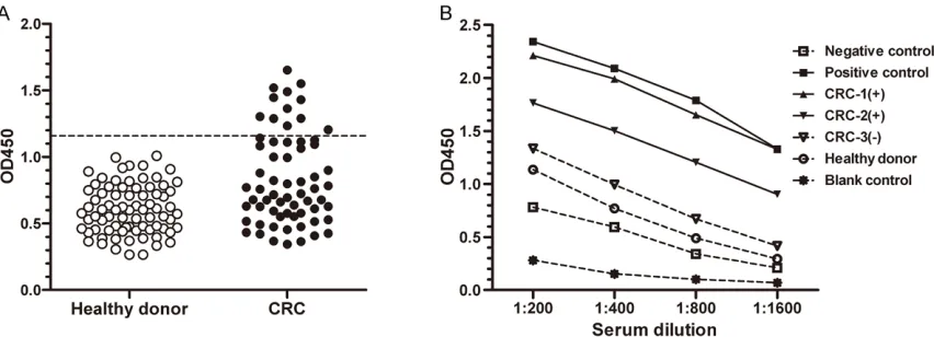
Figure 4. Anti-MAGE-D4 antibody in sera by ELISA. A. Detection of anti-MAGE-D4 antibody in the sera from 64 CRC patients and 77 normal donors. Three standard deviations above the mean absorbance in the sera from normal donors were used as cutoff (1.1593) for a positive result (dotted line). B. Sera titration curves of dilution series of
recombinant MAGE-D4 protein against four different concentrations. Sera from three CRC patients (▲, ▼, ▽) and
one healthy donor (○) were shown, together with a positive control (■) and a negative control (□). MBP protein (*) was used as a blank control. Patient 1 and 2 demonstrated the highest (▲) and the lowest (▼) MAGE-D4 antibody
titer among 12 seropositive CRC patients, respectively.
Prognostic impact of high MAGE-D4 protein expression
Prognostic impact of MAGE-D4 protein expres-sion in patients with CRC was analyzed as a continuous variable in a regression analysis. The result showed that mean overall survival (68.6 vs 122.2 months) was significantly short-er in patients with high MAGE-D4 protein expression than in those with low expression (P=0.030, Figure 3). Furthermore, Univariate analysis identified carcinoembryonic antigen (CEA) >5 ng/mL, distant metastasis and high
MAGE-D4 protein expression as significant prognostic factors (Table 1). In multivariate analysis, although distant metastasis exhibited the only independent prognostic factor, high MAGE-D4 protein expression tended to be as an independent prognostic factor (hazard ratio: 6.124; P=0.050; Table 1).
Serum antibody against MAGE-D4
[image:5.612.94.520.322.476.2]Table 2. Correlation among MAGE-D4 expression, serum antibody and clinicopathological parameters in CRC patients
Clinicopathological parameters
mRNA P
value
Protein P
value
Antibody P
value Positive/Total (%) High-expression/Total (%) Positive/Total (%) Gender
Male 34/42 (81.0) 0.319 13/17 (76.5) 0.443 10/47 (21.3) 0.490 Female 12/18 (66.7) 8/13 (61.5) 2/17 (11.8)
Age (year)
<56 22/28 (78.6) 0.744 8/12 (66.7) 1.000 3/32 (9.4) 0.055
≥56 24/32 (75.0) 13/18 (72.2) 9/32 (28.1)
Tumor location
Colon 28/36 (77.8) 0.803 17/24 (70.8) 1.000 6/32 (18.8) 1.000 Rectum 18/24 (75.0) 4/6 (66.7) 6/32 (18.8)
Tumor size (cm)
<5 23/27 (85.2) 0.158 10/13 (76.9) 0.691 7/41 (17.1) 0.742
≥5 23/33 (69.7) 11/17 (64.7) 5/23 (21.7)
CEA (ng/mL)a
≤5 25/31 (80.6) 0.451 10/13 (76.9) 0.691 4/25 (16.0) 0.751
>5 21/29 (72.4) 11/17 (64.7) 8/39 (20.5) Depth of tumor invasion
T1-T2 8/10 (80.0) 1.000 0/0 (0) - 2/18 (11.1) 0.483 T3-T4 38/50 (76.0) 21/30 (70.0) 10/46 (21.7)
Lymph node metastasis
N0 27/35 (77.1) 0.918 4/7 (57.1) 0.640 8/43 (18.6) 1.000 N1-3 19/25 (76.0) 17/23 (73.9) 4/21 (19.0)
Distant metastasis
M0 36/48 (75.0) 0.713 11/18 (61.1) 0.249 10/57 (17.5) 0.607 M1 10/12 (83.3) 10/12 (83.3) 2/7 (19.0)
TNM stage
І + II 27/35 (77.1) 0.918 4/7 (57.1) 0.640 8/43 (18.6) 1.000
III + IV 19/25 (76.0) 17/23 (73.9) 4/21 (19.0) Histological typea
Non-mucin-producing 41/54 (75.9) 1.000 20/27 (74.1) 0.207 10/54 (18.5) 1.000 Mucin-producing 5/6 (83.3) 1/3 (33.3) 2/10 (20.0)
Histological gradeb
G1 13/17 (76.5) 0.862 5/8 (62.5) 0.484 1/16 (6.2) 0.259 G2 20/27 (74.1) 14/18 (77.8) 6/28 (21.4)
G3 13/16 (81.2) 2/4 (50.0) 5/20 (25.0)
aNon-mucin-producing cancer includes tubular and (or) papillary adenocarcinoma, Mucin-producing cancer includes mucinous
cancer and signet-ring cell cancer. bHistological grade (G). G1, well differentiated; G2, moderately differentiated; G3, poorly
differentiated.
(18.8%) CRC patients, but not in every healthy controls (Figure 4A). The titration curves of selected MAGE-D4 antibody-positive and -neg-ative sera were illustrated in Figure 4B. Antibody against MBP protein failed to be detected in sera from MAGE-D4 seropositive patients (data not shown). Of 64 sera screened, 25 sera were collected from CRC patients whose tissues were also assessed for MAGE-D4
Associations among MAGE-D4 expression, se-rum antibody and clinicopathological param-eters in CRC
Associations among MAGE-D4 expression, serum antibody and clinicopathological param-eters including gender, age, tumor location and size, CEA level, depth of tumor invasion, Lymph node metastasis and distant metastasis, TMN and histological type and grade were statisti-cally evaluated. As shown in Table 2, No signifi-cant correlations were observed among MAGE-D4 expression, serum antibody and those clinical parameters.
Discussion
One of the major barriers to antigen-specific immunotherapy in CRC is the lack of well-defined immunogenic tumor antigens. It is urgent and challenging to identify and charac-terize tumor-specific antigens in CRC. In the present study, we characterized both expres-sion pattern and humoral immune response of MAGE-D4, a new member of the MAGE family, to assess its potential as a target for prognosis and immunotherapy of CRC.
Our results demonstrated that the expression frequency of MAGE-D4 mRNA in CRC tissues was significantly higher than that in adjacent non-tumor tissues (P=0.000). It is in disagree-ment with the report by Chung et al, they showed no differential expression of MAGE-D4 mRNA between CRC tissues and adjacent non-tumor tissues [26]. This discrepancy may result from different clinical samples, the heterogene-ity of gene expression in tumors and different research method. In general, RT-PCR analysis applied in present study turned out to be more sensitive than the cDNA microarray hybridiza-tion used by Chung et al [32]. Furthermore, MAGE-D4 protein was declared positive in all of CRC tissues, 70% of which exhibited high-expression. Previous studies have reported that CRC is a poor MAGE antigen expresser [13-18]. Our data explored that either mRNA or pro-tein of MAGE-D4 has a higher expression fre-quency as compared to other MAGE antigens in CRC, suggesting that MAGE-D4 may be a more promising target for CRC immunotherapy than other MAGE antigens, at least in Chinese. In analyzing MAGE-D4 expression in non-tumor tissue setting, we found that MAGE-D4 mRNA
was positive in a small portion of adjacent non-tumor tissues (15.0%, 9/60) and absent in nor-mal colorectal tissues. MAGE-D4 positivity in adjacent non-tumor tissues might be associat-ed with infiltrating tumor cells. Jeon et al [33] examined MAGE mRNA expression in 46 CRC tissues and matched normal mucosal tissues within 20 mm, 20 to 50 mm and more than 50 mm from tumors. They found that the MAGE expression rates were greatly decreased as the tissues collected far from tumor site. Therefore, when adjacent non-tumor tissues were collect-ed, their locations from tumor are an important issue. In addition, we also found that colorectal adenoma tissues showed no MAGE-D4 expres-sion by either RT-PCR or immunohistochemis-try. Many previous studies demonstrated colorectal adenoma as a precursor lesion for CRC [28, 34], thus MAGE-D4 seems not to be involved in transformation from benign to malignancy.
Taken together, MAGE-D4 expression analysis suggested that MAGE-D4 may be a CRC-specific antigen. However, heterogeneous intratumor expression of MAGE-D4 was observed, which may hamper the effectiveness of MAGE-D4-based immunotherapy for CRC. This phenome-non is also frequent in other MAGE antigens [35, 36]. The underlying reason is still largely unknown. Generally, most MAGE family genes belong to epigenetic-mediated regulation genes, by which the heterogeneity may be improved to a certain extent through epigenetic modulation [9, 37, 38]. Our previous study has shown that DNA methylation in MAGE-D4 pro-moter region is an important mechanism in regulation of MAGE-D4 expression, and treat-ment of 5-AZA-CdR, a DNA methyltransferase (DNMT) inhibitor, can enhance MAGE-D4 expression in MAGE-D4-negative glioma cells [39]. Whether the heterogeneous expression of MAGE-D4 in CRC is also related to DNA methyl-ation, it needs further investigation.
indepen-dent prognostic factor, suggesting that MAGE-D4 may have the potential value as a prognostic marker of CRC. However, the sample size is still small for longer period of follow-up. Further studies are needed to confirm its prog-nosis value by enlarging the sample size. It has been noted that overexpression of many tumor antigens may cause humoral immune response in patients with tumor burden [40-43]. So did patients with CRC [15, 28, 44]. Monitoring these antibodies in patients’ sera could have potential diagnostic and prognostic significance [45]. Considering MAGE-D4 overex-pression in CRC mentioned above, it is neces-sary to uncover a possibly immune response against MAGE-D4 in patients with CRC. Accordingly, we further tested MAGE-D4 serore-activity in CRC patients and healthy individuals. The results showed that sera antibody against MAGE-D4 was found in 18.8% (12/64) of patients with CRC and not in healthy individu-als. In our survey of 25 CRC patients where tumor and serum samples from the same patients were available, 3 of 21 CRC patients with MAGE-D4 mRNA-positive tumors had anti-bodies against MAGE-D4. Furthermore, the three sero-positive patients showed high-MAGE-D4 protein expression, suggesting that preferential, high expression of this antigen may give rise to the specific humoral immune response in patients with CRC. Although we have not tested antigen-specific CD8+ T cell
responses to MAGE-D4 in the present study, humoral immune response to MAGE-D4 in CRC patients may be a predictive of cellular immune response, resembling the case of MAGE-A1 and MAGE-A3 [46]. A HLA-A*25/MHC I-binding pep-tide of MAGE-D4 protein has been identified [20]. In a recent study in other cancer, we also found that MAGE-D4 protein can be recognized by CD8+ T cells in vitro and induce cytotoxic
reaction against glioma cell line which express-es MAGE-D4 (unpublished data). Clearly, fur-ther investigation will be needed to explore MAGE-D4-specific cytotoxic T cells in CRC. In conclusion, our findings demonstrate that MAGE-D4 is frequently expressed in CRC and show inherent immunogenicity. Hence, MAGE- D4 may be a potential target for prognostic marker and specific immunotherapy in CRC. The role of MAGE-D4 in CRC progression remains to be elucidated. Further studies of functional analysis should be done to clarify the
molecular mechanisms underlying the biologi-cal activities of MAGE-D4 in CRC.
Acknowledgements
We thank Ms Fang Chen from Guangxi Medical University and Ms Wen-Wen Guo from the People’s Hospital of Guangxi Zhuang Au- tonomous Region for excellent technical assis-tance. This work was supported by National Natural Science Foundation of China (No. 81060207, No. 81360371, No. 81360374), Natural Science Foundation of Guangxi (No. 2012GXNSFAA053114).
Disclosure of conflict of interest
All disclosures will be published if the manu-script is accepted.
Address correspondence to: Dr. Xiao-Xun Xie, Department of Histology & Embryology, School of Pre-clinical Medicine, Guangxi Medical University, 22 Shuangyong Road, Nanning, Guangxi 530021, China. Tel: 13807713891; Fax: 0086-5358872; E-mail: xiaoxunxie@gmail.com; Dr.
Shao-Wen Xiao, Department of Surgery, 1st Affiliated
Hospital, Guangxi Medical University, 22 Shuang- yong Road, Nanning 530021, Guangxi, China. Tel: 0086-771-5356506; Fax: 0086-771-5356506; E-mail: xsw57@yahoo.com
References
[1] Parkin DM. Global cancer statistics in the year 2000. Lancet Oncol 2001; 2: 533-543. [2] Ferlay J, Autier P, Boniol M, Heanue M,
Colom-bet M and Boyle P. Estimates of the cancer in-cidence and mortality in Europe in 2006. Ann Oncol 2007; 18: 581-592.
[3] Zafar SY, Malin JL, Grambow SC, Abbott DH, Kolimaga JT, Zullig LL, Weeks JC, Ayanian JZ, Kahn KL, Ganz PA, Catalano PJ, West DW and Provenzale D. Chemotherapy use and patient treatment preferences in advanced colorectal cancer: a prospective cohort study. Cancer 2012; 119: 854-862.
[4] Chi DD, Merchant RE, Rand R, Conrad AJ, Gar-rison D, Turner R, Morton DL and Hoon DS. Mo-lecular detection of tumor-associated antigens shared by human cutaneous melanomas and gliomas. Am J Pathol 1997; 150: 2143-2152. [5] Prins RM, Odesa SK and Liau LM.
Immuno-therapeutic targeting of shared melanoma-as-sociated antigens in a murine glioma model. Cancer Res 2003; 63: 8487-8491.
A and Boon T. A gene encoding an antigen rec-ognized by cytolytic T lymphocytes on a human melanoma. Science 1991; 254: 1643-1647. [7] Lucas S, De Plaen E and Boon T. MAGE-B5,
MAGE-B6, MAGE-C2, and MAGE-C3: four new members of the MAGE family with
tumor-spe-cific expression. Int J Cancer 2000; 87: 55-60.
[8] Inoue H, Mori M, Honda M, Li J, Shibuta K, Mi-mori K, Ueo H and Akiyoshi T. The expression of tumor-rejection antigen “MAGE” genes in human gastric carcinoma. Gastroenterology 1995; 109: 1522-1525.
[9] Wischnewski F, Pantel K and Schwarzenbach H. Promoter demethylation and histone acety-lation mediate gene expression of MAGE-A1, -A2, -A3, and -A12 in human cancer cells. Mol Cancer Res 2006; 4: 339-349.
[10] Duffour MT, Chaux P, Lurquin C, Cornelis G, Boon T and van der Bruggen P. A MAGE-A4 peptide presented by HLA-A2 is recognized by cytolytic T lymphocytes. Eur J Immunol 1999; 29: 3329-3337.
[11] Rosenberg SA, Yang JC and Restifo NP. Cancer immunotherapy: moving beyond current vac-cines. Nat Med 2004; 10: 909-915.
[12] Morse MA, Garst J, Osada T, Khan S, Hobeika A, Clay TM, Valente N, Shreeniwas R, Sutton MA, Delcayre A, Hsu DH, Le Pecq JB and Lyerly HK. A phase I study of dexosome immunother-apy in patients with advanced non-small cell lung cancer. J Transl Med 2005; 3: 9.
[13] Toungouz M, Libin M, Bulte F, Faid L, Lehmann F, Duriau D, Laporte M, Gangji D, Bruyns C, Lambermont M, Goldman M and Velu T.
Tran-sient expansion of peptide-specific lympho -cytes producing IFN-gamma after vaccination with dendritic cells pulsed with MAGE peptides in patients with mage-A1/A3-positive tumors. J Leukoc Biol 2001; 69: 937-943.
[14] Park MS, Park JW, Jeon CH, Lee KD and Chang HK. Expression of melanoma antigen-encod-ing genes (MAGE) by common primers for MAGE-A1 to -A6 in colorectal carcinomas among Koreans. J Korean Med Sci 2002; 17: 497-501.
[15] Gerhardt A, Usener D, Keese M, Sturm J, Schadendorf D and Eichmuller S. Tissue
ex-pression and sero-reactivity of tumor-specific
antigens in colorectal cancer. Cancer Lett 2004; 208: 197-206.
[16] Koketsu S, Watanabe T, Kazama S, Ishihara S, Komuro Y and Nagawa H. What types of colorectal cancer overexpress the MAGE pro-tein? Hepatogastroenterology 2004; 51: 1648-1652.
[17] Li M, Yuan YH, Han Y, Liu YX, Yan L, Wang Y and
Gu J. Expression profile of cancer-testis genes
in 121 human colorectal cancer tissue and ad-jacent normal tissue. Clin Cancer Res 2005; 11: 1809-1814.
[18] Morgan RA, Chinnasamy N, Abate-Daga D, Gros A, Robbins PF, Zheng Z, Dudley ME, Feld-man SA, Yang JC, Sherry RM, Phan GQ, Hughes MS, Kammula US, Miller AD, Hessman CJ, Stewart AA, Restifo NP, Quezado MM, Alim-chandani M, Rosenberg AZ, Nath A, Wang T, Bielekova B, Wuest SC, Akula N, McMahon FJ, Wilde S, Mosetter B, Schendel DJ, Laurencot CM and Rosenberg SA. Cancer regression and neurological toxicity following anti-MAGE-A3 TCR gene therapy. J Immunother 2013; 36: 133-151.
[19] Sasaki M, Nakahira K, Kawano Y, Katakura H, Yoshimine T, Shimizu K, Kim SU and Ikenaka K. MAGE-E1, a new member of the melanoma-associated antigen gene family and its expres-sion in human glioma. Cancer Res 2001; 61: 4809-4814.
[20] Kramer BF, Schoor O, Kruger T, Reichle C, Muller M, Weinschenk T, Hennenlotter J, Stenzl A, Rammensee HG and Stevanovic S.
MAGED4-expression in renal cell carcinoma and identifi -cation of an HLA-A*25-restricted MHC class I ligand from solid tumor tissue. Cancer Biol Ther 2005; 4: 943-948.
[21] Ito S, Kawano Y, Katakura H, Takenaka K, Ada-chi M, Sasaki M, Shimizu K, Ikenaka K, Wada H and Tanaka F. Expression of MAGE-D4, a novel MAGE family antigen, is correlated with tumor-cell proliferation of non-small cell lung cancer. Lung Cancer 2006; 51: 79-88. [22] Chong CE, Lim KP, Gan CP, Marsh CA, Zain RB,
Abraham MT, Prime SS, Teo SH, Silvio Gutkind J, Patel V and Cheong SC. Over-expression of MAGED4B increases cell migration and growth in oral squamous cell carcinoma and is associ-ated with poor disease outcome. Cancer Lett 2012; 321: 18-26.
[23] Germano S, Kennedy S, Rani S, Gleeson G, Cly-nes M, Doolan P, McDonnell S, Hughes L, Crown J and O’Driscoll L. MAGE-D4B is a novel marker of poor prognosis and potential thera-peutic target involved in breast cancer tumori-genesis. Int J Cancer 2012; 130: 1991-2002. [24] Oya H, Kanda M, Takami H, Hibino S, Shimizu
D, Niwa Y, Koike M, Nomoto S, Yamada S, Ni-shikawa Y, Asai M, Fujii T, Nakayama G, Sugi-moto H, Fujiwara M and Kodera Y. Overexpres-sion of melanoma-associated antigen D4 is an independent prognostic factor in squamous cell carcinoma of the esophagus. Dis Esopha-gus 2013; [Epub ahead of print].
[25] Takami H, Kanda M, Oya H, Hibino S, Sugimoto H, Suenaga M, Yamada S, Nishikawa Y, Asai M, Fujii T, Nomoto S and Kodera Y. Evaluation of MAGE-D4 expression in hepatocellular carci-noma in Japanese patients. J Surg Oncol 2013; 108: 557-562.
and Lin SR. Differential gene expression profile
of MAGE family in taiwanese patients with colorectal cancer. J Surg Oncol 2010; 102: 148-153.
[27] LH S, MK G and C W. TNM Classification of Ma -lignant Tumors. 7th edition. Oxford: Wiley-Blackwell; 2009. pp. 100-105.
[28] Luo B, Yun X, Fan R, Lin YD, He SJ, Zhang QM, Mo FR, Chen F, Xiao SW and Xie XX. Cancer testis antigen OY-TES-1 expression and serum immunogenicity in colorectal cancer: its rela-tionship to clinicopathological parameters. Int J Clin Exp Pathol 2013; 6: 2835-2845. [29] Thaker PH, Deavers M, Celestino J, Thornton A,
Fletcher MS, Landen CN, Kinch MS, Kiener PA and Sood AK. EphA2 expression is associated with aggressive features in ovarian carcinoma. Clin Cancer Res 2004; 10: 5145-5150. [30] He SJ, Gu YY, Yu L, Luo B, Fan R, Lin WZ, Lan
XW, Lin YD, Zhang QM, Xiao SW and Xie XX. High expression and frequently humoral im-mune response of melanoma- associated anti-gen D4 in glioma. Int J Clin Exp Pathol 2014; 7: 2350-2360.
[31] Zhou SF, Mo FR, Bin YH, Hou GQ, Xie XX and Luo GR. Serum immunoreactivity of SMP30 and its tissues expression in hepatocellular carcinoma. Clin Biochem 2010; 44: 331-336. [32] Wang Y, Barbacioru C, Hyland F, Xiao W,
Hunkapiller KL, Blake J, Chan F, Gonzalez C, Zhang L and Samaha RR. Large scale real-time PCR validation on gene expression measure-ments from two commercial long-oligonucle-otide microarrays. BMC Genomics 2006; 7: 59.
[33] Jeon CH, Kim DD, Lee HI, Oh HK and Chae HD. Melanoma-associated antigen (MAGE) expres-sion in the normal mucosa around colorectal cancer after curative resection: presence of undetectable free cancer cells. Int J Biol Mark-ers 2011; 26: 88-93.
[34] Bond JH. Clinical evidence for the adenoma-carcinoma sequence, and the management of patients with colorectal adenomas. Semin Gastrointest Dis 2000; 11: 176-184.
[35] Dhodapkar MV, Osman K, Teruya-Feldstein J, Filippa D, Hedvat CV, Iversen K, Kolb D, Geller MD, Hassoun H, Kewalramani T, Comenzo RL, Coplan K, Chen YT and Jungbluth AA. Expres-sion of cancer/testis (CT) antigens MAGE-A1, MAGE-A3, MAGE-A4, CT-7, and NY-ESO-1 in malignant gammopathies is heterogeneous and correlates with site, stage and risk status of disease. Cancer Immun 2003; 3: 9.
[36] Meek DW and Marcar L. MAGE-A antigens as targets in tumour therapy. Cancer Lett 2012; 324: 126-132.
[37] Weber J, Salgaller M, Samid D, Johnson B, Her-lyn M, Lassam N, Treisman J and Rosenberg SA. Expression of the MAGE-1 tumor antigen is up-regulated by the demethylating agent 5-aza-2’-deoxycytidine. Cancer Res 1994; 54: 1766-1771.
[38] Sigalotti L, Fratta E, Coral S, Tanzarella S, Dan-ielli R, Colizzi F, Fonsatti E, Traversari C, Alto-monte M and Maio M. Intratumor heterogene-ity of cancer/testis antigens expression in human cutaneous melanoma is methylation-regulated and functionally reverted by 5-aza-2’-deoxycytidine. Cancer Res 2004; 64: 9167-9171.
[39] Zhang QM, Shen N, He SJ, Xie S, Bi SQ, Luo B, Lin YD, Fu J, Zhou SF, Xiao SW and Xie XX. MAGED4 Expression in Glioma and Upregula-tion in Glioma Cell Lines with 5-Aza-2/-deoxyti-dine Treatment. Asian Pac J Cancer Prev 2014; 15: 3495-3501.
[40] Disis ML, Pupa SM, Gralow JR, Dittadi R, Men-ard S and Cheever MA. High-titer HER-2/neu
protein-specific antibody can be detected in
patients with early-stage breast cancer. J Clin Oncol 1997; 15: 3363-3367.
[41] Ueda R, Iizuka Y, Yoshida K, Kawase T,
Kawaka-mi Y and Toda M. Identification of a human
glioma antigen, SOX6, recognized by patients’ sera. Oncogene 2004; 23: 1420-1427. [42] Tammela J, Uenaka A, Ono T, Noguchi Y,
Jung-bluth AA, Mhawech-Fauceglia P, Qian F, Schnei-der S, Sharma S, Driscoll D, Lele S, Old LJ, Na-kayama E and Odunsi K. OY-TES-1 expression and serum immunoreactivity in epithelial ovar-ian cancer. Int J Oncol 2006; 29: 903-910. [43] Tureci O, Mack U, Luxemburger U, Heinen H,
Krummenauer F, Sester M, Sester U, Sybrecht GW and Sahin U. Humoral immune responses of lung cancer patients against tumor antigen NY-ESO-1. Cancer Lett 2006; 236: 64-71. [44] Scanlan MJ, Welt S, Gordon CM, Chen YT, Gure
AO, Stockert E, Jungbluth AA, Ritter G, Jager D, Jager E, Knuth A and Old LJ. Cancer-related se-rological recognition of human colon cancer:
identification of potential diagnostic and im -munotherapeutic targets. Cancer Res 2002; 62: 4041-4047.
[45] Lu H, Goodell V and Disis ML. Humoral immu-nity directed against tumor-associated anti-gens as potential biomarkers for the early di-agnosis of cancer. J Proteome Res 2008; 7: 1388-1394.