Original Article
Transforming growth factor-beta1 promotes Geistlich
Bio-Oss
®osteogenesis via inhibiting local
inflammation response in vivo
Chichi Li1, Wei Bi3, Dan Zhang2, Zhijian Sang3, Liqun Li1, Youcheng Yu3
Departments of 1Plastic Surgery, 2Respiratory Medicine, The First Affiliated Hospital of Wenzhou Medical
Univer-sity, Wenzhou, Zhejiang Province, PR China; 3Department of Stomatology, Zhongshan Hospital of Fudan University,
Shanghai, PR China
Received June 21, 2017; Accepted August 2, 2017; Epub September 1, 2017; Published September 15, 2017
Abstract: Inhibiting inflammation is helpful in relieving the absorption of alveolar bone and promoting periodontal bone regeneration. In a previous study, we showed that transforming growth factor-beta1 (TGF-β1)-induced Treg cells inhibit the absorption of tissue-engineered cartilage caused by endogenous IFN-γ and TNF-α. In this study, we investigated the effect of inhibiting local inflammatory responses on Geistlich Bio-Oss® osteogenesis promotion in vivo. TGF-β1+BMMSCs (bone marrow mesenchymal stem cells) were cultured in Geistlich Bio-Oss® medium, and biocompatibility was evaluated. Alveolar bone defects in New Zealand rabbits repaired by application of Geistlich Bio-Oss® were compared to the effects of added TGF-β1+BMMSCs. There was no significant difference between the untreated Geistlich Bio-Oss® medium-control group and the group treated with the addition of TGF-β1+BMMSCs. Pro-inflammatory cytokines IFN-γ and TNF-α delayed Geistlich Bio-Oss®-induced osteogenesis, but no significant differ -ence in osteogenesis was seen with the addition of TGF-β1+BMMSCs. Geistlich Bio-Oss® has good compatibility with TGF-β1+BMMSCs. However, the dual role of in vivo TGF-β1+BMMSCs in regenerating periodontal bone and limiting local inflammation is not clear.
Keywords:Transforming growth factor-beta1, interferon-γ, tumor necrosis factor-α, Geistlich Bio-Oss®
Introduction
It has been widely reported that periodontitis-induced tooth loss can be partly repaired by
implant treatment and bone graft technology.
However, the persistent periodontal pathogens
in the oral cavity can increase the incidence of
peri-implantitis and reduce alveolar bone resorption compared to periodontally healthy
patients, which leads to the instability of im-plants [1-3]. Therefore, the design of an effec -tive periodontal regeneration strategy that reduces alveolar bone loss has practical sig-
nificance for improving the quality of life of
patients with periodontitis and the long-term
success of implanted prostheses.
Infections are the leading causes of failing den -tal implants [4, 5]. At present, eradicating
pathogenic bacteria and pro-inflammatory fac -tors are the main surgical managements in
treating peri-implant inflammation. Restoring
original organizational structure contributes to
the stability of the dental implant by enhancing
bone regeneration or re-osseointegration [6-8].
However, the bone regeneration process after alveolar bone defect repair is not stable. In addition, the processes of peri-implant inflam
-mation and bone regeneration can be affected
by host immune regulation [9].
In our previous study [10], we demonstrated
that TGF-β1 can stimulate the newly forming regulatory T cells (Treg) cells and induce the dif
-ferentiation of BMMSCs. induced Treg (iTreg) cells, converted from CD4+ T cells, can sup
-press the inflammatory function of IFN-γ and TNF-α. Therefore, we hypothesized that TGF-β1
could promote bone generation by inhibiting
local inflammatory reaction.
In the present study, TGF-β1+BMMSCs were
cultured in Geistlich Bio-Oss® medium, and the
and TGF-β1+BMMSCs was evaluated by
detec-tion of TGF-β1+BMMSC cell growth. To investi
-gate the effect of inflammation on bone gener
-ation, TGF-β1+BMMSCs were used to inhibit the
peri-implant inflammation resulting from Gei-stlich Bio-Oss®. New Zealand rabbits with
alve-olar bone defects were treated with Geistlich Bio-Oss®, and the effect of addition of
TGF-β1+BMMSCs on subsequent osteogenesis was measured.
Materials and methods
New Zealand rabbits
Twenty-four male New Zealand rabbits (2.0 kg) were obtained from and maintained in the ani
-mal facility of the Ani-mal Laboratory of Shanghai
Medical College. All animal experiments were
performed under the institutionally approved animal research protocols of Zhongshan Hos-pital of Fudan University. All efforts were made to minimize both the suffering of the animals and the number of animals used.
Isolation of BMMSCs
Bone marrow cells were flushed out from the bone cavities of femurs and tibias of mice with 2% heat-inactivated fetal bovine serum (FBS;
Equitech-Bio) in PBS. A single-cell suspension
of all nucleated cells was obtained by passing the bone marrow cells through a 70-μm cell strainer. The single cells were then seeded at a density of 1×10⁶ cells per 10 cm culture dishes
and initially incubated for 48 h at 37°C under 5% CO2. To eliminate non-adherent cells, the
cultures were washed with PBS twice on the
second day. The attached cells were cultured for 16 days with alpha minimum essential medium (α-MEM) supplemented with 20% FBS, 2 mM L-glutamine, 55 μM 2-mercaptoethanol, 100 U/ml penicillin and 100 μg/ml streptomy
-cin (all from Invitrogen, Carlsbad, CA). To con
-firm the mesenchymal stem cell character of the cells, we performed a flow cytometric analy -sis to show that the BMMSCs were positive
for CD29, CD44 and CD90 and negative for
CD31, CD34 and CD45.
TGF-β1 gene transfection of BMMSCs
Second-passage BMMSCs were transfected using Lipofectamine 2000 (Gibco, USA) in
6-well plates in accordance with the protocol
provided by the manufacturer. Briefly,
Lipofec-tamine (10 μl) diluted in 250 μl of OptiMEM medium (Gibco) was mixed with 4 μg of purified pcDNA3.1 or pcDNA3.1-TGF-β1 (maintained by our team) diluted in 250 μl of OptiMEM medi
-um. The DNA-Lipofectamine mixtures were incubated for 20 min at room temperature and then added to each well at a final volume of 2 ml. Following incubation for 24 h at 37°C in a 5% CO2 incubator, the cells were sub-cultured
at a 1:20 dilution in fresh growth medium. G418 (400 μg/ml; Gibco) was added the follow
-ing day. High TGF-β1-express-ing clones were selected from neomycin-resistant BMMSCs via enzyme-linked immunosorbent assay (ELISA).
ELISA for TGF-β1 protein levels
The expression level of the TGF-β1 protein in cell culture medium was quantified using a commercial ELISA kit (ELISA; R&D Systems, USA) following the manufacturers protocol. TGF-β1 production was determined either after
activation with 0.2 M HCl, or without prior
acti-vation, to determine the concentration of active TGF-β1 in the medium. The culture medium was replaced with serum-free medium prior to the
assay, and the supernatant was then collected
at different time points for evaluation.
Adherence assay for TGF-β1+BMMSCs and
Geistlich Bio-Oss®
Transfected BMMSCs were divided into two groups: Group Geistlich Bio-Oss®+TGF-β1+BM-
MSCs (experimental group) and Group TGF-β1+BMMSCs (control group). One milligram of
Geistlich Bio-Oss® was slightly crushed by a sterile metal hammer and placed in a 6-well
plate. Then, 2×10⁶/ml TGF-β1+BMMSCs (3-5
passage) were seeded on the Geistlich Bio-Oss® and maintained in α-MEM at 37°C in a
humidified incubator of 5% CO2 for 4 h. Images of cells cultured for 3, 5 and 7 days were cap -tured by an inverted microscope.
Establishment of alveolar defect model in rabbit
Surgical methods: New Zealand rabbits were
anesthetized by 5 ml/kg of lidocaine. Incision location: Unilateral 1-2 cm critical-size defects were created at 60 degrees above the front direction in the mandible. Then, soft tissue was
dissected, and the bone exposed by gentle
retraction of the muscles. A dental implant
cavity. Drilling speed was controlled at 1000 rpm/min, and the drill diameter was 3.3 mm (Figure 1A). During drilling, 0.9% sterile physio-logical saline solution was used to cool the gap.
Alveolar defect rabbits were divided into 3 groups (group A, B and C). After that, Geistlich Bio-Oss® repair materials were fitted into the
gap, followed by slightly compaction and immer
-sion in blood, and then covered by a 10 mm× 10 mm Geistlich Bio-Gide membrane (Figure 1B). The periosteum, muscle and skin were sutured by layer. Following surgery, rabbits were fed with a standard laboratory diet and water. The weight of the rabbits was recorded daily,
and any changes in dietary habits or activity
were monitored closely. After 7 days, the stitch
-es were taken out.
Group A: Rabbits with alveolar defects were repaired by Geistlich Bio-Oss®(80 mg/hole)
and TGF-β1+BMMSCs (1×10⁶/hole).
Group B: Rabbits with alveolar defects were repaired by Geistlich Bio-Oss® (80 mg/hole),
Rabbits were sacrificed at 30, 60 and 90 days after surgery. Retrieved alveolar bone defect specimens were obtained by removing soft tis
-sues. The retrieved specimens were fixed in 10% neutral buffered formalin for subsequent
hard-tissue slicing analysis. Consecutive slicing
was performed using a hard tissue slicer (Leica, Germany) at a slice thickness of 30-40 μm and stained with Van Gieson’s picro-fuchsin meth -ods. Examination was conducted using an in- verted microscope.
Statistical analysis
SPSS 13.0 was used for statistical analyses. The data were expressed as the mean ± SEM. Analysis of variance (ANOVA) or Student’s t-test was used for two-treatment comparisons. A
P-value of less than 0.05 was considered to indicate a significant difference.
Results
Production of TGF-β1
The amount of TGF-β1 produced by the trans -duced cells was determined through ELISA
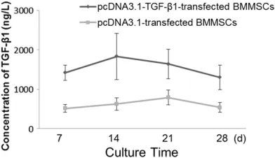
analysis of samples collected from the media at 7, 14, 21 and 28 days. The cells transduced with TGF-β1 synthesized 1419 ± 193, 1832 ± 588, 1640 ± 377 and 1300 ± 310 ng/l of active TGF-β1 at 7, 14, 21 and 28 days, respec
-tively. The maximum concentration of active TGF-β1 detected in the media was at 14 days
and gradually decreased with time. However,
even after 28 days, the transduced cells pro-duced significantly more TGF-β1 than the con-trol groups (Figure 2).
Adhesion between TGF-β1+BMMSCs and
Geistlich Bio-Oss®
[image:3.612.88.378.74.158.2]After 3 days of culturing, the medium was ch-anged for the first time, and we observed the
Figure 1. A. Establishment of the Alveolar Defect Model in Rabbit (φ3.3 mm). The top left corner shows soft tissues was removed. B. Geistlich Bio-Gide (left, 25×25 mm) and Geistlich Bio-Oss® (right, 0.5 g).
Figure 2. The synthesis of active TGF-β1 in trans -duced cells. The highest concentration of TGF-β1 was detected on day 14. The data are expressed as the means, and error bars represents the stan-dard deviation (SD), n = 5. The cell-conditioned me -dia were collected on the 7th, 14th, 21st and 28th day respectively, and then TGF-β1 concentrations in these media were determined by ELISA.
TGF-β1+BMMSCs (1×10⁶/ho-
le), and IFN-γ plus TNF-α (1:1,
200 ng/ml, 0,1 ml/hole).
[image:3.612.92.288.218.331.2]scattered distribution and varied morphology
of cells on the flask. After 7 days of culturing, adherent cells exhibited rod-like or short spin -dle morphology, and showed clear boundaries between cytoplasm and nuclei. In general, the
majority of cells possessed a single nucleus, and the nuclei were located near the center of
the cell expansion regions, though irregular pro-trusions were detected on the cell bodies. Most
of the cells showed an orderly arrangement along the long axis of the cell body, which was attached to the surface of the repair materials, and there was no significant difference com -pared with the control group (Figure 3). These
phenomena indicated good compatibility be- tween the repair material and the cells.
Status of animals after surgery
Within 3 days, rabbits with repaired alveolar
defects were eating less compared to the sham-operated group. After 3 days, they ate
normally compared to the sham-operated gr-
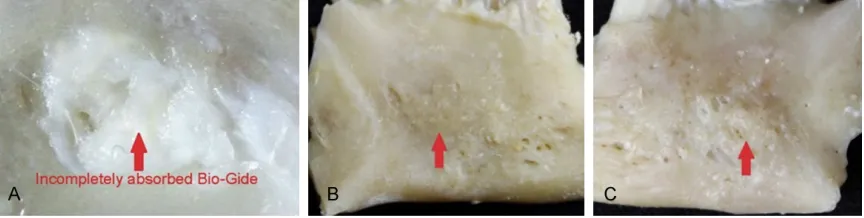
strip away (Figure 4A). There were no obvious differences in the animal specimens between experimental groups.
Sixty days after surgery, no white film was
observed on the repaired parts, which
indi-cates that the Geistlich Bio-Gide membrane has been completely absorbed. Geistlich Bio-Oss®, in small granular form, was mixed with the surrounding alveolar bone tissue and was
hard to cut and not easy to peel. There were no obvious differences in the animal specimens
between experimental groups (Figure 4B).
Ninety days after surgery, no white film was
observed attached to the repaired parts.
Geistlich Bio-Oss®, in fine granular form, had a
texture and color like the surrounding alveolar
bone and was hard to cut and could not be
stripped off. There were no obvious differences
[image:4.612.90.376.72.377.2]in the animal specimens between experimental groups (Figure 4C).
Figure 3. 2×10⁶/ml of TGF-β1+BMMSCs (3-5 passage) were seeded on the Geistlich Bio-Oss®. After 7 days of culturing, no significant differences were detected in the cells compared with the control group, indicating that the material has a good compatibility with the cells. (400×).
oup. The incision was dry, and
hemorrhagic and purulent
fluid was not detected. On
the 7th day, the sutures were removed, and wounds were
well healed. The postopera -tive masseter was much thin-ner than the contralateral
one. No significant differences were found between the ani -mals in each group.
Postoperative pathological analysis of rabbit alveolar
Thirty days after surgery, a white and tough film was
detected on the repair parts,
which indicated that the Ge-istlich Bio-Gide film was not fully absorbed. After removing the Geistlich Bio-Gide mem
-brane, we found that the Geistlich Bio-Oss® showed a granular shape, which was
similar to the original form. The clear boundary between Geistlich Bio-Oss® and alveo-lar bone tissue could be
Hard-tissue slices and Van Gieson’s Picro-Fuchsin stain
Thirty days after surgery, most Geistlich Bio-Oss® fell off during the tissue fixation and dehy -dration process so that only a little residue was
detected in the bottom of the defect parts. Therefore, it is difficult to observe the degree of ossification of the Geistlich Bio-Oss in tissue sections. Sixty days after surgery, Geistlich Bio-Oss® in each group showed a granular particle
shape, and its outer ossification and fusion was not obvious. There were no significant dif
-ferences in the degree of ossification in each group. On the 90th day, ossification was ob-served on the outer layer of Geistlich Bio-Oss®
particles in group A and C, and some of these
particles had merged with each other. In addi-tion, we detected that the newborn cells in-
creased significantly on the outer layer of Geistlich Bio-Oss® particles. In group B, more
significant ossification was detected on the outer Geistlich Bio-Oss® compared with that
on day 60, but most of the Geistlich Bio-Oss® appeared as single granular particles and
with-out obvious fusion (Figure 5). Discussion
Geistlich Bio-Oss® is a perfect material for bone repair, and in combination with mem-brane-guided bone regeneration techniques
can effectively treat alveolar bone defects and bone defects around dental implants [11-13]. After filling a bone defect with Geistlich Bio-Oss®, chemotaxis of bone cells is induced, pro
-moting proliferation, differentiation and secre
-tion of the extracellular matrix; eventually, the
graft material is replaced by new bone tissue, and the bone defect is repaired [14]. Given the
observed good biological compatibility and
guiding role in bone regeneration, Geistlich Bio-Oss® with membrane-guided bone regenera-tion techniques is a reliable combinaregenera-tion. BM-
MSCs are a group of cells with the ability of regeneration and differentiation. Because they
are easy to obtain, BMMSCs are widely studied.
BMMSCs can be directed to differentiate into a variety of tissue cells, including periodontal cells [15, 16]. The use of growth factors, such
as recombinant human platelet-derived growth
factor-BB (rhPDGF) or TGF-β1 with biocompati -ble matrices to promote tissue regeneration, represents a promising approach in the
disci-plines of periodontology and implantology [17]. TGF-β1 could affect the metabolism and growth of bone cells [18]. In these complex processes,
it is vital to create the most suitable tion environment to support tissue
regenera-tion [19]. In the present study, we transfected the TGF-β1 gene into BMMSCs. The level of TGF-β1 expression in transfected cells was
highest on day 14 and was maintained at this
level during the experimental period. The con
-tinuous high expression of the TGF-β1 protein is associated with a long protection time for
osteogenic processes. A previous study showed
that a concentration of 1-10 μg/l (1000-10,000 ng/l) of TGF-β1 can induce BMMSCs to differ -entiate into chondrocytes and thus promote
greater treatment efficiency.
Continuous inflammation, especially inflamma
-tion involving a variety of inflammatory factors,
[image:5.612.91.523.73.182.2]plays a leading the role in inducing implant and alveolar bone resorption. It is reported that
inflammatory T cells can down-regulate the runt-related transcription factor 2 (Runx-2) pathway in stem cells by secreting IFN-γ and up-regulate the TNF-α signal pathway by acti -vating caspase 8 and caspase 3, resulting in
apoptosis of transplanted cells [20, 21]. Our
previous study demonstrated that CD4+ T cells
can induce apoptosis in BMMSCs and tissue-engineered cartilage absorption by secreting
endogenous IFN-γ and TNF-α. However, TGF-β1
can induce CD4+ T cells to differentiate into
iTreg cells and then inhibit apoptosis of
BMMSCs and reduce tissue-engineered
[image:6.612.86.525.73.535.2]carti-lage absorption. Thus, TGF-β1 not only plays a
role in inducing cartilage but also promotes CD4+ T cells to differentiate into iTreg cells.
Therefore, we speculate that combining Gei-stlich Bio-Oss® and TGF-β1+BMMSCs achieves
dual functions in promoting bone guidance and controlling local inflammation. Suppressing local inflammation to provide environmental protection for the ossification of Geistlich Bio-Oss® promotes the formation and maturation
of new bone, which also has important clinical significance in preserving the height and thick
-ness of the alveolar ridg e.
In this study, we have established a rabbit
model of alveolar bone defects, and Geistlich Bio-Oss® with and without pro-inflammatory
cytokines and TGF-β1 complex were used to repair these defects. Up to 60 days after sur
-gery, no significant differences in repair were detected between groups. These observations indicate that pro-inflammatory cytokines and TGF-β1 had no significant effects on the ossifi
-cation of Geistlich Bio-Oss® at early stages and
suggest that stem cells cannot effectively pro -mote bone tissue regeneration. Ninety days
after surgery, tissue sections from the TGF-β1+BMMSCs+Geistlich Bio-Oss® group and the
Geistlich Bio-Oss® group showed bony uni- ons between nascent alveolar bone and newly
formed alveolar bone, reflecting an increased amount of new bone. However, these differenc
-es were not significant, which indicat-es that TGF-β1+BMMSCs did not effectively increase
bone formation induced by Geistlich Bio-Oss®.
In group B, pro-inflammatory cytokines signifi -cantly decreased the osteogenesis, indicating
that pro-inflammatory cytokines can cause inflammation and inhibit the osteogenesis of Geistlich Bio-Oss®. These results suggest that, compared to their in vitro effects, TGF-β1+BMMSCs did not effectively secrete TGF-β1
to inhibit inflammation in vivo.
Acknowledgements
Chichi Li and Wei Bi contributed equally to this
work. The authors gratefully acknowledge fund
-ing support from the National Natural Science Foundation of China (Grant No.81670956), the State Key Program (Grant No.14JC1490600)
and Shanghai Cooperative International Pro-
ject (Grant No.16520710400) of Shanghai Committee of Science and Technology, China.
Disclosure of conflict of interest
None.
Address correspondence to: Dr. Youcheng Yu, De- partment of Stomatology, Zhongshan Hospital of Fudan University, 180 Fenglin Road, Shanghai 200032, PR China. Tel: +86-15906493591; E-mail: 2003130823@163.com
References
[1] Corbella S, Weinstein R, Francetti L, Taschieri S, Del Fabbro M. Periodontal regeneration in aggressive periodontitis patients: a systematic review of the literature. J Investig Clin Dent 2016; [Epub ahead of print].
[2] Tolstunov L. Maxillary tuberosity block bone graft: innovative technique and case report. J Oral Maxillofac Surg 2009; 67: 1723-1729. [3] Sculean A, Nikolidakis D, Nikou G, Ivanovic A,
Chapple IL, Stavropoulos A. Biomaterials for promoting periodontal regeneration in human intrabony defects: a systematic review. Peri -odontol 2000 2015; 68: 182-216.
[4] Chrcanovic BR, Albrektsson T, Wennerberg A. Prophylactic antibiotic regimen and dental im-plant failure: a meta-analysis. J Oral Rehabil 2014; 41: 941-956.
[5] Robitaille N, Reed DN, Walters JD, Kumar PS. Periodontal and peri-implant diseases: identi-cal or fraternal infections? Mol Oral Microbiol 2016; 31: 285-301.
[6] Carlino P, Pepe V, Pollice G, Grassi FR. Immedi -ate transmucosal implant placement in fresh maxillary and mandibular molar extraction sockets: description of technique and prelimi -nary results. Minerva Stomatol 2008; 57: 471-483.
[7] Chen X, Hirt H, Li Y, Gorr SU, Aparicio C. Antimi -crobial GL13K peptide coatings killed and rup -tured the wall of streptococcus gordonii and prevented formation and growth of biofilms. PLoS One 2014; 9: e111579.
[8] Karaky AE, Sawair FA, Al-Karadsheh OA, Eimar HA, Algarugly SA, Baqain ZH. Antibiotic prophy-laxis and early dental implant failure: a quasi-random controlled clinical trial. Eur J Oral Im -plantol 2011; 4: 31-38.
[9] Koutouzis T, Catania D, Neiva K, Wallet SM. In -nate immune receptor expression in peri-im-plant tissues of patients with different suscep -tibility to periodontal diseases. J Periodontol 2013; 84: 221-229.
[10] Li C, Sun J, Gong Y, Ding X, Ruan H, Ye L, Yu Y. Transforming growth factor-beta1-induced Treg cells inhibit the absorption of tissue-engi -neered cartilage caused by endogenous IFN-gamma and TNF-alpha. Expert Opin Biol Ther 2014; 14: 573-581.
of the experimental setup. Clin Oral Implants Res 2013; 24: 329-335.
[12] Stavropoulos A, Karring T. Guided tissue regen -eration combined with a deproteinized bovine bone mineral (Bio-Oss) in the treatment of intrabony periodontal defects: 6-year results from a randomized-controlled clinical trial. J Clin Periodontol 2010; 37: 200-210.
[13] Liu HY, Zheng H, Hou XP, Zhong WJ, Ying XX, Chai SL, Ma GW. Bio-Oss((R)) for delayed os -seointegration of implants in dogs: a histologi -cal study. Br J Oral Maxillofac Surg 2014; 52: 729-734.
[14] Payer M, Lohberger B, Strunk D, Reich KM, Acham S, Jakse N. Effects of directly auto -transplanted tibial bone marrow aspirates on bone regeneration and osseointegration of dental implants. Clin Oral Implants Res 2014; 25: 468-474.
[15] Mao JJ, Giannobile WV, Helms JA, Hollister SJ, Krebsbach PH, Longaker MT, Shi S. Craniofa -cial tissue engineering by stem cells. J Dent Res 2006; 85: 966-979.
[16] Yamada Y, Ueda M, Hibi H, Nagasaka T. Trans -lational research for injectable tissue-engine-ered bone regeneration using mesenchymal stem cells and platelet-rich plasma: from basic research to clinical case study. Cell Transplant 2004; 13: 343-355.
[17] Wang Y, Zhou L, Li C, Xie H, Lu Y, Wu Y, Liu H. Bone marrow-derived cells homing for self-re -pair of periodontal tissues: a histological char -acterization and expression analysis. Int J Clin Exp Pathol 2015; 8: 12379-12389.
[18] Kaigler D, Avila G, Wisner-Lynch L, Nevins ML, Nevins M, Rasperini G, Lynch SE, Giannobile WV. Platelet-derived growth factor applica-tions in periodontal and peri-implant bone re-generation. Expert Opin Biol Ther 2011; 11: 375-385.
[19] Simon S, Smith AJ, Lumley PJ, Berdal A, Smith G, Finney S, Cooper PR. Molecular charac-terization of young and mature odontoblasts. Bone 2009; 45: 693-703.
[20] Singhatanadgit W, Salih V, Olsen I. Up-regula -tion of bone morphogenetic protein receptor IB by growth factors enhances BMP-2-induced human bone cell functions. J Cell Physiol 2006; 209: 912-922.