Original Article
Serum expression and significance of MicroRNA-30d-5p
in esophageal squamous cell carcinoma
Yanzhe Zhu1, Jiatao Liu1,2, Lulu Fan1, Fang Wang1, Lijia Bu1, Tai Ma1, Yingying Du1, Congjun Zhang1, Hua Wang1, Guoping Sun1
Departments of 1Oncology, 2Pharmacy, The First Affiliated Hospital of Anhui Medical University, Hefei 230022, Anhui Province, China
Received February 19, 2017; Accepted June 20, 2017; Epub August 1, 2017; Published August 15, 2017
Abstract: This study was aimed to assess serum microRNA-30d-5p (miR-30d-5p) expression in patients with esoph-ageal squamous cell carcinoma before and after operation, exploring its associations with clinical pathological parameters. A total of 30 esophageal cancer patients who underwent radical resection and were pathologically
confirmed with esophageal squamous cell carcinoma in the First Affiliated Hospital of Anhui Medical University,
from April to May in 2013, were enrolled, alongside 19 healthy controls. The expression levels of miRNA in serum from patients with esophageal squamous cell carcinoma before and after operation were assessed by microarrays and real-time PCR (RT-PCR). The associations of miR-30d-5p expression with clinical pathological parameters were determined. Serum hsa-miR-30d-5p levels in patients with esophageal squamous cell carcinoma (study group)
were significantly associated with the tumor depth of invasion, lymph node metastasis, tumor location and length,
histopathological type and degree of differentiation, as well as history of smoking and drinking (P<0.05). Moreover,
changes of serum miRNA levels were more prominent than that of thymidine kinase 1 (TK1). There were significant
differences in hsa-miR-30d-5p expression levels between the study and control groups (P<0.05). These results in-dicated that microRNA-30d-5p is a potential marker of esophageal squamous cell carcinoma, with high expression having a certain promoting role in the occurrence and development of esophageal cancer.
Keywords: MicroRNA-30d-5p, esophageal squamous cell carcinoma, microarray, real-time PCR, thymidine kinase 1
Introduction
Esophageal cancer is a common malignant tumor and comprises two pathological types, i.e. esophageal squamous cell carcinoma and adenocarcinoma. It is the main esophageal squamous cell carcinoma in China, accounting for about 90% of all esophageal cancers [1]. Esophageal cancer has high incidence in China,
with morbidity and mortality rankingfifth and
fourth, respectively, among various malignant tumors countrywide [2]. Male and female
mor-tality and morbidity in China all rank first in the world [3]. Due to the lack of specific early symp -toms or effective tumor markers, the patients are mostly diagnosed in the middle and late stages. Although treatment methods have pro-gressed greatly in recent years, most patients still succumb to local recurrence, disease pro-gression, distant metastasis, and resistance to adjuvant therapy; moreover, patient
progno-within 1 year after operation [4], and a 5-year survival rate of only 23% [5].
MicroRNAs (miRNAs) are recently described non-coding small RNAs with approximately 22 nucleotides. They regulate the expression of target genes by promoting target mRNA degra-dation or inhibiting translation, and play impor-tant roles in cell differentiation, proliferation, and apoptosis, as well as development, metab-olism, and immune regulation [6]. Silencing of coding genes by miRNAs can be achieved by the following three ways: 1) blocking the trans-lation of mRNAs; 2) mRNA degradation (similar to RNAi); 3) induction of DNA promotormethyl-ation. Currently, thousands of miRNAs have been described in human cells, and different
human organs and tissues have specific miRNA expression profiles. Increasing evidence clearly
cell proliferation, apoptosis and differentiation [7].
Although the miRNA expression profile of the
tumor tissue is associated with carcinogenesis and patient prognosis, current detection tech-niquesare complicated and cause important trauma, limiting their application for clinical dia- gnosis. Since 2008, scientists acknowledge that miRNA shave long-term stability in the cir-culating blood, and show resistance to RNase degradation; moreover, various treatment me- thods such as boiling, repeated freezing and thawing cycles, exposure to acid-base environ-ments, and long-term preservation do not de- crease serum miRNA levels, making them mo- re suitable tumor biomarkers than proteins [8]. Compared with tissues, the circulating blood can be obtained and assessed more easily, and is convenient for clinical application. The-refore, serum miRNAs can be used as valuable tumor markers, with high clinical applicability. Materials and methods
Clinical data
A total of 30 patients pathologically diagnos- ed with esophageal squamous cell carcinoma that underwent esophagectomy for esopha-
geal cancer in the First Affiliated Hospital of Anhui Medical University in April-May 2013
were enrolled. They included 26 malesand 4 females aged from 36 to 73 years, averaging 62 years old. Meanwhile, 19 healthy individuals in the same period were enrolled as the control group, including 12 males and 7 females of 45 to 74 years (average age of 61 years). All patients received surgery, whose pathological
confirmed for esophageal squamous carcino -ma. All the included patients had not received treatments like preoperative chemotherapy or radiation therapy. This study was approved by the Ethics Committee in our hospital, and informed consent was obtained from patients themselves for sample collection.
Experimental methods
Microarrays were used to assessserum mi- RNA expression levels in patients (3 cases) with esophageal squamous cell carcinoma before and after the operation; miRNAs (hsa-miR-30d-5p and hsa-miR-483-5p) with impor-tant changes were screened.
Preliminary real-time PCR (qRT-PCR) was used to verify the expresssion of the above miRNAs
of interest (9 cases). A new miRNA (onco-miR-NA) potentially related to esophageal squa-mous cell carcinoma was selected, with induc-tive or inhibiinduc-tive roles in tumors: hsa-miR- 30d-5p.
Then, sample size was increased. qRT-PCR was used to validate the changes of candidate onco-miRNAs in preoperative and postopera-tive serum samples from 27 cases with esoph-ageal squamous cell carcinoma. Furthermore, serum TK1 detection was performed for these 27 cases before and after the operation. Serum expression levels of candidate onco-miRNAs were compared between patients wi- th esophageal squamous cell carcinoma (27 cases) and the healthy control group (19 ca- ses).
Major instruments and reagents
Total Nucleic Acid Isolation Kit was used for
FFPE (Ambion); μParafloTM miRNA microfluidic chip was utilized for hybridization; laser scan-ning was performed, and data were analyzed with the Array-Pro image analysis software (Media Cybernetics).
Experimental procedure
Preparation of blood samples: 2 ml of periph-eral blood were obtained from each patient undergoing esophagectomy before and after operation, as well as from healthy controls. After centrifugation for 10 min, serum samples were collected and stored at -80°C until use. RNA extraction and quality control, and micro-array detection of miRNAs: Expression profile
chip for microRNAs (LC.Bio tech) was used to perform monochrome and real-time PCR to validate differences in the expressed genes. Serum RNA extraction: Total Nucleic Acid Isolation Kit for FFPE (Ambion, Cat. No. AM1975) was used, according to the manufac-turer’s instructions. RNA samples were stored at -80°C for later use.
Quality control of RNA samples: QC was per-formed for total RNA samples with a kit from LC.Biotech. Samples must have clear bandsas
detected by electrophoresis without
signifi-cant dispersion or smearing. Total RNA was assessed by Nanodrop; samples were
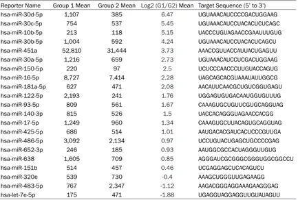
Table 1. Preoperative and postoperative microarray data for the 3 cases of esophageal cancer
Reporter Name Group 1 Mean Group 2 Mean Log2 (G1/G2) Mean Target Sequence (5’ to 3’) hsa-miR-30d-5p 1,107 385 6.47 UGUAAACAUCCCCGACUGGAAG
hsa-miR-30c-5p 754 537 5.45 UGUAAACAUCCUACACUCUCAGC
hsa-miR-10b-5p 213 118 5.15 UACCCUGUAGAACCGAAUUUGUG
hsa-miR-30b-5p 1,004 592 4.24 UGUAAACAUCCUACACUCAGCU
hsa-miR-451a 52,810 31,444 3.73 AAACCGUUACCAUUACUGAGUU
hsa-miR-30a-5p 1,216 659 2.73 UGUAAACAUCCUCGACUGGAAG
hsa-miR-150-5p 220 97 2.5 UCUCCCAACCCUUGUACCAGUG
hsa-miR-16-5p 8,727 7,414 2.28 UAGCAGCACGUAAAUAUUGGCG
hsa-miR-181a-5p 627 471 2.08 AACAUUCAACGCUGUCGGUGAGU
hsa-miR-122-5p 2,193 241 1.76 UGGAGUGUGACAAUGGUGUUUG
hsa-miR-93-5p 809 561 1.67 CAAAGUGCUGUUCGUGCAGGUAG
hsa-miR-140-3p 815 526 1.5 UACCACAGGGUAGAACCACGG
hsa-miR-17-5p 1,249 960 1.34 CAAAGUGCUUACAGUGCAGGUAG
hsa-miR-425-5p 686 514 1.01 AAUGACACGAUCACUCCCGUUGA
hsa-miR-486-5p 3,092 2,134 0.97 UCCUGUACUGAGCUGCCCCGAG
hsa-miR-652-3p 246 185 0.93 AAUGGCGCCACUAGGGUUGUG
hsa-miR-638 1,605 709 0.85 AGGGAUCGCGGGCGGGUGGCGGCCU
hsa-miR-151b 514 457 0.46 UCGAGGAGCUCACAGUCU
hsa-miR-320e 539 730 -0.4 AAAGCUGGGUUGAGAAGG
hsa-miR-483-5p 767 2,347 -1.12 AAGACGGGAGGAAAGAAGGGAG hsa-let-7e-5p 175 471 -1.88 UGAGGUAGGAGGUUGUAUAGUU
Microarray experiments: Microarray experi-ments were performed byLC Sciences, using
4-8 μg of total RNA per sample. Poly (A) tail was
added to the total RNA 3’ end by Poly (A) poly-merase, with an oligonucleotide tag attached
to the poly (A) tail for subsequent fluorescent labeling. On the microfluidic chip, each probe is composed of a chemically modified nucleotide
sequence complementary to the target microR-NA (from miRBase, http://www.mirbase.org/) and a spacer segment consisting of polyethyl-ene glycol. Hybridization was performed using
100 μL of 6×SSPE buffer containing 25% for -mamide at a hybridization temperature of 34°C. After hybridization, Cy3 was used for staining. A laser scanner (GenePix 4000B, Molecular Device) was used to collect hybrid-ization images, and the Array-Pro image analy-sis software (Media Cybernetics) was utilized for image digitalization. Background was
sub-tracted first, and LOWESS was employed for filtering (Locally-Weighted Regression) to nor -malize the signals.
Determination of serum TK1 concentration: 2 ml of peripheral blood were obtained from each patient undergoing esophagectomy before and after operation, as well as from healthy con-trols. After centrifugation for 10 min, serum
samples were collected and stored at -80°C until use.
Serum TK1 was analyaed by an ECL dot blot assay. The procedure was performed according to the manufacturer’s protocol (commercial kit; SSTK, Shenzhen, China) as described else- where.
Data analysis
All data were analyzed using the SPSS 11.0 software. Continuous data were presented as _
X ± S and categorical data as percentile. MiRNA expression levels in esophageal squa-mous cell carcinoma and their associations with clinicopathological features were assessed by paired t test. Preoperative and postopera-tive amounts of miRNA and TK1 inserumwere evaluated by Kolmogorov-Smirnov and Shapiro-Wilk tests. P<0.05 was considered statistically
significant.
Results
Microarray data for the 3 cases
cted, including hsa-miR-30d-5p (downregulat-ed) and hsa-miR-483-5p (upregulat(downregulat-ed). Preoperative and postoperative qRT-PCR of 9 cases with esophageal cancer
qRT-PCR validation was performed for the above two miRNAs of interest in 9 cases be- fore (Group 1) and after (Group 2) the opera- tion (Table 2). There was no obvious difference in mean hsa-miR-483-5p levels before and after the operation (P>0.05). In contrast, the trend of hsa-miR-30d-5p expresion before and after the operation was conistent with mic-
roarray data, with statistical significance (P= 0.047). Therefore, hsa-miR-30d-5p was select-ed for futhrer assessment as a new poten- tial onco-miR for esophageal squamous cell carcinoma.
Validation by qRT-PCR in 27 cases with esoph-ageal cancer before and after operation
Quantitative RT-PCR was used to validate the changes of hsa-miR-30d-5p in serum samples from 27 cases with esophageal squamous cell carcinoma before (Group 1) and after (Group 2) the operation (P<0.05) (Supplementary Figure 1). The qRT-PCR results were consistent with
microarray data, with statistical significance.
Associations of hsa-miR-30d-5p expression in cancer tissues with clinicopathological fea-tures of patients with esophageal squamous cell carcinoma
Paired t test was performed for the expres- sion of preoperative and postoperative hsa-miR-30d-5p as well as the histopathological parameters of the 27 cases. The results dem-onstrated that the expression ofserum hsa-miR-30d-5p in the patients with esophageal squamous cell carcinoma was correlated with clinicopathological features such as gender, age, depth of tumor invasion, lymph node metastasis, tumor location and length, patho-logical type and differentiation of tumor, to- gether with smoking and drinking history (P<0.05) (Table 3).
Associations of serum thymidine kinase 1 (TK1) with clinicopathological features in patients with esophageal squamous cell car-cinoma
Paired t test was performed to assess preop-erative/postoperative TK1 levels and clinico-2) miRNA expression levels in serum from pa-
tients with esophageal squamous cell carcino-ma in a scarcino-mall sample (3 cases). A total of 2555 miRNAs were detected, of which 143 had
sig-nal values ≥500. MiRNAs with significant
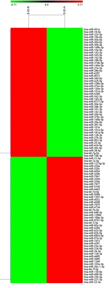
changes were screened, i.e. those with ratios of Group 1 to Group 2>2 (ratio of log2>1) or <0.5 (ratio of log2<-1) (Table 1). Then, chip data were imported into Cluster 3.0 for cluster an- alysis (Figure 1). Two miRNAs with the most
significant changes postoperatively were
[image:4.612.96.281.71.559.2]Table 2. Validation by qRT-PCR of serum miRNA levels in 9 patients before and after the operation
Assay Name Preoperative (_X ± S) Postoperative (_X± S) t P
has-miR-30d-5p 24.719±1.982 22.679±1.751 2.181 0.047 has-miR-483-5p 29.120±1.439 29.233±1.013 0.183 0.858
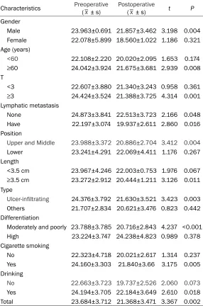
Table 3. Comparison of preoperative and postoperative hsa-miR-30d-5p in different groups
Characteristics Preoperative (_x ± s) Postoperative(_x ± s) t P
Gender
Male 23.963±0.691 21.857±3.462 3.198 0.004 Female 22.078±5.899 18.560±1.022 1.186 0.321 Age (years)
<60 22.108±2.220 20.020±2.095 1.653 0.174
≥60 24.042±3.924 21.675±3.681 2.939 0.008
T
<3 22.607±3.880 21.340±3.243 0.958 0.361
≥3 24.424±3.524 21.388±3.725 4.314 0.001
Lymphatic metastasis
None 24.873±3.841 22.513±3.723 2.166 0.048 Have 22.197±3.074 19.937±2.611 2.860 0.016 Position
Upper and Middle 23.988±3.372 20.886±2.704 3.412 0.004
Lower 23.241±4.291 22.069±4.411 1.176 0.267 Length
<3.5 cm 23.967±4.246 22.003±0.753 1.976 0.067
≥3.5 cm 23.272±2.912 20.444±1.211 3.126 0.011
Type
Ulcer-infiltrating 24.376±3.792 21.630±3.521 3.423 0.003 Others 21.707±2.834 20.621±3.476 0.823 0.442 Differentiation
Moderately and poorly 23.788±3.785 20.716±2.843 4.237 <0.001 High 23.224±3.747 24.238±4.823 0.989 0.378 Cigarette smoking
No 22.323±4.718 20.021±2.617 1.314 0.237 Yes 24.160±3.303 21.840±3.66 3.175 0.005 Drinking
No 22.663±3.723 19.737±2.526 2.060 0.073
Yes 24.194±3.705 22.184±3.649 2.610 0.018 Total 23.684±3.712 21.368±3.471 3.367 0.002
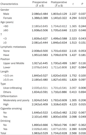
pathologic parameters in 27 patients. Interestingly, serum TK1 in patients with es- ophageal squamous cell carcinoma was associ-ated with gender, age, depth of tum- or invasion, pathological type, and degree of
dif-ferentiation, as well as histo-ry of smoking and drinking (P<0.05) (Table 4).
Comparison of preoperative and postoperative changes of hsa-miR-30d-5p and TK1 In patients with esophageal squamous cell carcinoma, preoperative and postope- rative differences between the two detection methods
were statistically significant
(P<0.05); more prominent changes were obtained for serumhsa-miR-30d-5p com-pared with TK1 (Table 5). Levels of hsa-miR-30d-5p in the study (27 cases with esophageal squamous cell carcinoma) and control (19 healthy individuals) groups
Serum expression levels of
hsa-miR-30d-5p were signifi -cantly different between the study and control groups (P<0.05). Preoperative and postoperative expression
levels were also significantly
different (t=2.916, P=0.007) (Table 6).
Follow-up deadline for all patients was January 31, 2017. There were 19/30 cases (63.3%) who died; the remaining patients were still alive (36.7%).
Discussion
In 2008, Mitchell [8] found that circulating miRNAs can be used as biomarkers of prostate cancer. They dem-onstrated that plasma miR-NAs remain highly stable after prolonged incubation at room tempera-ture and/or multiple freeze-thaw cycles. In addi-tion, some miRNAs are abundant in cells and
tissue-specific [9], thus having a biomarker
[image:5.612.92.378.196.628.2]Table 4. Comparison of serum TK1 before and after the operation in different groups
Characteristics Preoperative (_X ± S) Postoperative (_X± S) t P
Gender
Male 2.086±0.484 1.803±0.129 2.227 0.037
Female 1.388±0.389 1.140±0.322 4.294 0.023
Age (years)
<60 2.180±0.645 1.704±0.612 1.365 0.244
≥60 1.938±0.506 1.705±0.646 2.123 0.046
T
<3 1.839±0.627 1.499±0.588 2.323 0.043
≥3 2.081±0.444 1.846±0.634 1.513 0.151
Lymphatic metastasis
None 2.008±0.500 1.701±0.610 2.115 0.053
Have 1.951±0.583 1.709±0.678 1.427 0.181
Position
Upper and Middle 1.917±0.445 1.700±0.499 1.667 0.116
Lower 2.078±0.643 1.711±0.808 1.917 0.084
Length
<3.5 cm 1.840±0.527 1.620±0.619 1.752 0.100
≥3.5 cm 2.190±0.480 1.827±0.651 1.829 0.097
Type
Ulcer-infiltrating 2.035±0.511 1.701±0.541 3.057 0.006
Others 1.834±0.591 1.716±0.886 0.413 0.694
Differentiation
Moderately and poorly 1.924±0.543 1.792±0.609 1.305 0.206
High 2.242±0.409 1.318±0.625 4.123 0.015
Cigarette smoking
No 1.646±0.522 1.433±0.482 2.232 0.067
Yes 2.101±0.490 1.800±0.656 2.098 0.049
Drinking
No 1.890±0.666 1.760±0.796 0.967 0.362
Yes 2.029±0.461 1.677±0.551 2.390 0.029
Total 1.983±0.529 1.704±0.628 2.566 0.016
Table 5. Comparison of preoperative and postoperative changes between the two indicators
Normal Test _
X± S t P
PK-S PS-W
hsa-miR-30d-5p 0.200 0.253 2.316±3.573 2.927 0.007 TK1 0.200 0.535 0.278±0.563
Notes: K-S: Kolmogorov-Smirnov test; S-W: Shapiro-Wilk test.
These studies supported that circulating blood miRNAs satisfy the basic requirements of circu-lating tumor markers, with good application prospects. Currently, the relationships between tumors and circulating miRNA shave arou-sed
wide concerns. Studies ha- ve shown that miRNAs are in- volved in post-transcriptional regulation, controlling develop-ment and cell growth in various eukaryotes; therefore, they can be used as potential
biomark-ers of cancer, reflecting the
development of tumor lineage and differentiation stage [10]. Various miRNAs are consid-ered regulators of oncogene-sis, tumor suppression, cancer stem cells, and metastasis [11].
[image:6.612.90.373.577.633.2]squa-Table 6. Levels of hsa-miR-30d-5p in the study (27 cases with esophageal squamous cell carcinoma) and control (19 healthy individuals) groups
_
X± S t P
Control group 20.912±0.516
Preoperative study groupa 22.988±3.481 3.158 0.004
Postoperative study groupb 21.154±1.823 0.674 0.505
Notes: a: preoperative study group vs. control group; b: postopera-tive study group vs. control group.
mous cell carcinoma and 140 healthy controls, and 25 serum miRNAs in patients with esop- hageal squamous cell carcinoma showed an increasing trend compared with control values, as assessed by Solexa high-throughput se-
quencing. Quantitative RTPCR confirmed that 7
serum miRNAs (miR10a, miR-22, miR-140, miR-223, miR-133a and miR-127-3p) could be used as tumor markers for esophageal squa-mous cell carcinoma. Meanwhile, the 7 miRNAs could clearly distinguish patients with stage I/II esophageal squamous cell carcinoma from healthy controls. Thus, the 7 miRNAs could be used as serum tumor markers for the diagnosis of esophageal squamous cell carcinoma. In this study, microarray data showed that the- re were 2555 serum miRNAs in patients with esophageal carcinoma before and after opera-tion, with 143 miRNA sat high levels. Among them, a total of 10 miRNAs with more than 2 fold change between preoperative and postop-erative specimens were screened. A literature review revealed that two miRNA shave not been reported so far in esophageal squamous cell carcinoma: hsa-miR-483-5p and 30d-5p. Hsa-miR-30d is a precursor of hsa-miR-30d-5p, and hsa-miR-30d-5p is one of the stem-loop structures. Has-miR-30d has been widely studied, with roles in various tumors, regulating metastasis, apoptosis, proliferation, and differentiation in tumor cells [16]. Studies
have shown that hsa-miR-30d is significantly
down-regulated in central nervous system tumors [17] and lung squamous cell carcinoma compared with respective normal tissues [18]. Few studies have assessed hsa-miR-30d-5p, and demonstrated that hsa-miR-30d-5p is
sig-nificantly down-regulated in non-small cell lung
cancer (NSCLC), inhibiting the growth, motility and distribution of cells. In addition, cyclin E2 usually shows an increasing trend in NSCLC tis-sues, and may be a direct target of miR-30d-5p. This study demonstrated that the mir-
30d-5p/CCNE2 axis may contribute to pro-liferation and migration in lung cancer cells, suggesting that miR-30d-5p may serve as a potential therapeutic target for the treat-ment of NSCLC [19]. Furthermore, hsa-miR-30d-5 in the circulating blood is also relat-ed to diseases such as myocardial infarction [20], but reports assessing the relationship between hsa-miR-30d-5p in the circulating blood and tumors are scarce.
In this study, serum hsa-miR-30d-5p levels in patients with esophageal squamous cell were obviously associated with clinicopathological features such as age, gender, depth of tumor invasion, lymph node metastasis, tumor loca-tion and length, histological type and differen-tiation, TNM staging, and history of smoking and drinking. Thus, this miRNA should be con-sidered a useful marker for evaluating the prognosis of patients with esophageal cancer. Meanwhile, serum hsa-miR-30d-5p in patients with esophageal squamous cell carcinoma
was significantly higher than in healthy con
-trols, and significantly reduced in patients post -operatively; postoperative amounts were close to healthy control levels. Therefore, hsa-miR-30d-5p constitutes a potential circulating tu- mor markerin the clinical diagnosis and
moni-toring of postoperative efficacy.
Thymidine kinase 1 (TK1) is the key enzyme in the S phase of cell proliferation. Its concentra-tions are extremely low or undetectable in non-proliferating cells and healthy human serum, but increaseby 2~100 times in case of abnor-mal cell proliferation (such as in tumor cells) [21]. TK1, as a kinetic marker of abnormal
pro-liferation, has high sensitivity and specificity,
with an important prognostic value [22]. An analysis of patients with esophageal squamous cell carcinoma demonstrated that preoperative and postoperative changes of hsa-miR-30d-
5p and TK1 were statistically significant.
In-terestingly, associations of hsa-miR-30d-5p with various pathological characteristics were more pronounced than those of TK1. These
findings indicated that hsa-miR-30d-5p may
bea more sensitive reference index thanse- rum TK1 in predicting precancerous lesions, screening early malignant tumors, and perfor- ming early diagnosis.
esophageal squamous cell carcinoma were
significantly different pre- and post-operation.
In addition, hsa-miR-30d-5p expression was
significantly higher in patients with esophageal
squamous cell carcinoma than in the healthy
control group, with a significant decreasing
trend one month after the operation compared with preoperative values. Therefore, hsa-miR-30d-5p should be considered a potential tumor biomarker, with good clinical application pros-pect in the early diagnosis as well as patient prognosis in esophageal squamous cell car- cinoma.
Acknowledgements
This study was funded by National Natural Science Foundation of China (grants number. 81572430 and 81402040).
Informed consent was obtained from all indi-vidual participants included in the study. Disclosure of conflict of interest
None.
Address correspondence to: Dr. Guo-Ping Sun, De-
partment of Oncology, The First Affiliated Hospital of Anhui Medical University, 218 Jixi Road, Hefei
230022, Anhui Province, China. E-mail: sungp@ ahmu.edu.cn
References
[1] Hiyama T, Yoshihara M, Tanaka S, Chayama K. Genetic polymorphisms and esophageal can-cer risk. Int J Cancan-cer 2007; 121: 1643-1658. [2] Ferlay J, Shin HR, Bray F, Forman D, Mathers C,
Parkin DM. Estimates of worldwide burden of cancer in 2008: GLOBOCAN 2008. Int J Cancer 2010; 127: 2893-2917.
[3] Zhou MG, Wang XF, Hu JP, Li GL, Chen WQ, Zhang SW, Wan X, Wang LJ, Xiang C, Hu YS, Yang GH. [Geographical distribution of cancer mortality in China, 2004-2005]. Zhonghua Yu Fang Yi Xue Za Zhi 2010; 44: 303-308. [4] Mariette C, Balon JM, Piessen G, Fabre S, Van
Seuningen I, Triboulet JP. Pattern of recurrence following complete resection of esophageal carcinoma and factors predictive of recurrent disease. Cancer 2003; 97: 1616-1623. [5] Allum WH, Stenning SP, Bancewicz J, Clark PI,
Langley RE. Long-term results of a randomized trial of surgery with or without preoperative chemotherapy in esophageal cancer. J Clin On-col 2009; 27: 5062-5067.
[6] Schratt G. microRNAs at the synapse. Nat Rev Neurosci 2009; 10: 842-849.
[7] Shruti K, Shrey K, Vibha R. Micro RNAs: tiny se-quences with enormous potential. Biochem Biophys Res Commun 2011; 407: 445-449. [8] Mitchell PS, Parkin RK, Kroh EM, Fritz BR,
Wyman SK, Pogosova-Agadjanyan EL, Peter-son A, Noteboom J, O’Briant KC, Allen A, Lin
DW, Urban N, Drescher CW, Knudsen BS, Stire -walt DL, Gentleman R, Vessella RL, Nelson PS, Martin DB, Tewari M. Circulating microRNAs as stable blood-based markers for cancer
detec-tion. Proc Natl Acad Sci U S A 2008; 105:
10513-10518.
[9] Zen K, Zhang CY. Circulating microRNAs: a nov-el class of biomarkers to diagnose and monitor human cancers. Med Res Rev 2012; 32: 326-348.
[10] Braicu C, Calin GA, Berindan-Neagoe I. MicroR-NAs and cancer therapy-from bystanders to major players. Curr Med Chem 2013; 20: 3561-3573.
[11] George GP, Mittal RD. MicroRNAs: potential biomarkers in cancer. Indian J Clin Biochem 2010; 25: 4-14.
[12] Feber A, Xi L, Luketich JD, Pennathur A, Lan-dreneau RJ, Wu M, Swanson SJ, Godfrey TE,
Litle VR. MicroRNA expression profiles of
esophageal cancer. J Thorac Cardiovasc Surg 2008; 135: 255-260; discussion 260. [13] Mathe EA, Nguyen GH, Bowman ED, Zhao Y,
Budhu A, Schetter AJ, Braun R, Reimers M, Ku-mamoto K, Hughes D, Altorki NK, Casson AG, Liu CG, Wang XW, Yanaihara N, Hagiwara N, Dannenberg AJ, Miyashita M, Croce CM, Harris CC. MicroRNA expression in squamous cell carcinoma and adenocarcinoma of the esoph-agus: associations with survival. Clin Cancer Res 2009; 15: 6192-6200.
[14] Wijnhoven BP, Hussey DJ, Watson DI, Tsykin A, Smith CM, Michael MZ; South Australian
Oe-sophageal Research Group. MicroRNA profiling
of Barrett’s oesophagus and oesophageal ad-enocarcinoma. Br J Surg 2010; 97: 853-861. [15] Zhang C, Wang C, Chen X, Yang C, Li K, Wang J,
Dai J, Hu Z, Zhou X, Chen L, Zhang Y, Li Y, Qiu H, Xing J, Liang Z, Ren B, Yang C, Zen K, Zhang
CY. Expression profile of microRNAs in serum: a fingerprint for esophageal squamous cell car -cinoma. Clin Chem 2010; 56: 1871-1879. [16] Yang X, Zhong X, Tanyi JL, Shen J, Xu C, Gao P,
Zheng TM, DeMichele A, Zhang L. mir-30d reg-ulates multiple genes in the autophagy path-way and impairs autophagy process in human cancer cells. Biochem Biophys Res Commun 2013; 431: 617-622.
to identify circulating microRNA biomarkers. PLoS One 2013; 8: e66714.
[18] Gao W, Shen H, Liu L, Xu J, Xu J, Shu Y. MiR-21 overexpression in human primary squamous cell lung carcinoma is associated with poor patient prognosis. J Cancer Res Clin Oncol 2011; 137: 557-566.
[19] Chen D, Guo W, Qiu Z, Wang Q, Li Y, Liang L, Liu L, Huang S, Zhao Y, He X. MicroRNA-30d-5p inhibits tumour cell proliferation and motility by directly targeting CCNE2 in non-small cell lung cancer. Cancer Lett 2015; 362: 208-217. [20] Ward JA, Esa N, Pidikiti R, Freedman JE, Ke-aney JF, Tanriverdi K, Vitseva O, Ambros V, Lee R, McManus DD. Circulating cell and plasma
microRNA profiles differ between non-ST-seg -ment and ST-seg-ment-elevation myocardial in-farction. Fam Med Med Sci Res 2013; 2: 108.
[21] Zhang F, Li H, Pendleton AR, Robison JG, Mon-son KO, Murray BK, O’Neill KL. Thymidine ki-nase 1 immunoassay: a potential marker for breast cancer. Cancer Detect Prev 2001; 25: 8-15.