Original Article
Positive regulation of placentation by L-amino
acid transporter-1 (lat1) in pregnant mice
Xiaojie Wang1, Wenping Luo2,3,4, Dongmei Tan1, Hao Liang1, Qian Zhang1, Na Tian1, Ke Cao1, Yi Tan1, Jing Ma5, Ruiyu Han5
1Laboratory Animal Center, Chongqing Medical University, Chongqing, China; 2Stomatological Hospital of
Chongq-ing Medical University, ChongqChongq-ing, China; 3Chongqing Key Laboratory of Oral Diseases and Biomedical Sciences,
Chongqing, China; 4Chongqing Municipal Key Laboratory of Oral Biomedical Engineering of Higher Education,
Chongqing, China; 5Key Laboratory of Family planning and Health Birth, National Health and Family Planning
Commission, Hebei Research Institute for Family Planning, Shijiazhuang, China
Received October 10, 2016; Accepted November 27, 2016; Epub September 1, 2017; Published September 15, 2017
Abstract: Placenta plays multi-functions in embryo-uterine dialogue through facilitating gas and nutrient exchange, providing an immunological barrier between the fetus and mother and secreting hormones and growth factors to regulate pregnancy. The successful formation and development of placenta requires invasion and differentiation of trophoblast cells, and any defects would result pregnancy related diseases such as intrauterine growth retardation (IUGR), preeclampsia (PE). Lat1 (L-type amino acids transporter 1) is a major Na+ independent transporter of large
neutral amino acids, including several essential amino acids. It has been showed that amino acid was fundamental regulator on cell function and energy metabolism in early embryonic development. It has been reported that Lat1
mRNA expressed in zygote, blastocyst during the pre-implantation stages and trophoblast giant cells (TGCs) in post-implantation placenta in mouse. Little is known the role of lat1 on placentation. Our research was to explore the effects of lat1 on the placentation in mouse. The expression of lat1 was detected from day 9 to 18 of pregnancy in placenta. The effects of lat1 on placentation were assessed with inhibitor of leucine transport 2-aminobicyclo-(2, 2, 1)-haptane-2-carboxylic acid (BCH) treatment by uterine horns injection on day 8 (D8) of pregnancy. The protein of lat1 was mainly localized in the cytoplasm of maternal decidual cell, spongiotro-phoblast cell (Sp) and labyrinth (Lab). Inhibition of lat1 transportation activity by uterine horns injection with BCH in vivo results in disorder of pla-cental anatomical structure in mid-late pregnancy. These results suggest that lat1 might play an important role in mouse placentation progress.
Keywords: Placenta, L-amino acid transporter-1 (lat1), mice
Introduction
The placenta is the first formed organs during mammalian embryogenesis. It is the site of gas, nutrient exchange and waste products between the mother and the fetus. The placen-ta also forms immunological barrier to pro- tect fetus from maternal immunological rejec-tion. Besides, it can secrete hormones which can change the physiology of mother and re- gulate maternal adaptions to pregnancy [1-4]. Although there are some small differences be- tween human and mouse placenta in archi- tecture, it is thought to be quite similar in the molecular mechanism underlying placental de- velopment because they belong to discopla-
with human carcinogenesis. Over expression of lat1 is characteristic of many primary human cancers and may be involved with tumor pro-gression by changing intracellular amino acid or signaling transduction [12-14]. Well-control- led trophoblast invasion at maternal-fetal inter-face is a critical event for the normal develop-ment of placenta which was similar to tumor invasion and shared comparable gene and pro-tein regulator [15]. Lat1 has showed high ex- pression in trophoblast giant cells (TGC’s) on D10 and D12 and the highest levels of Lat1
mRNA appeared in trophoblast giant cells (TGC’s) on D8 [16]. Our previous study discov-ered that lat1 was expressed in mouse uterus during early phase placentation and promoted ectoplacental cones (EPCs) outgrowth in vitro, these data suggested that lat1 may participate in the placentation of mouse. In the present study, we explored the expression of lat1 in mouse placenta from D9 to D18; furthermore, we examined the effects of lat1 on placenta-tion in vivo.
Materials and methods
Animals and treatments
Adult Kun-Ming female mice, aged 6-8 weeks and weighing 20-25 g, were purchased from Chongqing Medical University and raised in a constant photoperiod (12 h light: 12 h dark-ness). The mice were fed with standard food and ad libidum water. All animal procedures were approved by the Ethics Committee of Chongqing Medical University. The Guidelines for the Care and Use of Animals in Research were followed. Virgin female mice were mated with fertile males of the same strain to induce pregnancy. The appearance of a vaginal plug on the next day morning was designated day 1 of pregnancy (D1). Pregnant mice were sacrificed
were sacrificed on D16 at 9-10 a.m. and pla-centa tissues were collected for the subse-quent experiments. There were at least 10 mice sacrificed in every group.
Immunocytochemistry
Placenta tissues from D9 to D18 of pregnancy mouse were fixed in 4% paraformaldehyde for 24 h, dehydrated, and then embedded in paraf-fin. Sections (5 µm) were cut, dehydrated and rehydrated with graded alcohol/water mixtures. Endogenous peroxidase activity was inhibited by 3% H2O2 for 30 minutes at room tempera-ture. Antigen was retrieved by citric acid buffer (PH 6.0, contain 1.8% 0.1 M Citric acid and 8.2% 0.1 M sodium citrate). After washes in PBS, non-specific binding was blocked in 10% normal goat serum for 1 h at room temperature followed by incubation with rabbit anti-lat1 pri-mary antibody (1:500, sc-134994, Santa Cruz Biotechnology) overnight at 4°C for 14-18 h. After three washes in PBS, the sections were incubated with goat anti-rabbit IgG (ZB-2301, Zhongshan Biotechnology, Zhongshan, China) for 60 min at room temperature and then the sections were subsequently incubated with horseradish peroxidase-conjugated goat anti-rabbit IgG (ZB-2301, Zhongshan Biotechno- logy) for 40 min at room temperature. The sec-ondary antibody was detected with 3, 3’-diami-nobenzidine solution (ZLI-9033, Zhongshan Biotechnology). For some sections, primary antibody was replaced with normal rabbit IgG (2 µg/ml IgG instead of primary antibody) to serve as negative controls.
Semiquantitative RT-PCR
GCCATCC-3’, Reverse primer: 5’-CTCTCAGCT- GTGGTGGTGAA-3’).
Western blot analysis
Total protein samples were extracted from mouse placenta with IP cell lysis buffer (Be- yotime Biotechnology, Beijing) contained 1% phenylmethylsulfonyl fluoride (PMSF, Beyotime Biotechnology, Beijing) from D9 to D18 of preg-nancy. The concentration was obtained with the Bradford assay. Protein samples (30 μg) were separated on a 12% sodium dodecyl sul-fate (SDS)-polyacrylamide gel, and then trans-ferred to polyvinylidene fluoride (PVDF) mem-brane (Hybond-C, Amersham Bio-sciences, Pis- cataway, NJ). Membranes were blocked with 5% Albumin Bovine V (BIOSHARP) in tris-buff-reaction mixture containing 4 µl MgCl2, 25
mM; 2 µl Reverse Transcription 10× Buffer; 2 µl dNTP Mixture, 10 mM; 0.5 µl Recombinant RNasin® Ribonuclease Inhibitor, 15 U AMV Re- verse Transcriptase (High Conc.), and 0.5 µg Random Primers (A3500, Promega). The PCR was performed in a total volume of 25 µl containing 12.5 µl GoTaq® Green Master Mix (M7122, Promega), 0.5 µM primers and 1 µl cDNA and was carried out over 22 cycles for
β-Actin employed as an internal control and 25 cycles for Lat1. The thermal cycling condi-tions were as follows: 94°C for 30 s, 55-59°C for 30 s, and 72°C for 30 s. The primers used in this study include Lat1 Mus (NM_011404.3) (Forward: 5’-CTTTGTACAGCGGCCTCTTC-3’, Re- verse: 5’-CAGGACATGACACCCAAGTG-3’) and
[image:3.612.90.523.71.381.2]β-Actin (Forward primer: 5’-AGCCATGTACGTA-
ered saline-tween 20 (TBST, 0.05% Tween 20) for 1.5 hr at room temperature, and incubated with rabbit lat1 (1:400) and rabbit anti-gapdh (1:1000) primary antibody at 4°C over-night for 14-18 h. After washed with TBST three times incubated with HRP-labeled goat anti-rabbit IgG (1:2000, A0208, Beyotime) for 2 h at room temperature, and then the membranes were washed with TBST for three times and 1× TBS for one time, and then were subjected to enhanced chemiluminescence.
Hematoxylin and eosin (H&E) staining
Mice with BCH treatment in vivo were sacrifi- ced on D16 and placenta tissues were fixed in 4% papraformaldehyde for 24 h, dehydrated, and then embedded in paraffin. Serial sections of 5 µm were cut, dehydrated and rehydrated with graded alcohol/water mixtures and stain- ed with hematoxylin (C0107, C0190, Beyotime Biotechnology) for 30 min and eosin (C0107, C0190, Beyotime Biotechnology) for 5 min re- spectively and then rehydrated.
Statistical analysis
Each experiment was performed at least three times. One-way analysis of variance followed by a least significant-difference test was used for
statistical comparisons among multiple groups. Significant differences were assigned at P< 0.05, and highly significant differences were assigned at P<0.01.
Results
Expression pattern of lat1 in the mouse pla-centa
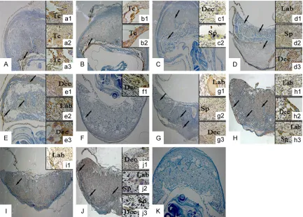
[image:4.612.92.518.73.269.2]Immunohistochemical staining was shown in Figure 1. Lat1 was mainly located in maternal decidual, spongiotrophoblast and labyrinth and continuously expressed in cytoplasm of degrad-ed decidual cells and trophoblast cells locatdegrad-ed in the placenta. On D9 and D10, lat1 highly expressed in decidual cells and trophoblast cells which were located in the maternal-fetal interface. On D11, lat1 expressed in spongio-trophoblasts, trophoblasts and degraded de- cidual cells. On D12, D13, D15, D16, and D18, lat1 expressed in the degraded decidual cells and trophoblast cells in three layers of placenta (the outer maternal layer, junctional region, innermost labyrinth) which were fully formed on D12. But on D14, lat1 was mainly expressed in the degraded decidual cells, and on D17 lat1 had the highly expression in the trophoblast cells of spongiotrophoblast.
Discussion
In the present study, we have investigated the
Lat1 mRNA and protein spatio-temporal expres-sion in mouse placentation. From D11 the spongiotrophoblast has been formed, and the three layers of placenta were fully formed on D12. On D14, the Glycogen cells of spongiotro-phoblast began to invade into decidual zone and the chorionic trophoblast began to differ-entiate into two labyrinth cell types on D15 when the maternal blood spaces expand ra- pidly, and the labyrinthine volume fraction in- creased continually until D18 accompanied with decreased junctional zone and decidua basalis [17, 18]. The pattern expression of lat1 in placenta showed coordinately with placenta development. We found that lat1 was mainly located in maternal decidual, spongiotropho-blast and labyrinth and continuously express- ed in cytoplasm of degraded decidual cells and trophoblast cells located in the placenta. Amino acid transport is critical for fetal growth, not only as nutrients but also as regulators for cell motility and function during implanta-tion and placentaimplanta-tion [19-22]. Although it was known as twenty amino acid transport systems, the knowledge about them was not understood. System A has been intensively studied in both human and mouse placenta. In human it has been demonstrated that the system A amino acid transporter activity in the microvillus
mem-Expression level of lat1 in the mouse placenta
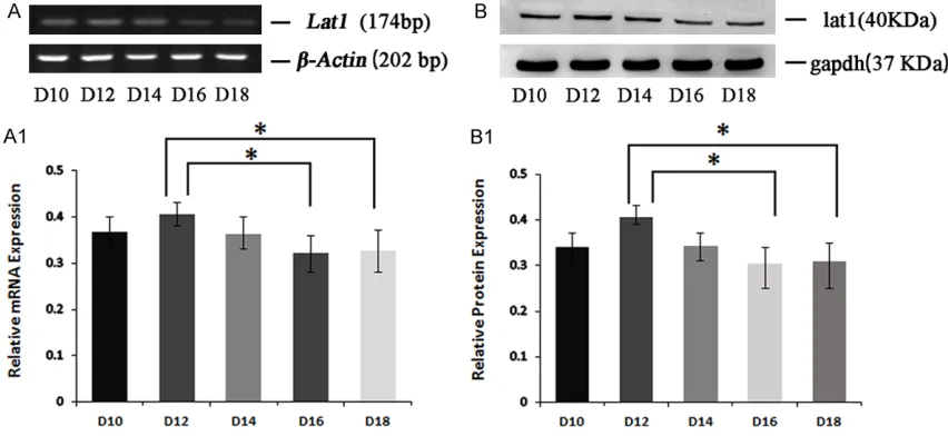
Expression of Lat1 mRNA and protein in mouse placenta from D10 to D18 of pregnancy was examined by semi-quantitative RT-PCR and Western blot (Figure 2A and 2A1). The placen-tal toplacen-tal proteins of D10, D12, D14, D16 and D18 of pregnancy were detected by Western Blot. (Figure 2B and 2B1) The peak level of
Lat1 mRNA in placenta was detected on D12, which had statistics difference compared with that on D16 and D18 (P<0.05). The similar ten-dency was present in protein levels.
Morphological effects of BCH on placentation in vivo
[image:5.612.90.526.71.231.2]To examine the effects of lat1 on placentation, mice were injected with different concentration BCH on day 8 of pregnancy by uterine horns and the placenta was collected on D16. The H&E staining showed that the morphological structure of placenta was disorder, and the placental three layers were not readily discern-ible (Figure 3). Compared with blank control group, placenta with 0.25 μg treatment show- ed maternal decidual zone and spongiotropho-blast thicker, accompanying with a larger cell density and loses of the sponge sample struc-ture, while the labyrinth became thinner as well as differentiated aberrantly. The 0.125 μg and NH4OH treatments did not interfere with the placentation process.
Figure 3. H&E staining showed morphology effects of BCH on D16 placentation after uterine horns injections on D8. A-E: 0.25 μg BCH, 0.125 μg BCH, 1 mol/L NH4OH, normal saline and normal pregnancy with any treatment, 40 times
lat1 expression was one of the most signifi- cant predictors of outcome, independent of all other variables, which suggested a key role for lat1 in the biological behavior of tumor [24]. Trophoblast giant cells share the character of highly proliferative and invasive phenotype with cancer [15]. Chrostowski et al. reported that Lat1 mRNA and protein were detected in all stages of pre-implantation mouse embryos and D8, D10 and D12 three post-implantation development stages. Lat1 was localized to in- vasive phenotype trophoblast giant cells at D8, which indicated that lat1 might be involved in trophoblast giant cells aggressive phenotype. Lat1 played a key role in mouse trophoblast invasion by mTOR pathway, and active trans-port of amino acids is required for success- ful placentation in mouse trophoblast stem in vitro [16, 25]. In this study, we found that the level of Lat1 mRNA and protein in placenta were significantly increased on D12 while de- creased on D16 and D18 (P<0.05). These re- sults coincided with Chrostowski data and also implied lat1 might assist with the function of trophoblast giant invasive phenotype before placenta maturity fully.
The placenta is composed of many trophoblast cell types: 1) the outer maternal layer, which is composed of uterus decidual cell and mater-nal vasculature bring s blood to/from implan- tation site; 2) the junctional region including Sp and TGCs that provide structural support and enable invasion to the uterus; 3) the inner-most labyrinth that is the main site of sub-stance exchange and consisted of multinucle-ated syncytiotrophoblasts that control fetal-maternal transport and sinusoidal trophoblast giant cells that have endocrine functions and act as hematopoietic signaling centers [5, 26]. Placenta mediates concentrative transport of
normal physiological activity, so we only chose BCH injection to interfere partly in uterine on D8. Furthermore, BCH was injected in mouse uterine by uterine horns which resulted in the differentiation of placental morphological structure was disturbed and three placental layers were not readily discernible accompa-nied with enlarged spongiotrophoblast thick-ness and decreased labyrinth thickthick-ness. Thus, we speculate lat1 could regulate the placenta-tion by participating in labyrinth trophoblast cell differentiation.
It has been proved that the labyrinth pheno-types are severe dysfunctional associated with these mutations in many signaling pathways such as Tsc-1, EGFL7, Notch2, Mash2 and so on [32-34]. Amino acid regulation depended on intracellular signaling pathways, such as P13K/Akt/mTOR, Wnt, and STAT3, whether the effects of lat1 on placental formation were col-laborated with these signaling factors would need further research.
Conclusion
Overall, our study provided some lines of evi-dence lat1 were actively involved in placenta-tion progress in vivo. Thus, we could speculate that inhibiting the activity of lat1 can inhibit the placentation progress and lat1 could regulate the placentation by participating in labyrinth trophoblast cell differentiation. But lat1 gene defects can result in gestational diseases such as IUGR and PE directly or indirectly? The role of lat1 in pathophysiological role in human pla-centa awaits further investigations.
Acknowledgements
Iwai K and Minato N. 4F2 (CD98) heavy chain is associated covalently with an amino acid transporter and controls intracellular traffick-ing and membrane topology of 4F2 heterodi-mer. J Biol Chem 1999; 274: 3009-3016. [11] Prasad PD, Wang H, Huang W, Kekuda R,
Rajan DP, Leibach FH and Ganapathy V. Human LAT1, a subunit of system L amino acid transporter: molecular cloning and transport function. Biochem Biophys Res Commun 1999; 255: 283-288.
[12] Kim DK, Ahn SG, Park JC, Kanai Y, Endou H and Yoon JH. Expression of L-type amino acid transporter 1 (LAT1) and 4F2 heavy chain (4F2hc) in Oral squamous cell carcinoma and its precursor lesions. Anticancer Res 2004; 24: 1671-1675.
[13] Kim DK, Kanai Y, Choi HW, Tangtrongsup S, Chairoungdua A, Babu E, Tachampa K, Anzai N, Iribe Y and Endou H. Characterization of the system L amino acid transporter in T24 human bladder carcinoma cells. Biochim Biophys Acta 2002; 1565: 112-121.
[14] Yanagida O, Kanai Y, Chairoungdua A, Kim DK, Segawa H, Nii T, Cha SH, Matsuo H, Fukasawa Y, Tani Y, Teketani Y, Uchino H, Kim JY, Inatomi J, Okayasu I, Miyamoto K, Tekeda E, Goya T and Endou H. Human L-type amino acid trans-porter 1 (LAT1): characterization of expression in tumor cell lines. Biochim Biophys Acta 2001; 1514: 291-302.
[15] Lala PK, Lee BP, Xu G and Chakraborty C. Human placental trophoblast as an in vitro model for tumor progression. Can J Physiol Pharmacol 2002; 80: 142-149.
[16] Chrostowski MK, McGonnigal BG, Stabila JP, Padbury JF. LAT1 expression in pre and post Implantation embryos and placenta. Placenta 2009; 30: 270-276.
[17] Coan PM, Ferguson-Smith AC and Burton GJ. Developmental dynamics of the definitive mouse placenta assessed by stereology. Biol Reprod 2004; 70: 1806-1813.
[18] Coan PM, Ferguson-Smith AC and Burton GJ. Ultrastructural changes in the interhaemal membrane and junctional Placental adapta-tions, fetal growth and programming 85 zone of the murine chorioallantoic placenta across gestation. J Anat 2005; 207: 783-796. [19] Gardner DK and Lane M. Amino acids and
ammonium regulate mouse embryo develop-ment in culture. Biol Reprod 1993; 48: 377-385.
[20] Devreker F, Hardy K, Van den Bergh M, Vannin AS, Emiliani S and Englert Y. Amino acids pro-mote human blastocyst development in vitro. Hum Reprod 2001; 16: 749-756.
[21] Lane M and Gardner DK. Nonessential amino acids and glutamine decrease the time of the (No.31301021), Project Supported by Program
for Innovation Team Building at Institutions of Higher Education in Chongqing in 2016 and Project Supported by Chongqing Municipal Key Laboratory of Oral Biomedical Engineering of Higher Education.
Disclosure of conflict of interest
None.
Address correspondence to: Jing Ma, Key Labora- tory of Family planning and Health Birth, National Health and Family Planning Commission, Hebei Research Institute for Family Planning, Shijiazhuang 050071, Hebei, China. Tel: +86-311-87041896; Fax: +86-311-87046164; E-mail: mj5658700@126. com
References
[1] Norwitz ER, Schust DJ and Fisher SJ. Implan- tation and the survival of early pregnancy. N Engl J Med 2001; 345: 1400-8.
[2] Cross JC, Simmons DG and Watson ED. Cho- rioallantoic morphogenesis and formation of the placental villous tree. Ann NY Acad Sci 2003; 995: 84-93.
[3] Davis J and Glasser SR. Placental changes in rats after fetectomy. Acta Anat 1968; 69: 542-608.
[4] Fowden AL, Sferruzzi-Perri AN, Coan PM, Constancia M and Burton GJ. Placental effi-ciency and adaptation: endocrine regulation. J Physical 2009; 587: 3459-3472.
[5] Rossant J and Cross JC. Placental develop-ment: lessons from mouse mutants. Nat Rev Genet 2001; 2: 538-548.
[6] Cross JC, Werb Z and Fisher SJ. Implantation and the placenta: key pieces of the develop-ment puzzle. Science 1994; 266: 1508-1518. [7] Lague MN, Detmar J, Paquet M, Boyer A,
Richards JS, Adamson SL and Booerboom D. Decidual PTEN expression is required for tro-phoblast invasion in the mouse. AM J Physiol Endocrinol Metab 2010; 299: E936-946. [8] Watson ED, Cross JC. Development of
struc-tures and transport functions in the mouse placenta. Physiology (Bethesda) 2005; 20: 180-193.
[9] Kanai Y, Segawa H, Miyamoto K, Uchino H, Takeda E and Endou H. Expression cloning and characterization of a transporter for large neutral amino acids activated by the heavy chain of 4F2 antigen (CD98). J Biol Chem 1998; 273: 23629-23632.
212.
[32] Guillemot F, Nagy A, Auerbach A, Rossant J and Joyner AL. Essential role of Mash-2 in ex-traembryonic development. Nature 1994; 371: 333-336.
[33] Gasperowicz M, Rai A and Cross JC. Spatio- temporal expression of Notch receptors and ligands in developing mouse placenta. Gene Expr Patterns 2013; 13: 249-254.
[34] Lacko LA, Massimiani M, Sones JL, Hurtado R, Salvi S, Ferrazzani S, Davisson RL, Campagnolo L and Stuhlmann H. Novel expression of EGFL7 in placental trophoblast and endothelial cells and its implication in preeclampsia. Mceh Dev 2014; 133: 163-176.
pression is highly correlated with Gleason score in prostate cancer. Mol Clin Oncol 2013; 1: 274-280.
[25] Chrostowski MK, McGonnigal BG, Stabila JP, Padbury JF. Role of the L-amino acid trans- porter-1 (LAT-1) in Mouse Trophoblast Cell Invasion. Placenta 2010; 31: 528-534. [26] Chhabra A, Lechner AJ, Ueno M, Acharya A,
Van Handel B, Wang Y, Iruela-Arispe ML, Tallquist MD and Mikkola HK. Trophoblasts regulate the placental hematopoietic niche through PDGF-B signaling. Dev Cell 2012; 22: 651-659.
[27] Cleal JK and Lewis RM. The mechanisms and regulation of placental amino acid transport to the human foetus. J Neuroendocrinol 2008; 20: 419-426.